The Layers of the Abdominal Wall
Between the skin and the parietal peritoneum lie several layers of fascia and muscle. A proper appreciation of these layers, and the direction of their fibres, is important when making surgical incisions for entry to the abdominal cavity.
1.3.1 The skin
The skin, or common integument, varies in thickness between species and bodily location. The abdominal skin is very thick (4-5 mm) in the ox but is quite delicate and thin (1-3 mm) in the other domestic species. Hair grows from the skin in all of the species but is much less in the pig. In all species there is much less hair on the ventral abdomen than elsewhere. Most of the hair of the sheep has a specific structure and is termed wool. In all species except the pig a principal function of the hair/wool is to reduce heat loss; the pig relies on a large amount of subcutaneous fat for this function.
The domestic species vary in regard to the number and distribution of the mammary glands. The mare has only two mammary glands, and these are located either side of the midline on the ventral abdomen in a prepubic position. The cow usually has four mammary glands, collectively known as the udder; it is located mainly ventral to the caudal abdomen but with its caudal part ventral to the pelvis. The udder is suspended by strong elastic tissue extending essentially from the linea alba and the symphyseal tendon.
There are seven pairs of mammary glands in the sow, although only 8-10 are usually functional depending on litter size. In this species the mammary tissue extends in the body wall from the axilla to the level of the stifle.
The udder of small ruminants comprises two glands and is situated in the inguinal region. In the bitch there are usually five pairs of mammary glands; in the cat there are generally four pairs.
1.3.2 The subcutaneous fascia
Superficial fascia: In the pig this layer is adipose over most of its area and functions as an insulating layer promoting heat retention.
However, in most other species this adipose tissue is not complete except in the inguinal region. In horses and cattle the cutaneous muscle is well developed in the superficial fascia layer and serves to twitch the skin to dislodge flies.Deep fascia: In the horse and ox the deep fascia is developed as a thick sheet of fibroelastic tissue covering most of the external abdominal oblique muscle, the ribs and the tuber coxae. This is termed the yellow elastic tunic providing support for the abdominal contents and contributing to the suspensory apparatus of the udder in the cow.
1.3.3 The rectus abdominis muscle (Figures 1.1 and 1.2)
Origin: The ventral surfaces of the sternal ribs and sternum.
Insertion: The cranial border of the pubis with the prepubic tendon. The prepubic tendon is the tendon of insertion of the two rectus abdominis muscles, although most of its fibres extend between the iliopubic eminences.
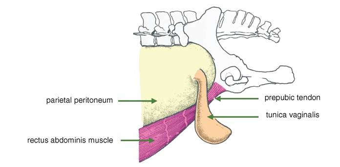
Figure 1.1 Lateral view of inguinal region of horse showing the rectus abdominis muscle. The peritoneum of the vaginal tunic is strongly reinforced by fusion with the internal spermatic fascia (derived from the transverse abdominal muscle).
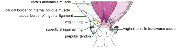
Figure 1.2 Ventral view of the inguinal region of horse showing the rectus abdominis muscle. The left side of the diagram shows the relative positions of the superficial inguinal ring and the vaginal ring. On the right side of the diagram the vaginal tunic is shown wending its way from the deep inguinal ring, through the inguinal canal and out through the superficial inguinal ring.
Structure: The left and right muscles are separated longitudinally by the linea alba, a band of fibrous tissue extending from the xiphoid cartilage to the prepubic tendon. A series of three to six transverse tendinous inscriptions cross each muscle belly, but the resulting muscle segments are not correlated with the nerve supply.
Species variations: In the ox there is wide separation of the medial borders of the rectus abdominis muscles. caudally. In the immature animal the linea alba is perforated by the umbilicus.
1.3.4 External abdominal oblique muscle (Figures 1.3-1.5)
Origin: The lateral surfaces of the ribs caudal to the fourth rib and the lumbodorsal fascia.
Insertion: The linea alba and prepubic tendon.
Structure: Most of the muscle fibres run caudoventrally. At its origin it consists of muscle fibres but towards its insertion caudoventrally it becomes a tendinous
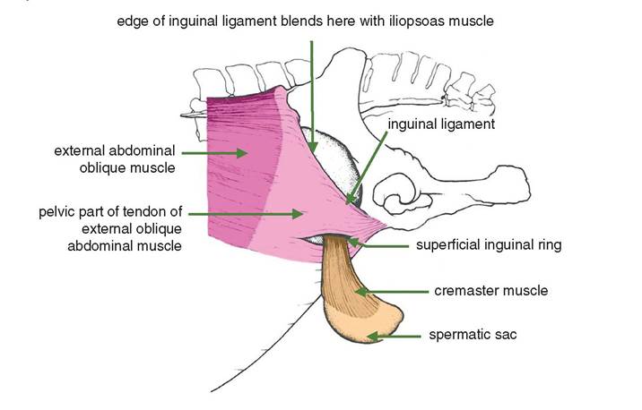
Figure 1.3 Lateral view of the inguinal region of the horse left external oblique abdominal muscle. The spermatic sac is seen emerging from the left superficial inguinal ring.The spermatic sac contains the testicle and the spermatic cord (See Figure 15.4).For a definition of the spermatic sac see Section 16.4
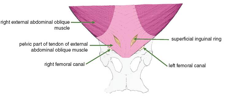
Figure 1.4 Ventral view of inguinal region of the horse showing the left and right external abdominal oblique muscles. The arrows indicate the left and right femoral canals, providing exit for the femoral arteries and veins.
aponeurosis. Towards its insertion in the prepubic tendon there is a slit in the aponeurosis; this is the superficial inguinal ring. The slit divides the tendon into an abdominal part cranially and a pelvic part caudally. The caudal edge of the pelvic part of the tendon is the inguinal ligament.
Species variations: The external abdominal oblique muscle of the dog and pig is mainly muscular almost to the dorsal edge of the rectus abdominis muscle. In
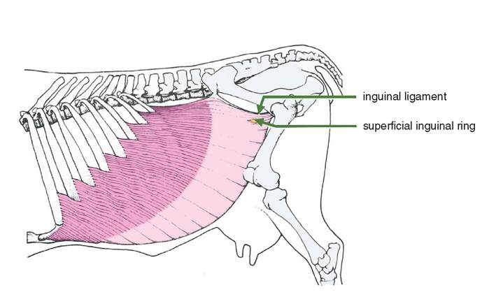
Figure 1.5 Lateral view of the abdomen of the ox showing the left external abdominal oblique muscle.
ruminants there is no origin from the Iumbodorsal fascia, but there is an insertion on the tuber coxae.
In the ox the aponeurosis of this muscle is extensive. In the horse the external abdominal oblique muscle is very large and inserts onto the femoral fascia, linea alba, tuber coxae and the prepubic tendon.1.3.5 Internal abdominal oblique muscle (Figures 1.6-1.8)
Origin: Tuber coxae and lumbodorsal fascia.
Insertion: Linea alba (except for the most caudal part), last rib and cartilages of the caudal ribs.
Structure: This is a sheet of muscle and tendon with the fibres running cranioven- trally. It is muscular at its origin and becomes tendinous ventrally. In the male a slip of the internal abdominal oblique muscle passes through the inguinal canal on the lateral aspect of the vaginal process and becomes the cremaster muscle (see Section 16.4).
Species variations: The fibres of this muscle run almost ventrally in the dog. In carnivores the tendinous portion divides to pass dorsally and ventrally to the rectus abdominis muscle in the cranial third of the abdomen; it passes only ventrally in the caudal two-thirds of the abdomen. In the ox the internal abdominal oblique is quite substantial, being the largest flank muscle in this species; its tendon passes both ventrally and dorsally to the rectus abdominis. In the horse the internal abdominal oblique muscle originates only from the tuber coxae, and its tendon passes ventrally to the rectus abdominis. See Figures 1.10a-c for a summary of the species variation of the sheath of the rectus abdominis.
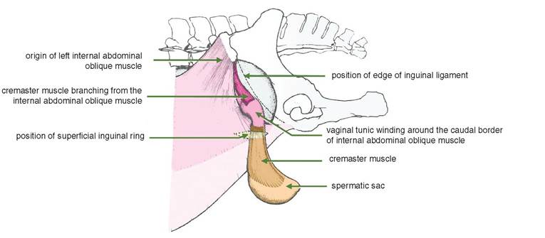
Figure 1.6 Lateral view of inguinal area of horse showing the internal abdominal oblique muscle. The left external abdominal oblique muscle has been removed although the position of the left superficial inguinal ring is shown. The mid-section of the left cremaster muscle has been excised to expose the vaginal tunic.
id="Picutre 7" class="lazyload" data-src="/files/uch_group31/uch_pgroup304/uch_uch7240/image/image007.jpg">
Figure 1.7 Ventral view of inguinal region showing internal abdominal oblique muscle.
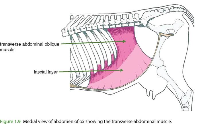
1.3.6 Transverse abdominal muscle (Figure 1.9)
Origin: The medial surfaces of the ventral parts of the caudal ribs and the deep layers of the lumbodorsal fascia.
Insertion: The linea alba.
Structure: Again this muscle is sheet-like, although its fibres run ventrally and transversely to the longitudinal axis. Caudally the muscle thins out to only a fascial layer.
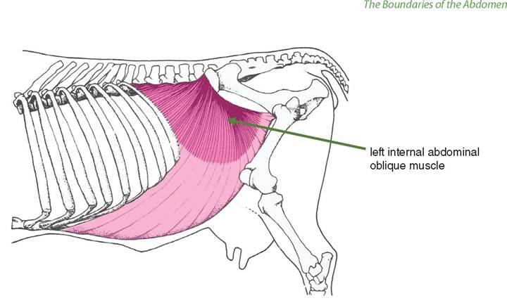
Figure 1.8 Lateral view of abdomen of ox showing left abdominal oblique muscle. The external abdominal oblique has been removed.
Species variations: In the dog the cranial two-thirds of the tendon pass dorsally to the rectus abdominis with the caudal third passing ventrally.
1.3.4 Retroperitoneal fascia
This tissue layer is equivalent to the superficial fascia but less defined. Its significance is due to its large fat content in the adult pig, fat ponies and beef breeds of cattle. Where the fascia is minimal the peritoneum is closely applied to the transverse abdominal muscle. The falciform ligament (see Sections 3.3 and 3.4) is a fold of peritoneum attached to the liver. It is a remnant of the peritoneum that contained the umbilical vein of the foetus; it attaches to the abdominal wall at the umbilicus.
1.3.5 Parietal peritoneum
This peritoneal layer lines the whole abdominal wall. It is a largely transparent and delicate layer that is reflected as mesenteries that are continuous with the visceral peritoneum that covers the abdominal viscera. The peritoneum comprises an outer layer of simple squamous epithelium called the mesothelium and is supported by a layer of loose connective tissue.
1.4