The Tissue Layers of the Testes and Scrotum (Figure 16.2)
1) The innermost layer, covering the seminiferous tubules, is the tunica albuginea. This is a tough white fibrous tissue that is continuous with the mediastinum testis, a cord of connective tissue lying centrally and longitudinally in the testes.
2) The next layer, lying over the tunica albuginea, is the visceral vaginal tunic. This fibrous layer is continuous with the visceral peritoneum through the vaginal ring. The vaginal tunic is essentially a diverticulum of the peritoneal cavity.
3) The parietal peritoneum is continued through the vaginal ring as the parietal vaginal tunic. Together with the visceral vaginal tunic it forms a flask-like sac that is reflected over the testis and epididymis distally and covering the testicular vessels and ductus deferens proximally. The testicular blood vessels are suspended within the cavity of the vaginal tunic by a fold of the parietal peritoneum called the mesor- chium; it is attached to the dorsal body wall. The mesorchium is actually a wide sheet with a thickened cranial border containing the pampiniform plexus, the testicular artery and the lymphatic vessels. Medially there is a fold in the mesorchium that contains the ductus deferens. The caudal edge of the mesorchium is continuous with the parietal layer of the vaginal tunic. It is important to realise that the cavity of the vaginal tunic is continuous with the peritoneal cavity via the vaginal ring.
4) Three fascial layers derived from the muscles of the abdominal wall are next. They are the external spermatic fascia (external abdominal oblique muscle), the cremasteric fascia and muscle (internal abdominal oblique muscle) and the internal spermatic fascia (transverse abdominal muscle). The internal spermatic fascia is closely adherent to the outer layer of the peritoneum of the vaginal process (the vaginal tunic). The entire assembly of the cremaster muscle and fascia, the internal spermatic
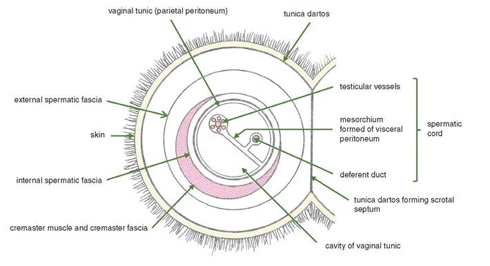
Figure 16.2 Diagrammatic cross section through the neck of the scrotum.
The vaginal tunic is strongly reinforced by the closely adherent and much thicker internal spermatic fascia. In life the only potential space is the cavity of the vaginal tunic. All other layers shown in the diagram are closely apposed to their neighbours. Note that this diagram does not show a cross section of a testis.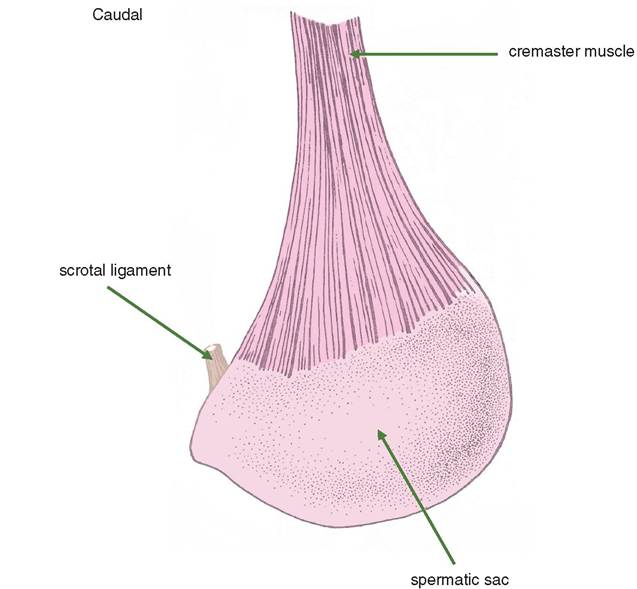
Figure 16.3 Lateral view of the right spermatic sac of the horse. The sac is hanging as it would in a standing horse in which the skin, dartos and external spermatic fascia have been dissected away during castration. The spermatic sac is a thick membrane. It consists of the vaginal tunic strongly reinforced by fusion with the thick internal spermatic fascia distally and the cremaster muscle proximally.
fascia and the vaginal tunic is called the spermatic sac (Figures 16.3 and 16.4). When a ‘closed' castration is performed, the whole sac is dissected free of the outer layers of the scrotum. The cavity of the spermatic sac is continuous with the peritoneal cavity so that an ‘open' castration gives access to the lumen of the peritoneal cavity with a risk of infection and requiring closure and control of haemorrhage with an emasculator.
5) The penultimate layer is the tunica dartos, comprising mainly fibroelastic tissue and smooth muscle. The dartos gives rise to the midline scrotal septum. The scrotal ligament attaches the dartos to the spermatic sac. The external spermatic fascia is loosely adherent to the dartos and is easily separated from the cremasteric fascia when performing a closed castration.
6) The outermost layer is the thin and elastic skin. In the horse there are a few hairs, whereas the scrotum of the sheep is covered with wool ventrally. Sebaceous and sweat glands are present, and there is a pigmented midline present in the horse.
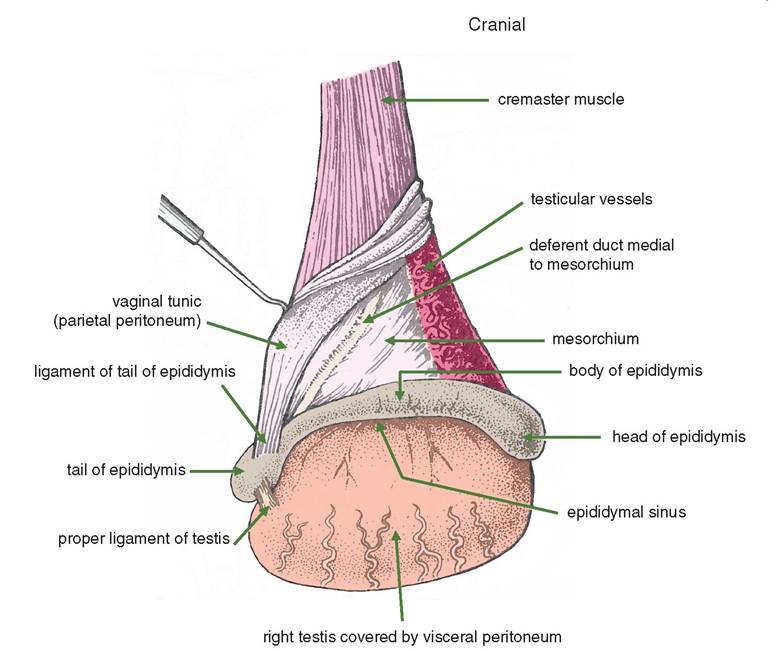
Figure 16.4 Lateral aspect of the contents of the right spermatic sac of the horse. The most ventral part of the spermatic sac as shown in Figure 16.3 has been incised so as to enter the lumen of the vaginal tunic.
16.5