Abdominal Distention
Geoffrey W. Smith
Ruminal Bloat
Ruminal bloat is uncommon in calves younger than 5 weeks of age because of the relatively undeveloped state of the neonatal rumen. Causes of ruminal bloat in calves include ruminal putrefaction, obstruction of the cardia or esophagus, and vagal indigestion (Box 20.3).
Ruminal bloat must be differentiated from abomasal bloat, which is much more common in young calves. However ruminal bloat can occur as a consequence of inadequate esophageal groove closure in young calves. The esophageal groove is a continuation of the lower esophagus, passing the medial wall of the reticulum and terminating in the reticulo-omasal orifice. The closure reflex is maintained by the suckling activity of the calf, and the groove is not properly closed if the calves
■ BOX 20.3
Causes of Abdominal Distention
Obstruction
Foreign body (hairballs in calf) Malformation (atresia coli, recti, ani) Intussusception
Volvulus, torsion, or strangulation
Uroperitoneum
Ruptured bladder (uncommon) Torn or necrotic urachus, ureter
Peritonitis
Generalized infection Devitalized bowel
Perforated gastric or intestinal ulcer Severe umbilical infection
Gas and Fluid Accumulation in Abomasum, Intestinal Tract
Intolerance to diet
Ileus
Gastric, abomasal, duodenal ulceration Necrotizing enterocolitis
Ruminal bloat
Miscellaneous
Hemoperitoneum Ruptured umbilical vessels Ruptured spleen or liver Congenital tumor
Ascites
Severe liver or renal failure
Severe hypoproteinemia
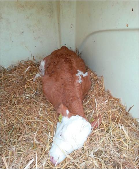
FIG. 20.2 Classic appearance of a rumen drinker calf. The calf is extremely depressed from systemic acidosis with abdominal distention. Note the front legs extended caudally, which can often be seen in these cases.
drink out of buckets.
It may also not close if milk temperatures are variable, the calves are under stress, the calves are fed too low to the ground, or milk flow rates are too high.91 Failure of esophageal groove closure leads to rumen drinking, where greater than normal amounts of milk enter the ruminoreticulum instead of the abomasum. Prolonged retention of milk in the rumen results in anaerobic bacterial fermentation and production of lactic acid,92 leading to both a ruminal and a systemic (metabolic) acidosis.93 Clinical findings in ruminal drinker calves include refusal of milk, poor suckle reflex, recurrent ruminal bloat, and splashing sounds on the left side of the distended abdomen in young calves (Fig. 20.2). Calves are frequently depressed and dehydrated and may die due to the severity of the metabolic acidosis. Treatment primarily involves rumen lavage with a stomach tube to siphon off the fermented milk remaining in the rumen, along with fluid therapy to correct the acidosis. Although ruminal drinker syndrome has been described in calves representing multiple breeds, it appears to be most common in Simmental calves from Germany and Switzerland.94 Reducing the volume of milk fed per feeding, feeding from nipples rather than buckets, and introducing calf starter to promote ruminal development help prevent the condition. Antimicrobial therapy may also help affected calves by killing the putrefactive gut flora.Bloat is occasionally observed as a complication of severe bronchopneumonia in calves as a consequence of swollen mediastinal lymph nodes compressing the esophagus or compression or inflammation of the vagus.95 Relief of rumen distention is important for return of rumen function. Chronic ruminal bloat may be relieved by placement of a rumen fistula. Correct placement of the trocar, as described by Dirksen and Garry,95 reduces the risk of inducing peritonitis. The rumen must be bloated so that it lies firmly against the body wall as the trocar is screwed into place.
The site for the trocar is shaved and scrubbed, a small skin incision is made, and the trocar is quickly and forcefully screwed into the belly wall and rumen. After removal of the stylet, the outer rim of the trocar is kept under constant outward tension so that the ruminal wall is held tightly against the parietal peritoneum by the last ridge of the screw. To fix the trocar in this position, gauze soaked in antibiotic should be wrapped around the stem of the trocar between the outer rim and the body wall.95Abomasal Ulcers
Abomasal ulcers are usually asymptomatic in young calves, but if perforation occurs, peritonitis and shock rapidly develop. It is often difficult to determine the underlying cause of abomasal ulcers; however, several possibilities have been identified, including trauma to the mucosa from the addition of coarse roughage feeds, pica resulting from enteritis, abomasal bezoars, environmental and physical stress, hyperacidity, vitamin E deficiency, lactic acidosis, mycotic infection, and low immune status associated with copper deficiency.96-98 Inconsistency of milk fed to calves (varying osmolality or total solid levels) and/ or inconsistency of feeding schedules also appear to be significant risk factors. Several bacteria, including E. coli and Sarcina and Clostridium species, have also been isolated from various calves with ulcers.99 Although no definitive infectious component has been identified in cattle, some authors have hypothesized that a bacterial component is likely in some cases.100 This could be similar to humans, in which Helicobacter pylori and gastric ulcers routinely occur together.
Four categories or ulcers are described in calves , :
Type 1: Nonperforating ulcer—The ulcer does not perforate the abomasal wall, and intraluminal hemorrhage is minimal. (Fig. 20.3, A)
Type 2: Nonperforating ulcer with severe blood loss—The ulcer does not perforate the abomasal wall but erodes a major vessel in the submucosa, resulting in severe intraluminal hemorrhage.
Type 3: Perforating ulcer (Fig. 20.3, B) with local peritonitis— The ulcer perforates the abomasal wall, and abomasal contents leak into the peritoneal cavity or omental bursa. Peritonitis is localized by fibrin deposition, and the abomasum becomes adhered to the peritoneum, omentum, or surrounding viscera.
Type 4: Perforating ulcers with diffuse peritonitis—The ulcer perforates the abomasal wall, and abomasal contents quickly leak into and spread throughout the peritoneal cavity, resulting in diffuse peritonitis.
Abomasal ulcers can be very difficult to diagnose antemortem in calves. Clinical signs are vague and include sudden death, abdominal distention, pain on abdominal palpation, expiratory grunt, drooling saliva, bruxism, and melena. Less commonly, a syndrome of chronic abdominal pain is observed after abomasal perforation.101 Absence of inflammatory changes suggests that the gut is unlikely to be perforated or necrotic. Severe hypoproteinemia is common with diffuse peritonitis, presumably because of the combination of poor colostral uptake and loss of protein into the abdominal exudate. Obtaining peritoneal fluid from normal calves is difficult; if peritonitis is suspected, collection of abdominal fluid is facilitated by locating pockets of peritoneal fluid via abdominal ultrasound.
In general, treatment is limited to calves with bleeding or deep, nonperforated ulcers. Therapy can include blood transfusions, antibiotic administration, changes to the diet, oral administration of antacids, and administration of histamine H2-receptor antagonists. Aluminum hydroxide/magnesium hydroxide—Al(θH)3∕Mg(OH)2—antacids neutralize acid in the abomasum and require three doses per day. Antacids are an inexpensive option for treatment of cattle with ulcers and
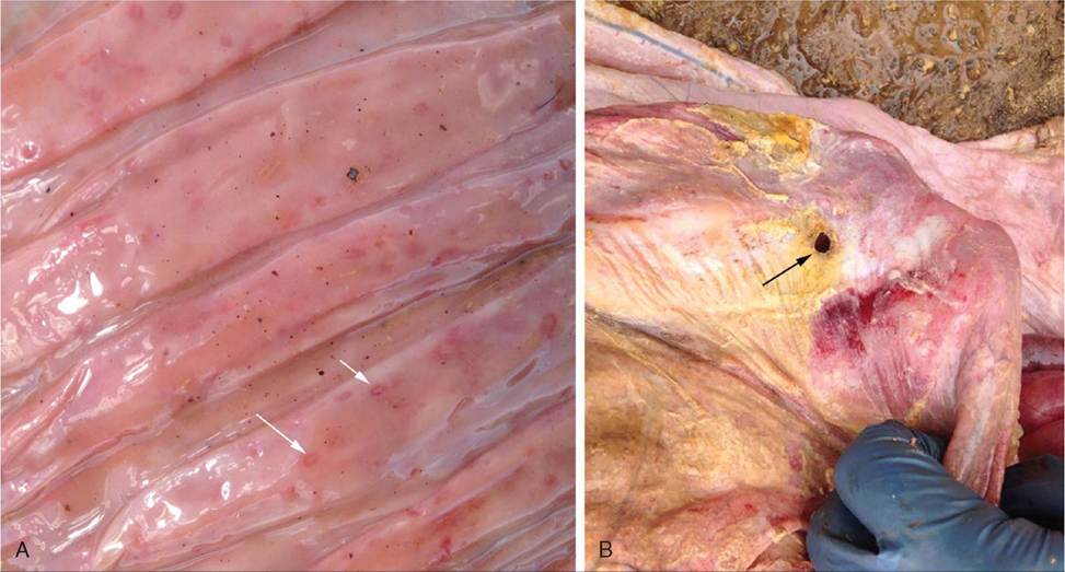
FIG. 20.3 Abomasal ulcers in calves can range from small reddish erosions (A) to perforated lesions that lead to diffuse peritonitis (B).
have a potential therapeutic advantage. Al(OH)3 directly absorbs pepsin, thus decreasing the proteolytic action of pepsin in the stomach.102 Also, Al(OH)3 and Mg(OH)2 bind bile acids, thus protecting against ulceration caused by bile reflux.102 Ahmed and colleagues found that healthy calves given 25 to 50 mL of oral antacid had a transient increase in abomasal luminal pH.103 This effect appeared to be dose related, and the extent of the acid neutralization was increased when not given with milk replacer.
Histamine H2-receptor antagonists reduce acid secretion of parietal cells by selective and competitive antagonism of histamine at H2 receptors on the parietal cells. Cimetidine and ranitidine are synthetic H2-receptor antagonists that inhibit basal as well as pentagastrin- and cholinergic-stimulated gastric acid secretion. Daily oral administration of cimetidine at 10 mg/ kg for 30 days in veal calves was found to aid healing of abomasal ulcers.104 Oral administration of cimetidine (50 to 100 mg/kg q8h) and ranitidine (10 to 50 mg/kg q8h) caused a dosedependent increase in abomasal luminal pH, and ranitidine had greater potency than cimetidine.105 Omeprazole, a proton pump inhibitor, has also been studied in calves. Once-daily oral administration of omeprazole (4 mg/kg) to calves increased the mean 24-hour luminal pH by 1.3 pH units; however, this increase in pH was less pronounced with subsequent daily treatments.106
Surgical repair of ulcers can be performed on selected cases. Calves need to be in an operable condition and have isolated ulcers. Surgery needs to be performed early in the disease process. Typically ulcers are diagnosed during a laparotomy and either they can be resected or the serosa can be inverted with a mattress suture. One study reported a survival rate of 74% following surgical repair, with survival being defined as calves living at least 3 months past surgery date or dying because of another disease.107
Abomasal Displacement
Abomasal displacement is rare in neonatal ruminants.
Clinical signs include reduced appetite, poor weight gain, recurrent tympany (left side), and diarrhea. An association of left-sided abomasal displacement with pneumonia in calves suggests that altered vagal function may be involved in the pathogenesis of the condition.108,109 Typically, left-sided abomasal displacement in calves occurs between 6 and 14 weeks of age, but younger calves may be affected. Displacement of the abomasum is diagnosed by auscultation and percussion; affected animals may have a hypochloremic metabolic alkalosis. Correction can be attempted by rolling the calf on its back or by surgery.Abomasal Bloat
A number of reports on a complex syndrome known as abomasal tympany, abomasal bloat, braxylike disease, and abomasitis have 98110111
been published. ,, Ihis syndrome in young calves is characterized by anorexia, abdominal distention (often bilateral), bloat, and often sudden death within 48 hours. In mild cases clinical signs are described as diarrhea, watery fluid in the abomasum, and depression. Hyperglycemia (10.5 to 28 mmol/L) accompanied by glucosuria (3 to 6 mmol/L) was also reported in those calves. Severely affected calves show perceptible dehydration, colic, prominent abdominal distention, diarrhea, and recumbency. Systemic acidosis, as evidenced by low blood pH, low serum bicarbonate concentration, and base deficit, was also reported for calves with abomasal bloat.112 Calves typically exhibit abdominal distention on the right side of the abdomen or potentially on both sides as the abomasum fills up with gas and occupies the majority of the abdominal cavity. At necropsy most of these calves present with abomasal tympany, forestomach and abomasal edema, hemorrhage, mucosal inflammation, erosion and necrosis, and occasionally mural emphysema. Emphysematous bullae are present in the stomach walls of these calves.111
Abomasal bloat most commonly occurs in dairy calves and seems to have a sporadic occurrence, with some farms having multiple outbreaks at times.98 Abomasal bloat has been identified on farms using conventional milk replacers, “accelerated growth” milk replacers, and both pasteurized and unpasteurized whole milk. No single diet type or feeding method emerged as a conspicuous risk factor for abomasal bloat in this survey. Other risk factors that have been reported for abomasal bloat include high-osmolality milk replacers or oral electrolyte solutions, improper mixing of milk replacers, feeding a large volume of milk in a single daily feeding, cold milk (or milk replacer), not offering water to calves, erratic feeding schedules, and FPT.98
A bacterial etiology is often mentioned in association with abomasal bloat. The most frequently incriminated bacterial pathogens are Clostridium perfringens along with Campylobacter and Sarcina species.111,113 These pathogens have also been associated with abomasal bloat in small ruminants.114,115 Additional bacterial pathogens isolated from calves affected with abomasal bloat include alpha streptococci, other Streptococcus species, and E. coli. C. perfringens is most frequently seen as type A, E, or C, producing beta toxin.112 Beta toxin damages the intestinal microvilli, mitochondria, and terminal capillaries in the mucosa. Progressive necrosis of the mucosa follows, and a large number of gram-positive bacilli invade those areas. C. perfringens type A was identified as the causative agent in a case report of 24 dairy calves dying from severe acute abomasal disease.116 In another report, Sarcina-like bacteria were reported to contribute to the development of abomasal bloat in goat kids.114 Histologic evidence of Sarcina ventriculi and Sarcina maxima was detected in the superficial mucosa, but no bacteria could be cultured from the lesions.117 Vatn and colleagues reported that evidence of Sarcina could be identified in the abomasal wall of all lambs with bloat; however, the bacteria could be isolated in only one case.115 Intraruminal inoculations of C. perfringens type A into healthy dairy calves resulted in 118
anorexia, depression, bloat, diarrhea, and death.118 Ihe authors assumed that esophageal groove dysfunction allows abnormal amounts of milk to enter the abomasum, where bacteria like C. perfringens find a suitable environment for proliferation and invasion of the abomasal mucosa, resulting in abomasitis.
Experimental induction of abomasal bloat in calves was achieved by drenching Holstein calves younger than 10 days of age with a carbohydrate mixture containing milk replacer, corn starch, and glucose mixed in water to provide a meal with excessive fermentable carbohydrate.119 The authors suggested that the syndrome of abomasal bloat in calves is multifactorial and proposed that the pathophysiology primarily involves excess fermentation of high-energy GI contents in the abomasum (from milk, milk replacer, or high-energy oral electrolyte solutions), along with the presence of fermentative enzymes (produced by bacteria), leading to gas production and bloat. This process would be accelerated by anything that slowed abomasal emptying or caused GI ileus. Ultimately the exact etiology of abomasal bloat is unknown, but it likely involves both bacteria that produce gas and something that slows down abomasal emptying.
Treatment generally involves placing the calf in dorsal recumbency and inserting a needle or catheter into the abomasum to relieve the gas.120 Attempting to deflate the bloat in a standing calf is often unrewarding, since this approach generally fails to completely drain the abomasum and carries a high risk of inducing peritonitis.120,121 The best results occur when the calf is turned upside down and the abomasum is deflated with a 14-gauge, 50-mm needle inserted into the highest point of the abdominal wall between the umbilicus and xiphoid.120 With this technique, 20 of 21 calves with abomasal tympany were successfully managed without complications. Repeated paracentesis carries a high risk of inducing peritonitis; if after paracentesis the calf's condition deteriorates or tympany recurs, a right flank laparotomy is performed to correct a possibly torsed abomasum.122 Intravenous fluids are administered to correct dehydration and electrolyte and metabolic derangements. Antibiotic therapy may also be indicated in these calves (most likely parenteral procaine penicillin or oral β-lactam antibiotics to target Clostridium bacteria).
Control of abomasal bloat problems on a dairy farm generally begin with a thorough evaluation of the nutrition program to see if any changes need to be made. This evaluation would include what type of milk or milk replacer is being fed, volume fed at each feeding, feeding schedule, temperature of milk fed to calves, how the milk replacer is mixed, how feeding equipment is sanitized, and possibly even a water analysis in some cases. Recording the total solids of the milk replacer and potentially even measuring osmolality will provide additional information about the density and mixing consistency of the milk replacer being fed. Anecdotally, focusing on controlling abomasal bloat through nutritional approaches and minimizing feeding practices that slow abomasal emptying is often much more successful than trying to control the problem by instituting C. perfringens vaccination.123 However, specific data on the efficacy of clostridial vaccines to control bloat have not been published.
Abomasal bloat is a significant problem in artificially raised lambs. Feeding systems that allow lambs to drink large quantities of milk replacer at infrequent intervals and housing lambs on litter are predisposing factors.124,125 Proliferation of Lactobacilli, E. coli, and C. perfringens has been implicated in the disease process.125,126 Fermentation of sugars contained in milk replacer produces carbon dioxide, distending the abomasum.127 Lambs may die within hours because of acute abdominal tympany compromising vascular return and respiration. Early treatment of bloated lambs with oral doses of antibiotics is sometimes an effective treatment. Addition of 0.1% formalin (37% formaldehyde) to milk replacer reduces the incidence of the condition.126
Intestinal Atresia
Intestinal atresia is the most common cause of abdominal distention of calves in the first week of life. Typically calves are born normally but develop progressive abdominal distention shortly after birth. Signs of mild colic are occasionally observed. The spiral loop of the ascending colon is usually the site of atresia.128 Other congenital abnormalities may also be present (18% of cases).128,129 Historically there was some thought that pregnancy diagnosis by palpating the amniotic sac before 40
130
days of gestation may cause colonic atresia in cattle130; however, recent studies have not supported that theory.131,132 An autosomal recessive inheritance in Holstein cattle has also been proposed.133 Surgical repair of atresia ani that can be treated with a perineal incision and rectal pull-through carries a good prognosis. However, surgical repair by resection of the distended proximal blind end and anastomosis of the proximal segment of intestine to the descending colon or use of a colostomy has a guarded to poor prognosis.128,129,134
Intussusception
Intussusception occurs most commonly in the jejunum, but the frequency of ileocecal and colon intussusceptions appears to be higher in calves than adults.135 Commonly there is a history of diarrhea. Clinical signs may include intermittent colic, absence of feces, and melena; however, these are inconsistent. The inconsistency of clinical signs and inability to perform a rectal examination make the diagnosis more difficult in calves than adults.135 Abdominal ultrasound may be useful. The prognosis following surgical correction is strongly influenced by the duration of the condition prior to correction.
Twisting of the intestinal mass around the cranial root of the mesentery is a rare event but occurs more frequently in calves than adults.135 Clinically the condition is characterized by a sudden onset of severe colic (kicking at the abdomen, dropping to the ground) that rapidly progresses (abdominal enlargement, tachycardia, tachypnea, reduced or absent fecal
■ TABLE 20.6
Common Enteropathogens in Dairy Calves (Comparative Frequency of Isolation From Diarrheic and Normal Calves Is Shown)
| Pathogens | Overall % Positive | % Positive Among Diarrheic Calves | % Positive Among Healthy Calves | p-Value | Odds Ratio | ||
| Bovine norovirus | 29.1 | 44.7 (89∕199)a | 16.3 (40/245)a | 0.042 | 2.0 (1.002-3.9)b | ||
| Cryptosporidium | 15.1 | 33.7 (67/199) | 0.0 (0/245) | 0.0007 | 173.0 (8.9-3365.1) | ||
| parvumc | |||||||
| Bovine coronavirus | 20.9 | 31.7 (63/199) | 12.2 (30/245) | 0.0034 | 2.7 (1.4-5.1) | ||
| Bovine rotavirus | 12.2 | 27.1 (54/199) | 0.0 (0/245) | 0.0025 | 79.9 (4.7-1369.5) | ||
| group A | |||||||
| Nebovirus | 0.9 | 21.6 (43/199) | 1.6 (4/245) | 0.0001 | 16.7 (4.0-68.8) | ||
| Salmonella spp. | 4.1 | 9.0 (18/199) | 0.0 (0/245) | 0.0056 | 80.6 (3.6-1803.7) | ||
| Bovine enterovirus | 20.3 | 5.0 (10/199) | 32.7 (80/245) | bgcolor=white>+ | +/- | ++++ | |
| Salmonella species | ++ | + | + | ||||
| Campylobacter faecalis | + | ||||||
| Campylobacter coli | ++ | -- | |||||
| Campylobacter jejuni | ++ | +/- | |||||
| Clostridium perfringens | |||||||
| Type A | + | - | + | + | |||
| Type C | + | ++ | |||||
| Clostridium sordellii | + | ||||||
| Viral | |||||||
| Rotavirus | ++ | ++ | +++ | +++ | |||
| Coronavirus | +++ | + | + | +++ | |||
| Bovine virus diarrhea | + | +++ | + | ||||
| Bovine torovirus (Breda virus) | +++ | + | + | ||||
| Bovine norovirus | + | + | + | + | |||
| Nebovirus | + | + | + | ||||
| Parvovirus | +/- | + | |||||
| Astrovirus | |||||||
| Parasitic | |||||||
| Cryptosporidium species | ++++ | ++ | + | ||||
| Eimeria species | ++ | + |
-, Negative finding; +, positive finding.
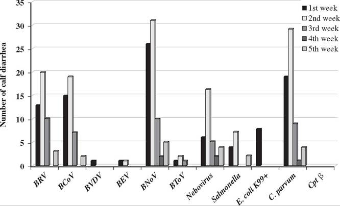
FIG. 20.4 Age distribution of diarrheic calves whose feces were positive for one or more enteric pathogens. Animals of 0 to 4 weeks of age are classified on a weekly basis after birth based on the information provided by submitting veterinarians. These data were compiled from submissions to the Iowa State University Veterinary Diagnostic Laboratory and represent approximately even numbers of beef and dairy calves. BCoV, Bovine coronavirus; BEV, bovine enterovirus; BNoV, bovine norovirus; BRV, bovine rotavirus; BVDV, bovine viral diarrhea virus; C. parvum, Cryptosporidium parvum; Cpt β, Clostridium perfringens beta toxin; E. coli K99+, Escherichia coli K99+. (From Cho Y-I et al.: Case-control study of microbiological etiology associated with calf diarrhea. Veterinary Microbiology, 166(3^):375-385, 2013.)
Continued feeding may result in more nutrients presented to the small intestine than the damaged villi can absorb164,165; excess nutrients are fermented in the large intestine, promoting bacterial overgrowth149,166 and generation of organic acids and other deleterious compounds. The osmotic effect of the unabsorbed nutrients drags water into the gut and contributes to the diarrhea.160 Marked inflammation is a feature of salmonellosis and clostridiosis. This contributes to diarrhea by increasing mucosal pore size and hydraulic pressures within the intestinal wall, by destroying absorptive cells, and by increasing prostaglandin production, which in turn stimulates secretory 149160
mechanisms within the enterocytes.149,
On an individual animal basis, diarrhea is significant because of fluid and electrolyte losses. As long as the neonate can compensate for these losses, it will remain fairly bright and continue to suck. If the losses exceed intake, systemic effects of dehydration (salt and water loss) or acidosis are seen. Fluid is lost preferentially from the vascular compartment,167,168 and cardiovascular collapse results. Acidosis has several causes, including fecal loss of bicarbonate, endogenous synthesis of L-lactic acid in response to dehydration and poor tissue perfusion, and D-lactic acid production through bacterial fermentation of undigested or malabsorbed milk within the GI tract.169-174 Acidosis contributes to the calf's malaise by increasing vascular resistance, impairing cardiac function by direct effects, and inhibiting the action of catecholamines. Esophageal groove function may be compromised in acidotic calves, promoting ruminal drinking with the consequences of further production of D-lactic acid in the rumen and subsequent ruminal acidosis.174,175
The neonate becomes depressed, loses its suck reflex, and becomes weak; if the disease progresses, recumbency and coma may develop. One cause of death is believed to be heart failure as a result of myocardial potassium imbalance due to the combined effects of potassium losses into the GI tract and the redistribution of potassium from the cells to extracellular fluid as a result of acidosis.176-178 Hypothermia will also contribute to cardiac failure. In cases of enterotoxigenic E. coli, cryptosporidia, rotavirus and coronavirus infections, correcting the fluid, electrolyte, and acid-base imbalances restores the neonate's ability to walk and suck. A residual degree of malaise may persist, which can be attributed to inflammation within the gut wall and damage to the integrity of the mucosal barriers allowing invasion of enteric microbes or their toxins. If malabsorption persists, cachexia can develop—particularly if milk continues to be withheld as part of therapy—and death from malnutrition or hypoglycemia may occur.
Salmonella are invasive and release endotoxins in the systemic circulation. Clostridia produce exotoxins. Both endotoxins and exotoxins have profound systemic effects, which are often directly responsible for malaise, microcirculatory failure, and cardiovascular collapse. Correcting fluid and electrolyte disturbances in these infections will aid the neonate but will not overcome the effects of toxemia or bacteremia.
Etiology
Bacteria
ESCHERICHIA COLI. E. coli are part of the normal flora of the bovine GI tract. Pathogenic strains of E. coli possess virulence attributes that are involved in the pathogenesis of disease. Virulence attributes include adhesins, enterotoxins, and cytotoxins. Pathogenic strains of E. coli may be shed by adult cattle with transmission to neonates by the fecal-oral route. Sick neonates amplify environmental contamination via prolific fecal shedding.
ENTEROTOXIGENIC E. COLI. Enterotoxigenic E. coli (ETEC) possess two virulence factors: fimbriae (pili) and enterotoxins. F5 (K99) and/or F41 fimbriae mediate adherence, and ther- molabile (LT) and thermostable (STa and STb) enterotoxins stimulate a secretory response by intestinal crypt cells. Expression of fimbriae is influenced by the pH, with expression occurring at a pH of 6.5 or greater.179 The distal small intestine is the initial site of colonization because the pH of intestinal fluid increases as it moves caudally, reaching this threshold at the ileum.151 Bicarbonate-containing oral replacement fluids may favor the proliferation of ETEC, expression of the K99 antigen, and secretion of STa.151 Oral electrolyte solutions containing acetate are recommended for treatment of ETEC diarrhea.151
Although some bovine-origin ETEC produce LT, most strains that cause diarrhea in neonatal calves produce STa heat-stable enterotoxin.180 The STa enterotoxin and F5 antigen are plasmid-mediated virulence factors. Susceptibility to ETEC is age dependent according to the binding specificity of pili antigens to immature enterocytes.181 Disease is typically observed in calves younger than 3 days of age, but concurrent infection with rotavirus may extend this window to 7 to 14 days of age.182,183 Intestinal cells of calves older than 2 days of age acquire natural resistance to F5 adhesion.181 Despite this, F5-positive E. coli have been isolated from healthy 4- to 12-week-old calves, and F5-positive ETEC are shed in feces for several weeks following experimental infection of newborn calves.184 A recent worldwide study has shown that the prevalence of F5-positive ETEC appears to be decreasing.185
ATTACHING AND EFFACING E. COLI AND SHIGA TOXINPRODUCING E. COLI. Attaching and effacing E. coli (AEEC) and Shiga toxin-producing E. coli (STEC) have also been identified as causes of diarrhea and dysentery in calves.186,187 Disease is mediated by cytotoxic damage to the intestinal mucosa. Lesions may be observed in the ileum, cecum, and descending colon.188 AEEC (verotoxin- or HeLa toxinproducing) induce a mucohemorrhagic colitis, with petechial or ecchymotic hemorrhages in the wall of the colon and rectum.189-191 E. coli that carry this toxin often belong to O serogroups 5, 26, 111, 118, and 145.190,192 Naturally occurring outbreaks have been reported in 2-day-old to 4-week-old calves.193 Diarrhea and enteritis has been associated with naturally occurring E. coli 0157:H7 in 1- to 3-week-old calves in Argentina, the United Kingdom, and Korea.194-196 The most common clinical sign is diarrhea, but dysentery, dullness, reluctance to move, weight loss, anorexia, abdominal pain manifested by bruxism, and dehydration are seen in some cases. The feces of affected calves can appear normal, but more commonly feces are watery yellow with blood.
STEC serotypes associated with dysentery in calves include O5:H-, O26:H11, O111:H-, and O113:H21197; these serotypes may produce Shiga toxins, those that are immunologically similar to the Shiga toxin produced by Shigella dysenteriae (STx1) and those that are immunologically distinct from S. dysenteriae Shiga toxin (STx2).198 Bovine STEC produce either STx1 or STx2, or both.199 AEEC that cause disease and do not produce enterotoxins or Shiga toxin is referred to as enteropathogenic E. coli (EPEC).
The prevalence of AEEC and STEC in calves and the incidence of disease caused by these strains are not clearly defined, as most diagnostic laboratories do not routinely screen for AEEC and STEC. A recent meta-analysis investigated the prevalence of STEC in 8053 isolates from calves in 19 countries included in 61 publications. The average prevalence of STEC isolated from healthy animals was 19.4% and from diseased animals was 18.2%. Moreover there was a significant decrease of STEC from pre-1990 to the present.185 Similarly, in a study aimed at determining the clinical significance and prevalence of AEEC in Swiss cattle, fecal swabs of 93 cattle from two farms with calf diarrhea and of 54 cattle from two similar farms without clinical problems were screened for AEEC by PCR and colony-blot hybridization. On average, 21% of all cows were positive for AEEC by PCR, with no differences between farms with and without diarrhea problems. By contrast, AEEC were detected by PCR in 60% of animals younger than 2 years from farms with diarrhea problems, whereas only 32% of comparable control animals from farms without clinical problems had AEEC.
The relevance of AEEC and STEC has increased in recent years due to the zoonotic potential of the organisms. Cattle are a major reservoir of STEC and enterohemorrhagic E. coli (EHEC), including E. coli 0157:H7, which is the EHEC serotype most often associated with hemorrhagic colitis and hemolytic uremic syndrome in humans.196,200
SALMONELLA. There are more than 2600 reported serotypes of Salmonella, yet fewer than 2% of these account for approximately 80% of the disease reported in livestock.201 In cattle, more than 95% of salmonella associated with disease is in serogroups B, C, D, and E. Salmonella induces a wide spectrum of disease in cattle of all ages, ranging from inapparent subclini- cal infections to acute fulminant bacteremia, endotoxemia, and death. The variable manifestations of disease reflect the tissue trophisms of different Salmonella serotypes and the influence of challenge dose and host immunity. Common clinical signs associated with “salmonellosis” include fever, diarrhea, anorexia, depressed mentation, and dehydration. Many of the clinical signs are associated with endotoxemia. Systemic signs of endotoxemia include fever, tachypnea, tachycardia, scleral injection, leukopenia or leukocytosis, and weakness. Some serotypes, particularly Salmonella typhimurium, have a tendency to induce severe inflammation of the bowel mucosa, resulting in dysentery and passage of fibrin and mucosal casts. Fluid, electrolyte, and protein loss may progress rapidly and become life threatening if not corrected. With severe disease, animals rapidly become emaciated due to the catabolic state induced by release of tumor necrosis factor (TNF)-α. Sequelae occasionally observed following invasive Salmonella infections in neonates include septic osteoarthritis and meningitis.
Immunity to Salmonella changes rapidly during the first 3 months of life. At 2 weeks of age the LD50 for some virulent strains is 105,202 at 6 to 7 weeks is 107, and at 12 to 14 weeks is 1010.203 In contrast, administration of 1010 Salmonella to 24- to 28-week-old calves failed to induce clinical signs of disease.203 The numbers cited reflect the influence of age on immunity but should not be interpreted as absolute. Different age predilections, manifestations of disease, and virulence are observed between Salmonella serotypes and between different strains of the same serotype.204,205 Although adults may serve as carriers and a source of infection of S. dublin infection for neonates, disease in adults is less common in mature cattle compared to calves. In contrast, S. typhimurium tends to manifest disease in an epidemic manner, causing illness in all age-groups. Environmental stressors such as heat, cold, and inadequate nutrition often play a role in herd disease outbreaks via compromising host immunity. Environmental conditions also influence the dynamics of Salmonella in the environment and subsequently the challenge dose encountered. Salmonella may proliferate in manure packs when they become wet; warmth promotes this process.
Calves on endemically infected farms may be infected in utero.206 Salmonella challenge frequently occurs during the first 24 hours of life.207 Exposure may occur via contaminated colostrum or milk, surface contamination of teats and udder, personnel, equipment, or the environment. Chronically infected carriers may shed 2.5 ? 108 Salmonella in milk per day (25 kg of milk containing 105 Salmonella/mL).208 Feeding utensils and personnel often play a significant role in transmitting Salmonella between calves.209 Salmonella infects the salivary glands and is shed in saliva and nasal secretions.210,211 Adequate cleaning and disinfection of feeding and medicating utensils is necessary to remove Salmonella contamination. Salmonella is sensitive to most disinfectants, but removal of contaminating organic debris is imperative, as the activity of disinfectants is reduced by the presence of organic matter.212
Flys, birds, and rodents may also play a role in disseminating Salmonella in feed, the environment, and between calves.213,214
CLOSTRIDIA. Although Clostridia are not commonly considered a major pathogen causing NCD, they are recognized as a cause of enteritis and abomasitis. In contrast to the other enteric pathogens, the disease tends to be more sporadic. Prerequisites for disease include the presence of the organism in the GI tract, sufficient carbohydrate or protein nutrients to support bacterial growth, and reduced GI motility that allows segmental overgrowth of bacteria within the GI tract.215
Clinical disease is associated with rapid bacterial overgrowth within the GI tract and subsequent exotoxin release. Passage of soluble carbohydrates or protein into the small intestine may induce rapid replication of Clostridia and elaboration of toxins from the vegetative state. Neonates are more susceptible due to the presence of trypsin inhibitors, which attenuate the digestion of clostridial exotoxins, in colostrum and milk for up to the first 10 to 21 days after parturition. Disease can occur in older ruminants if bacterial or plant-derived trypsin inhibitors are present in the feed.215
Management practices that increase the risk of clostridial- associated enteritis and abomasitis in beef calves include calf separation or environmental conditions that delay or interrupt normal nursing patterns.215 Poor milk hygiene, intermittent feeding of large volumes of milk, and milk replacers with higher carbohydrate or protein concentrations contribute to increased risk in dairy calves. Variables associated with delayed abomasal emptying include feeding large volumes in single feedings, feeding with an esophageal tube, high caloric content, and 123215
high osmolality milk.123,215
C. perfringens (see also Chapter 32) is the most important cause of clostridial enteric disease in calves. Although limited tissue invasion by C. perfringens does occur, most local and systemic lesions result from the effects of potent exotoxins. Some types of C. perfringens (mainly type A) are consistently recovered from the intestinal tracts of animals and from the environment, whereas others (types B, C, D, and E) are less common in the intestinal tracts of animals and can occasionally be found in the environment in areas where disease produced by these organisms is enzootic.216 All five genotypes produce alpha toxin; type B also produces beta and epsilon toxin, type C produces beta toxin, type D produces epsilon toxin, and type E produces iota toxin. Multiplex PCR is used to identify C. perfringens genotypes from anaerobic culture of samples.
C. perfringens type A has been associated with acute hemorrhagic abomasitis in neonatal calves. Clinical signs include acute abdominal distention, colic, depression, and sudden death. Onset of clinical signs is rapid; affected animals become anorexic, depressed, or restless. Signs of abdominal discomfort are observed in approximately half of the cases and include treading on the spot and kicking at the abdomen. On physical examination, splashing and metallic sounds are heard on succussion of the distended abdomen; passage of a stomach tube fails to relieve the distention. Fecal output is reduced, and melena may be observed. Gross pathology may include abomasal ulcers, abomasitis, and abomasal tympany.112,118 Trace mineral deficiencies of copper and/or selenium may also be involved in the pathogenesis of the condition.217 A decreased prevalence of abomasal tympany and ulceration was reported in neonatal calves from herds having a history of these problems following implementation of a C. perfringens vaccination program.217,218 Enterotoxemia caused by C. perfringens type A has been described in 2- to 4-month-old calves, with the condition observed more often in beef than dairy calves.219 The disease is characterized by a high case fatality rate, sudden deaths, lesions of necrotic and hemorrhagic enteritis of the small intestine, and, most often, an absence of other clinical signs.220
C. perfringens type B is not commonly associated with neonatal diarrhea in calves. C. perfringens type C infections are most frequently observed in neonates younger than 10 days of age, reflecting the trypsin inhibitors attenuating the digestion of beta toxin.215,221 Vigorous, healthy calves develop hemorrhagic, necrotic enteritis and enterotoxemia, often accompanied by evidence of abdominal pain and neurologic signs that may include frenzied bellowing, aimless running, tetany, and opisthotonus. Death may be peracute, occasionally without other clinical signs, but may also follow a clinical course of several days.
Clostridium difficile has recently emerged as a pathogen in both human and veterinary medicine. Toxins released cause epithelial cell death and activation of the enteric nervous system, resulting in a malabsorptive and secretory diarrhea.222 The role of C. difficile as a primary pathogen has not been established, as it has been detected in both healthy and diarrheic calves223 and experimental infection has not induced disease.224
CAMPYLOBACTER SPECIES. The clinical significance of Campylobacter spp. in calf scours is inconclusive. Campylobacter spp. are part of the normal intestinal flora. Experimental challenge studies have demonstrated the capacity of Campylobacter jejuni to cause enteritis in calves., However, there is a paucity of convincing reports that demonstrate a causal association in naturally occurring cases.228
Viruses
Intestinal viruses multiply within enterocytes. As the epithelial cells are destroyed, villous atrophy develops. The various agents cannot be readily separated on clinical grounds. Diarrhea can vary in severity from soft to watery feces.
ROTAVIRUS. Rotaviruses are the most common cause of neonatal diarrhea in calves.229,230 Affected calves are generally 5 days to 2 weeks of age, although disease can occur at 24 hours, particularly in colostrum-deprived calves (see Fig. 20.4).231,232 This age predilection is thought to exist because many cows secrete antirotavirus antibody in their colostrum, which confers local protection against rotavirus attack until antibody levels in milk decline 48 to 72 hours postpartum.233,234 Resistance to infection is not age dependent, but age-dependent resistance to clinical disease has been demonstrated.235 Age restriction may be related to acquired immunity, as neutralizing antibodies increase with age and virus exposure. In addition, the expression of intestinal mucins and the rate of epithelial cell replacement and fluid absorption are also age dependent and have been shown to affect rotavirus infection and disease expression.236
Mature villous enterocytes of the small intestine are the main target for rotavirus.159 Infected enterocytes are rapidly shed and replaced with immature squamous and cuboidal cells from the crypts, resulting in villous blunting.159,237 Intestinal secretions are increased due to the compensatory hyperplasia of crypt cells and enterotoxigenic activity of the viral nonstructural protein NSP4.162,163 Both increased secretory load and impaired absorption due to villus hypoplasia contribute to the diarrhea. It is thought that virulent strains replicate more quickly and infect a larger area of epithelium. Difference in rotavirus replication rates in the gut and age-dependent differences in the rate of enterocyte loss and natural replacement rate may explain the differences in clinical outcome. Concurrent infection with ETEC has also been shown to cause clinical signs at a later age than a single infection of either agent alone.238
Rotavirus of calves, lambs, kids, pigs, foals, mice, and children are morphologically identical. They are classified by the antigenic properties and/or sequence of the genes encoding the viral capsid proteins. Viral protein (VP) 6 is used to separate them into nine antigenically distinct serogroups, A to FI. Rotaviruses from serogroups A, B, and C have been isolated from cattle, and serogroup A is the most common cause of diarrhea in calves. Group B rotaviruses have been isolated from calves and adult cattle, but there is less information regarding their significance and prevalence in cattle.239-243 Group
0.0 O Jr 0 Jl
B rotaviruses are more common in lambs than calves.244 Group C rotaviruses have been isolated only from adult cattle.242 Each serogroup may be classified further by G-types (glycoprotein) and P-types (protease-sensitive protein), which correlate to the serotype and/or genotype of capsid proteins VP7 and VP4, respectively.245 A range of both serotypic and genotypic diversity and virulence has been reported within serogroup A.235,246-248 Rotavirus is shed in the feces of infected animals, and transmission is primarily through the fecal-oral route. Clinical signs occur 1 to 3 days after infection and last for 5 to 9 days. Virus excretion commences with the onset of clinical signs and continues for 3 to 7 days.235,249 Adult cows can be subclinically infected and intermittently shed the virus during pregnancy and especially at parturition.250-252 It is likely that this is the most common source of infection, with carrier cows infecting their calves and then these calves infecting other calves.253 Calves from carrier cows have a significantly higher risk of clinical disease, and the birth of calves from known carrier cows have been associated with the beginning of an outbreak. Recovered calves can become reinfected and shed virus.254
The environment may be an important source of infection. Rotaviruses can survive in fresh water for more than 2 weeks at 23o C and for months in water or soil at less than 5o C.255 They are also stable in feces and effluent for up to 9 months and therefore are likely to remain in calving areas from year 256
to year.256
CORONAVIRUS. BCV commonly causes diarrhea in calves 5 days to 1 month of age.138,143,257,258 Disease can occur within 24 hours in colostrum-deprived calves and has also been recorded in calves up to 5 months of age.259 Respiratory infections are common in older calves and may be important in the epizootiology of enteritis.259
Calves may be infected with coronavirus by the oral or respiratory route.249 Fecal shedding commences 3 days after infection and persists for up to a week; nasal shedding can be detected 2 days after infection and persists for 2 weeks. Once infected, calves initially excrete high levels of virus and are potent sources of contamination. Infection persists for weeks in apparently recovered calves, and they excrete low levels of virus for weeks.260 Subclinical infection is common. Disease is more common in the winter months, and coronavirus survives in the environment from year to year.
Calves may be infected by virus shed by persistently infected cows.261 Coronavirus has been detected in the feces of more than 70% of clinically normal cows.252 The rate of virus excre- 250262 tion increases at parturition and in the winter months.250,262 Calves born to carrier animals are at a significantly increased risk of developing diarrhea.250
The pathology of coronavirus is often more severe than rotavirus, resulting in a mucohemorrhagic enterocolitis. The virus infects both the small and the large intestine. In the spiral colon there is widespread destruction of the cells of the colonic ridges.156,158 Virus replication occurs in the surface epithelium, especially in the distal half of the villi, resulting in stunting and fusion of the villi. Immature cells replace epithelial cells, and in severe infection there can be areas of complete desquamation. Intestinal secretions continue, and absorption is impaired by reduced surface area. Undigested lactose accumulates in the intestinal lumen, often resulting in a secondary bacterial overgrowth, fermentation, lactate production, and an osmotic imbalance that draws fluid into the intestinal lumen. Most infections are self-limiting because the virus rarely attacks crypt epithelial cells.261 In response to infection, the mitotic rate of crypt cells increases, producing immature cells that are more resistant to virus infection and that migrate up the villi to replace the damaged cells.
In experimental challenge studies, diarrhea develops 48 hours after infection. Calves are initially depressed and anorexic for the acute phase and may become dehydrated and pyrexic in a severe infection.261 Severe infections can result in death due to dehydration, acidosis, shock, and cardiac failure. Respiratory signs are generally mild. Rhinitis, sneezing, and coughing may occur. Lesions may be found in the lungs but clinical signs of pneumonia are rare, except when secondary infection occurs.
BOVINE VIRAL DIARRHEA VIRUS. BVDV occasionally causes diarrhea and thrombocytopenia in young calves outside the confines of the persistently infected disease model.145,146 Colostral antibodies generally protect young calves from BVDV infection, but disease may occur due to FPT or the introduction of novel BVDV strains with new cattle or viral mutation in persistently infected homegrown cattle. BVDV is also thought to exacerbate infections due to other pathogens.263 It has also been implicated in necrotic enteritis, an acute enteritis of 7- to 12-week-old beef calves reported in the UK.264 Affected calves usually show oral ulcerations, particularly on the hard and soft palate. The buccal papillae are often blunted, and the tips may be ulcerated.265 Some variants of the virus produce intestinal bleeding, petechiation, ecchymosis, or prolonged bleeding from venipuncture sites secondary to thrombocytopenia.145,266-268 Hematologic findings often include leucopenia and thrombocytopenia. The disease must be differentiated from other causes of enteritis that are complicated by bovine papular stomatitis infection. Bovine papular stomatitis (see Chapter 32) is common in neonatal calves. It produces oral lesions that are hyperemic and red, with a central white area of necrosis and often a raised rim of proliferating epithelial cells. These lesions often involve the mucosa around the molars. They are usually of little consequence and their importance lies in the fact that they may be confused with BVDV One feature that helps identify BVDV ulcers is that they lack the zones of epithelial proliferation seen in bovine papular stomatitis.
BOVINE TOROVIRUS (BREDA VIRUS). Bovine torovirus has been detected worldwide269-272 and has recently been implicated as an important cause of calf diarrhea.141,142 Initially known as Breda virus, it is part of the Coronaviridae family. It has been relatively infrequently reported because it is difficult to recognize by electron microscopy (EM) and it cannot as yet be grown in cell culture, which has precluded the development of routine immunospecific diagnostic tests.141 Laboratory studies using PCR have implicated it as the sole pathogen isolated in 25% to 30% of fecal samples from calves with diarrhea younger than 6 weeks of age.141,142 It is also found in the feces and nasal secretions of asymptomatic animals,141,143 implicating that the epizootiology is likely to be similar to that of rotavirus and coronavirus, with asymptomatic carriers acting as a reservoir of infection within a herd.250 It is mainly a disease of calves younger than 3 weeks of age, with diarrhea commencing as early as 1 to 3 days after birth,271,272 but clinical signs have been observed up to 10 months of age.142,273 Clinically it produces mild to moderate diarrhea in calves under both experimental and field conditions.272,274 The virus infects the distal half of the ileum, jejunum, and colon, resulting in necrosis of the crypts and villous enterocytes.275,276 Clinical signs develop 24 to 72 hours after experimental infection.272 It has also been isolated from the respiratory tract of cattle and associated with respiratory signs in calves at 1 month and 4 to 6 months of age.277
OTHER VIRUSES. Norovirus, nebovirus, torovirus, astrovirus, enterovirus, kobuvirus, adenovirus, parvovirus, and picobirna- 278283 virus have all been associated with neonatal calf diarrhea.2'8-283 The pathogenicity and contribution of these viruses to field outbreaks is uncertain. In a larger case control study, nebovirus and norovirus were isolated in higher frequency from diarrheic calves, suggesting they may play a role in neonatal scours in the United States.284 The lack of readily available diagnostic assays for detection of these viruses in veterinary diagnostic laboratories limits their routine detection. As with rotavirus and coronavirus, the different viruses have been isolated from both healthy and diseased calves. Some of the viruses such as norovirus and nebovirus do induce lesions in experimental infections of gnotobiotic calves. Others such as kobuvirus, astrovirus, enterovirus, and torovirus have not been demonstrated to or inconsistently induce pathology in gnotobiotic
283 y p gy g
calves.283
Protozoa
CRYPTOSPORIDIUM. Four species of Cryptosporidium have been identified in cattle: Cryptosporidiumparvum, Cryptosporidium andersoni, Cryptosporidium bovis, and Cryptosporidium ryanaeC8 Conventional understanding has been that C. parvum is mostly found in preweaned calves, and numerous studies have shown 286289 a significant correlation between its occurrence and diarrhea.286-289 C. andersoni is seen in low prevalence in asymptomatic adult cattle, and the two other common species, C. bovis and C. ryanae, usually infect weaned calves and yearlings and are 287288290
generally nonsymptomatic.287,288,290
However, similar to the situation in humans, it is becoming apparent that the distribution of Cryptosporidium spp. varies between geographic regions.291 In China, where preweaned calves are mostly infected with nonpathogenic Cryptosporidium spp., C. parvum is starting to appear in dairy calves associated with concentrated animal feeding operations.292 However, in Sweden, studies have shown C. bovis to be the dominant species 293294
in dairy calves, even in this age-group,, and in suckler beef herds all four species were detected in calves younger than 3 months of age.295
C. parvum was initially thought to be a single species that affected both humans and a broad range of animals. Later, it was recognized that two distinct genotypes were present: type 1, which was found in human sources; and type 2, which was considered to be zoonotic and could be isolated from bovines and other farm animals such as sheep and goats.296,297 The most recent molecular work recognizes that C. parvum genotype 1 isolates are Cryptosporidium hominis, a human-specific pathogen that is responsible for the majority of cases of cryptosporidiosis in humans in the United States.298 Calves generally become infected between 1 and 4 weeks of age and display clinical signs for 4 to 14 days. Animals of all ages can be infected, but diarrhea is mainly associated with calves before weaning.299 Cryptosporidial infections are asymptomatic in cattle older than 4 months of age. C. parvum mainly infects the distal small intestine, but lesions are also found in the cecum and colon and occasionally the duodenum.300 The parasite invades the superficial cells of the mucosa in the intestine but is surrounded by an invagination of the host cell membrane and remains extracytoplasmic. Parasitic invasion of the mucosa leads to epithelial destruction and mild to moderate villus atrophy, with microvillus shortening and destruction. This leads to impaired nutrient digestion and malabsorption diarrhea. Increased mucosal prostaglandin secretion promotes crypt secretion of chloride and bicarbonate and inhibits villus sodium chloride absorption.301
Affected calves often show no sign other than diarrhea but can show depression, dehydration, and anorexia.302 Pyrexia and tenesmus have been noted.303,304 Variable levels of morbidity have been reported, and mortality is generally low.302,303,305 Other pathogens can be involved and are likely to contribute to the severity of the disease. Affected calves can take 4 to 6 weeks to recover. Cryptosporidiosis occurs less frequently in suckler calves at pasture, but when these calves are affected, outbreaks were reported to be more severe than those found in dairy calves, with mortality rates of up to 30%.306 High mortality rates have been attributed to lack of herd immunity in seasonal calving herds where the transmission cycle is broken. Neutralizing antibodies in colostrum and milk reduce infectivity by immobilizing the parasite, blocking invasion, and inhibiting adhesion to host cells or direct cytotoxicity to Cryptosporidium sporozoites.307 High mortality rates have also been associated with concurrent low levels of selenium, inadequate nutrition, presence of concurrent enteric infections, and specific management practices.306
Transmission is fecal-oral by ingestion of an encysted, sporulated oocyst. Transmission can be direct from host to host, by ingestion of contaminated food or water, and probably mechanically via flies.308 A study of oocyst shedding in experimentally infected neonatal calves demonstrated a prepatent and a patent period ranging from 3 ± 6 and 4 ± 13 days, respectively.286 The parasite is capable of autoinfection, sporulating within the intestine and immediately infecting adjacent cells, which can result in protracted clinical illness and relapses. The ability to autoinfect results in huge parasite burdens following very small infective doses. Calves 1 week to 4 months of age are most likely to be actively shedding significant numbers of oocysts, with peak shedding occurring between 1 and 3 weeks of age.286,299,309,310 Infected calves can shed in excess of 106 oocysts/g of feces.286,311 C. parvum oocysts have also been isolated from adult cows, with herd prevalence ranging from 7% to 100%.299,312-314 Mean shedding intensity reported for adult cows has ranged from 3 to 900 oocysts/g of feces.314-316 It is likely that carrier cows are a source of infection for young calves.
The most critical factor affecting environmental oocyst survival is the temperature. Drying of oocysts has been shown to dramatically reduce their viability and infectivity in mice.317,318 Oocysts can enter watercourses and groundwater by direct contact with cows or from runoff of rain or irrigation water from pastures and manure storage areas.306,319 Cryptosporidium oocysts have been shown to survive in water for at least 12 weeks at 4o C.320 Oocysts are resistant to chlorination of water and most disinfectants.306 They have also been shown to survive in silage.321 Wildlife may be a significant reservoir for C. parvum and act as a method of amplification and infection in the environment.312,322,323
Cryptosporidium spp. cause diarrhea and sometimes death in 3- to 30-day-old lambs. Protracted infections and mortality are most common in lambs infected in the first few days of life, as age resistance is seen after about 3 weeks of age.169,324-326 Cryptosporidiosis has also been described in goats, where it affected 5- to 20-day-old kids, signs lasted from 3 to 7 days, relapses were not uncommon, and there was a moderate 327
mortality rate.327
People working with diarrheic neonates should be warned of the risk of zoonotic disease. An outbreak of cryptosporidiosis has been described in caregivers in a veterinary hospital treating diarrheic calves. Affected people suffered from watery diarrhea, cramping, flatulence, and headache.328 One person became infected as a result of handling soiled clothing.
GIARDIA. Giardia is often found in diarrheic calves in association with other pathogens, but its relevance as a pathogen in its own right is unclear. Several authors have documented cases of diarrhea where Giardia infection has been implicated as the causative agent either by itself or in conjunction with C. parvum and rotavirus.329-331 Affected calves are at least 2 weeks old, and often older than 1 month of age, with infection often becoming chronic and lasting for several months.309,329,332-334 Giardia has a prepatent period of 7 to 8 days, and the delayed interval between birth and infection likely relates to high levels of colostral protection against Giardia but low protective levels in milk.335 Many calves were shown to have a poor specific immune response to the infection, accounting for the chronicity of the infection.
The significance of Giardia as a primary pathogen has been questioned by the observation of similar or lower rates of infection in calves with diarrhea compared to asymptomatic calves.309,336 Treatment of affected calves with fenbendazole reduces the duration but not the number of diarrhea episodes.331
COCCIDIOSIS. Thirteen species of Eimeria have been reported in cattle.337 Eimeria bovis and Eimeria zuernii have historically been the most common pathogenic species, but there are increasing reports of Eimeria alabamensis causing disease.338-340 Transmission is fecal-oral. Infected animals pass unsporulated oocysts in their feces that sporulate and become infective. The sporulated oocysts are protected from the environment by a double cyst wall.341 Moist, temperate, cool conditions favor sporulation, and oocysts can survive for several years. Sporulated oocysts can resist freezing to -8o C for several months but are destroyed by high temperatures and dry conditions within a few weeks.342 Under optimal conditions, sporulation can occur within a few days. The prepatent period of the two main pathogenic species is 15 to 20 days, and the patent period is around 11 days. E. alabamensis has a prepatent period of only 8 days and a patent period of 5 days.
Calves start shedding at about 1 month of age and shed for 3 to 4 months. E. bovis and E. zuernii schizonts first reproduce in the lower small intestine and then produce second-generation schizonts and gamonts in the cecum and colon, where they attack crypt cells.337 These latter stages induce both local and more extensive lesions.
Outbreaks of disease in calves and lambs are often related to overcrowded and confined conditions. Up to 95% of infections are subclinical, causing decreased growth rates that are often unnoticed.343 Clinical disease can be chronic or acute and is generally found in calves ages 3 weeks to 6 months, although animals of 2 years of age or older may be affected. In beef cattle the most common reports of clinical disease are associated with weaning stress.344 Clinical signs may include diarrhea, ill thrift, increased susceptibility to pneumonia, tenesmus, increased mucus in feces, and hematochezia. Pyrexia, dehydration, and anemia may also be observed. The disease is usually self-limiting without reinfection. Chronic disease is often underdiagnosed.343 Calves appear weak and listless with pasty feces, drooping eyes, and a staring coat. Fecal oocyst count is low or negligible. Disease results from continual reinfection due to a heavily contaminated environment.
Nutritional Diarrhea
Producers often express the opinion that scours is caused by calves consuming too much milk. However, there is no documented research in healthy calves to support this. Calves fed 16% to 20% of body weight per day or allowed ad libitum access to milk have not developed problems with diarrhea.345,346 However, in studies where calves are also infected with enteric pathogens, the diarrhea and depression were exacerbated by feeding normal amounts of whole cows milk in the early stages. Villous atrophy as a result of attack by an enteropathogen reduces the ability of the calf to digest nutrients,161,165 and this predisposes to GI overload with fermentation of milk in the large intestine. Deliberate underfeeding of healthy calves also predisposes to diarrhea.
Studies in Scotland have shown that poor clotting ability of milk is associated with diarrhea and abdominal distention in calves 1 to 3 weeks of age in beef suckler herds.347-349 Milk should clot within 7 minutes when incubated with rennet; the milk from the affected cows took at least 1 hour to clot and in some cases took more than 24 hours. Diarrhea may be caused by the rapid passage of undigested milk through the bowel or be secondary to infection by enteric pathogens facilitated by the conditions created in the bowel. Milks with poor clotting ability were shown to have low ultrafilterable calcium levels and low total magnesium levels.349 Calves responded to treatment with 30 mL of 1 molar solution of calcium chloride (CaCl) administered three times daily orally (PO) and relapsed when this treatment was stopped. The majority of the milk samples clotted when 100 μL of 1 mol/L CaCl solution was added prior to the addition of rennet. The exact cause of the impaired clotting ability was not determined. The diet of one group of affected cows was shown to be low in calcium.347,348 After a mineral mixture containing additional calcium was added to the diet of these cows, the clotting time was reduced to less than 12 minutes, treatment of the calves was stopped, and there was no recurrence of clinical symptoms.347
Calves seem to experience more problems with diarrhea when fed certain milk replacers. One study showed that calves performed well on milk replacers containing soy protein when healthy, but that during an outbreak of salmonellosis there was better weight gain and less mortality in calves fed whole milk.350
Establishing an Etiologic Diagnosis
An etiologic diagnosis is useful in selecting specific diagnostic and preventative regimes for bacterial infections. Establishing an etiologic diagnosis for viral infections will allow establishment of specific control methods and development of an appropriate vaccination strategy. Diagnosis of salmonellosis, cryptosporidiosis, and giardiasis can have public health implications. Once an agent has been identified, one of the major problems is in interpreting whether or not that agent is responsible for diarrhea in the individual or herd, since most agents can also be found in a percentage of normal calves (see Table 20.6).
Sample Collection
Appropriate selection of diagnostic specimens is required to achieve a meaningful diagnosis. Best results are obtained when fresh samples and specimens are collected from calves early in the course of disease. To establish causality, a fresh necropsy is informative, as it provides an opportunity to relate the presence of pathogens to a disease process. The quality of the information gathered is to a large extent determined by the quality of the samples submitted to the diagnostic laboratory. Autolysis and bacterial invasion of gut mucosa begin within 5 minutes of death. Autolysis is a common cause of poor tissue sections for histopathology; this may reflect prolonged postmortem interval or poor tissue preparation, handling, or transport. To avoid autolysis, formalin needs to distribute into the lumen of intestinal sections, hence intestinal specimens should be no longer than an inch long and the tissue to formalin ratio should be no greater than 1 to 10.
Diagnostic Tests
Bacterial Pathogens
ESCHERICHIA COLI. E. coli is a normal inhabitant of the GI tract. Isolation of E. coli from fecal samples or gut contents is therefore of no significance unless the isolates are demonstrated to possess virulent attributes that are consistent with the clinical and or pathologic presentation. Virulence attributes include adhesins, enterotoxins, and cytotoxins. Enterotoxigenic E. coli adhere to enterocytes in the jejunum and ileum.351 On gross pathology, enterotoxigenic E. coli is associated with fluid- distended loops of bowel without enteritis.352 Calves infected with enterotoxigenic E. coli have a mild inflammatory reaction in the small intestinal wall and some villous atrophy. In fresh specimens, sheets of gram-negative bacilli can be seen adhering to the small intestinal wall.351 Definitive diagnosis of entero- toxigenicity rests on demonstration of the ability of the E. coli to dilate intestinal loops.353 Enterotoxigenic E. coli can also be identified by the presence of F5 (K99) using antigen-specific immunoassays, including latex agglutination,354 ELISA,355 fluorescent antibody,356 slide agglutination,356 multiplex PCR,199
y,,,
real-time PCR (RT-PCR),357 and rapid dipstick tests. A potential limitation of immunoassays is the specificity of the antibodies used, as strains of enterotoxigenic E. coli using non-F5 fimbriae will not be detected by these tests.187,358
Attaching and effacing E. coli (AEEC) and Shiga toxinproducing E. coli (STEC) mediate disease by cytotoxic damage to the intestinal mucosa. The confirmatory diagnosis of AEEC is made by microscopic examination of the small intestine and colon. A distinct histologic appearance occurs at the attachment sites, where clusters of gram-negative rods attach and form a scalloped appearance to the epethelial cells.193,359 Diagnosis of E. coli infection may be achieved using phenotypic differentiation of pathogenic strains from nonpathogenic normal flora E. coli via bioassays or immunoassays for toxins and fimbriae. Immunoassays have been developed to identify the presence of STx1 and STx2 in feces as a presumptive test for the detection of STEC in cattle feces.360-362 An alternative approach to identifying and differentiating ETEC, AEEC, and STEC is to use PCR to identify virulence-associated genes commonly found in these E. coli strains (F5, F41, enterotoxin, intimin, STx1, and STx2).199 The significance of STEC, EPEC, and AEEC in bovine enteritis is unknown due to a lack of appropriate assays for routine detection and because of the widespread presence of verotoxin producing E. coli strains in healthy cattle that complicate the interpretation of detecting fecal shedding in sick animals.363-365 Demonstration of verotoxin in cultures from bovine enteritis is not sufficient to imply a causative association.
CLOSTRIDIUM SPECIES. Clostridium perfringens has been associated with enterotoxemia and hemorrhagic abomasitis in calves.216,220 C. perfringens are normal flora of the GI tract, hence isolation of C. perfringens from feces is not in itself diagnostic. Pathogenic strains of C. perfringens produce exotoxins, five of which (α, β, ε, l, and enterotoxin) are involved in the pathogenesis of disease.216 Production of specific toxins can only be demonstrated in a proportion of cases.366 Isolation of toxin-positive C. perfringens from intestinal contents does not confirm a clinical diagnosis of bovine enterotoxemia, as almost as many C. perfringens isolates from normal calves produce toxin and toxin production cannot be demonstrated in as many as 40% of affected calves.367
A fresh necropsy is required to definitively diagnose clostridial enteritis. Observing many gram-positive bacilli in the mucosa associated with hemorrhagic enteritis is suggestive of clostridial enterotoxemia. Quantitative bacterial counts of intestinal contents at the site of the lesion have proven to be one of the most reliable methods for diagnosing enterotoxemia.220 A C. perfringens count greater than 106∕mL of intestinal contents is 220
consistent with a diagnosis of enterotoxemia.220 Demonstrating the presence of C. perfringens toxins or the capacity to produce toxins provides support for the diagnosis. Tests for detecting toxins or the bacteria’s capacity to produce toxins include bioassays, immunoassays, western blot, and PCR.368 The basis of the bioassay is to demonstrate protection of mice using antitoxin. C. perfringens enterotoxin is produced during sporulation. In vitro detection of enterotoxin production capacity by a C. perfringens isolate using western blot or immunoassays requires sporulation to occur. In vitro techniques to induce sporulation are not 100% efficient, so detection of enterotoxin using these methods are less sensitive than PCR is at detecting the genes required to produce enterotoxin.369
SALMONELLA SPECIES. Salmonellae are capable of causing disease in cattle of all ages. Neonatal infections are common. The classic pathologic lesion is fibrinous or fibrinonecrotic to ulcerative enteritis.370 The severity of lesions is usually greatest in the distal small intestine and proximal large bowel. Hypertrophy of the mesenteric lymph nodes is a common finding.371 Serosal hemorrhages may be observed in the small and large intestine. Septic infarcts in the kidneys and inflammation of the gallbladder are less common findings. Pneumonia is a common finding with S. dublin infections, and gangrenous necrosis of distal extremities may also be observed.372 Bacteremia is a feature of neonatal salmonellosis and may manifest as osteomyelitis and/or meningitis.
Isolation of salmonella from feces of calves with diarrhea is consistent with a diagnosis of salmonellosis but in itself does not necessarily establish causality, as salmonella may be isolated from the feces of apparently healthy calves.373 Isolation of salmonella from tissues at necropsy is indicative of invasive salmonellosis. A definitive diagnosis of salmonellosis is based on the clinical presentation, pathologic lesions, and isolation of salmonella from tissues at necropsy.
There are numerous methods for isolating and detecting the presence of salmonella. These include direct culture, enrichment cultures, PCR (both conventional and real-time), immunoseparation, and immunoassays.
The process of directly inoculating tissues or other samples onto selective plating media, except in the case of acute infections, is usually nonproductive. Typically, with subclinical infection the number of salmonellae shed in feces is low relative to the high number of other bacteria. Fecal samples should be inoculated into selective enrichment media for optimal recovery of Salmonella. Selective-enrichment broths are formulated to selectively inhibit other bacteria while allowing Salmonella to multiply to levels that may be detected after plating. Internal organs that are normally sterile do not need to be inoculated on selective media; rather, they should be inoculated onto nonselective (blood agar) or weakly selective (MacConkey agar) media.
A number of rapid detection methods have been developed to expedite the detection of salmonella. These methods include electrical conductance and impedance, immunologic techniques, nucleic acid-based assays, and PCR. These methods generally take 24 to 52 hours to screen for or detect and identify salmonella. The majority of these tests, particularly the enzyme- linked immunologic techniques, require 105 cells/mL for reliable results. Accordingly, all of these tests involve a pre-enrichment stage, and some also involve a selective enrichment culture.374 When salmonella is causing disease, clinically affected calves may shed 109 salmonella/gram of feces.375 Detection of salmonella in clinical samples when it is the inciting cause of the disease process is not normally difficult when multiple samples are collected from a representative sample of the affected population.
Viral Enteropathogens
Viruses are usually identified by direct examination of the feces, immunoassays, or fluorescent antibody examination of intestinal mucosa. Molecular techniques involving PCR and RT-PCR have been described for most pathogens but are not routinely available in all diagnostic laboratories. Electron microscopic examination of feces is not a sensitive means of detecting virus particles, but it has the advantage that many different types of viruses can be detected, including those such as parvovirus that are not recognized as common causes of diarrhea. The recent development of relatively inexpensive immunoassay diagnostic test kits makes these an attractive option; limited test-specific data regarding test sensitivity and specificity limit the application of some of these tests.
CORONAVIRUS. Coronavirus replication occurs in the epithelial cells of the distal half of the villi of the lower small intestine and colon. Infected cells die, slough, and are replaced by immature cells. In the small intestine these changes result in stunting and fusion of adjacent villi, and in the large intestine they lead to atrophy of the colonic ridges. On histopathology the tall columnar epithelial cells are replaced by cuboidal and squamous epithelial cells, and in severe infections there may be areas of complete desquamation.376 Virus is shed in respiratory secretions and feces. There are several methods for detecting bovine coronavirus in feces. These include isolation of the virus in cell culture,377 EM,378 immunoelectron microscopy,259 immunoassays252,355,379-382 and molecular techniques including dot blot hybridization assays,383 and conventional and real-time PCR.384-387 Isolation of bovine coronavirus using cell culture techniques is not often performed in diagnostic laboratories, as the technique is difficult and requires viable virus (fresh samples or samples shipped on dry ice).388 EM has been used as a standard diagnostic method for bovine coronavirus. Although the intact virion of bovine coronavirus is fairly characteristic in appearance, it is not uncommon for the identifying surface projections of the virus to be lost during sample preparation or storage, making it difficult to properly identify virus particles by EM.
Numerous ELISAs have been described for the detection of BCV antigen in feces. A number of companies have developed commercial kits using this technology. The use of monoclonal antibodies rather than polyclonal antibodies is reported to increase the sensitivity and specificity of bovine coronavirus ELISAs.382 The limit of detection for ELISAs range from 104 to 105 virions/mL of feces.
A one-step RT-PCR assay, targeting a 730-bp fragment of the nucleocapsid gene of bovine coronavirus, and a nested PCR assay, targeting a 407-bp fragment of the nucleocapsid gene, have been developed to detect bovine coronavirus. Compared to an antigen capture ELISA, the limit of detection for the RT-PCR and nested PCR was 103 and 10 virions/mL, respectively, compared to 105 virions/mL for the ELISA.389,390
Two quantitative RT-PCR methods have been used for detection of coronavirus in feces: TaqMan and SYBR Green.386,387 Detection levels achieved using the TaqMan assay for coronavirus have been in the order of 101 to 109 RNA copies and were shown to be 1 log more sensitive than gel-based RT-PCR. The SYBR Green chemistry assay had similar detection levels but was unable to differentiate between different coronaviruses like the TaqMan assay.
ROTAVIRUS. Bovine rotavirus infects enterocytes of the intestinal villus; infected cells are predominantly in the distal third to half of the villus. The age at time of infection influences the distribution of the virus in the GI tract and the number of virions shed in feces. In experimental challenge studies, infection of 1-day-old calves resulted in a uniform distribution of virus throughout the small intestine.391 Challenge of 10-day- old calves led to a patchy distribution of the virus, with maximal viral load observed in the mid small intestine.391 Villus stunting is more pronounced in young calves.
Methods for detection of rotavirus include cell culture, fluorescent antibody staining, EM, immunoelectron microscopy, immunoassays, electrophoretic procedures, and conventional and real-time PCR.239-243,354,355,380,392-395 Bovine rotavirus is difficult to isolate in cell cultures because of the cytotoxic nature of feces and fecal filtrates and because the virus is inconsistent in production of cytopathic effects.393 The fluorescent antibody technique is simple, rapid, and specific, but rotaviral antigen is usually difficult to detect within 24 to 72 hours after the onset of diarrhea because rotavirus-infected epithelial cells are rapidly shed from the tips of the villi.396 Comparative studies evaluating methods of detecting rotavirus in feces give good agreement between antigen capture assays (ELISA, latex agglutination) and EM.354-380-393-397 Direct immunoflorescence testing of fecal samples gives good agreement (90%) with electron microscopic examination for rotaviruses when samples are collected during the 24 hours following the onset of diarrhea398 but poor agreement (33%) for field specimens submitted to a diagnostic laboratory.393
RT-PCR detection for rotavirus has been shown to be both highly sensitive and specific when the correct primers and probes are identified.395 RT-PCR assays have the ability to increase the sensitivity of detection by up to 100-fold when compared to one-step RT-PCR.399
BOVINE VIRUS DIARRHEA. Bovine viral diarrhea virus rarely causes diarrhea in neonatal calves.146 Sporadic disease may be observed in persistently infected calves. Pathologic lesions include ulceration of the oral cavity, particularly on the hard and soft palate, and blunting of the buccal papillae.265 Erosions may be observed in the esophagus, and necrosis of Peyer's patches may be observed in the ileum. Thrombocytopenia has been observed with BVD type II infections. Outbreaks of neonatal disease have been observed with this strain. Petechial and ecchymotic hemorrhages are a feature of this condi- tion.145,267,268 Hematologic findings often include leucopenia and thrombocytopenia.
Several options are available for the detection of BVD, including virus isolation,400,401 RT-PCR,402 immunohistochem- istry,403 and antigen capture ELISA.404 Immunohistochemistry and antigen capture ELISA are used in most commercial laboratories (see Chapter 32 for more on this). Maternal antibodies reduce the sensitivity of the ELISA in young calves.402
BOVINE TOROVIRUS (BREDA VIRUS). Bovine torovirus produces cytolytic infections of villi and crypt enterocytes in the small and large intestine.405 Bovine torovirus does not grow in tissue culture, cell culture, or embryonated eggs.406 Therefore the large-scale preparation of reference antisera and antigens for the development of diagnostic tests has been precluded. Torovirus is capable of causing diarrhea in cattle, with disease observed most frequently in calves younger than 3 weeks of 143 272 274 276 407 408
age.143,272,2z4,2z6,40z,408 Like other enteric viruses, bovine torovirus has been detected in feces of normal calves; therefore detection of the virus in feces from diarrheic cattle cannot be interpreted as causal. The lack of diagnostic reagents has limited the study of bovine torovius, leaving questions about its epidemiology and relative importance in calf diarrhea.142 Diagnostic methods that have been used to detect bovine torovirus include EM, immunofluorescence, antigen capture ELISA, and RT-PCR142,272
Protozoa
EIMERIA SPECIES. Eimeria spp. are host specific. E. bovis affects primarily the mucosa of the cecum and the proximal part of the large intestine, whereas E. zuernii affects the mucosa of the cecum as well as the entire large intestine, including sometimes the rectum.409 The clinical signs of bovine coccidiosis are associated with the final stages of the eimerian life cycle and commence shortly prior to oocyst shedding. Gross lesions in the cecum and large intestine range from semiliquid contents with little or no blood and few areas of epithelial sloughing to extensive hemorrhage and large areas of epithelial sloughing and necrosis of the mucosa.409 The serosal surface is often reddened opposite the affected mucosal area and the submucosa and external muscular layers thickened by edema.
Oocysts usually can be recovered 2 to 4 days after the onset of diarrhea.410 Oocysts can be identified microscopically either by direct smear or preferably following flotation or centrifugation methods. The sensitivity of coproscopical methods is reduced in diarrheic feces due to dilution411 and particularly in severe E. bovis or E. zuernii infections when large amounts of blood, tissue, or mucus are shed. Oocysts become trapped within tissue and fibrin and are thus obscured in conventional coproscopy.411,412 Oocyst counts of 5000 per gram of feces or greater are considered significant in cattle.341 Identification of oocysts in feces is not diagnostic for clinical coccidiosis, as the parasite is frequently detected in small numbers in the feces of healthy cattle.413 When investigating scour problems, multiple samples should be collected for oocyst counts to provide an indication as to the level of infection within the group. The potential for discord between clinical signs and fecal shedding limits the diagnostic utility of a single sample from an individual animal.
GIARDIA. Giardia infection is not associated with changes in intestinal villus height or crypt depth. However, transmission EM has been used to demonstrate a reduction in microvillus surface area.414 Diagnostic methods for detection of Giardia include direct microscopy, immunomagnetic separation, fluorescent antibody staining,330,415 ELISA,416 and PCR.417,418 When using direct microscopy, fecal samples should be examined within 24 hours of collection. Trophozoites and cysts are concentrated 309 419 420 via density gradient centrifugation or filtration.309,419,420
CRYPTOSPORIDIA. Cattle are commonly infected with four Cryptosporidium spp.: C. parvum, C. bovis, C. ryanae, and C. andersoni. C. parvum is of most concern due to the potential of some subtypes to infect humans.291 No matter which PCR tool is used in genotyping or subtyping Cryptosporidium, all broadly specific tools have the problem of detecting only the dominant genotype in the specimen because of the inherent nature of exponential amplification by PCR, and consequently mixed infections are difficult to detect.291 Cryptosporidium infections are mainly concentrated in the distal small intestine, but lesions may also be found in the cecum and colon, and occasionally in the duodenum.421 The pathologic findings associated with Cryptosporidium are a mild to moderate villous atrophy, villous fusion, and changes in the surface epithelium with infiltration of mononuclear cells and neutrophils in the lamina propria.421
Calves infected with C. parvum usually develop diarrhea in 72 to 96 hours; diarrhea is observed for 4 to 23 days,422 during which time oocysts are excreted in feces. C. parvum and C. andersoni have morphologically distinct oocysts and differ genetically.423 C. bovis and C. ryanae, which are morphologically identical to C. parvum, are mainly responsible for diarrhea in weaned calves,289,424-426 but they have also been found in all age-groups of cattle.295,427 Oocysts are stable in feces for many days at room temperature.428 Laboratory methods for the diagnosis of cryptosporidial infections include microscopic examination of fecal smears or fecal preparations, immunoassays, and PCR. Cryptosporidia oocysts are small (4 to 6 μm in diameter) and easily missed on a fecal smear. Because fecal smears do not concentrate the oocysts, this technique is less sensitive than fecal flotation. Concentration of the protozoa is achieved by salt429 or sugar flotation. Special stains may be used to facilitate detection of cryptosporidia during microscopic examination. Differential staining techniques are useful to distinguish cryptosporidium oocysts from other fecal components (especially some yeasts) of similar size and shape.430-433
A number of immunoassays have been developed for the detection of cryptosporidia. The detection threshold of the different methods have been reported to be 3 ? 105 oocysts/g for a monoclonal antibody-based antigen capture ELISA, compared with 1 ? 106 oocysts/g detected by examination of acid-fast stained fecal smears and 1 ? 103 oocysts/g detected by indirect immunofluorescence.434 The detection threshold may be further enhanced by using a combination of immuno- magnetic separation coupled with immunofluorescent microscopy. With this combination it is possible to detect as few as 10 oocysts/g.435 A number of dipstick immunoassays have also been developed. The detection threshold for this technology is reported to be 1 ? 103 oocysts/g.436 This technology offers the potential for rapid, cost-effective detection of cryptosporidia in fecal specimens.
Molecular techniques have been described for detection and typing of cryptosporidia.296,425 The capacity to differentiate the different genotypes makes this approach useful for epidemiologic studies of cryptosporidia.425
Multiplex RT-PCR
Several multiplex RT-PCR assays have been developed for the simultaneous detection of the major enteric pathogens.437 The multiplex quantitative RT-PCR offers significant advantages over individual pathogen testing in that 96 fecal samples can be tested at the same time within 4 hours. This offers increased efficiency through decreased cost, labour, and turnaround time. Another major advantage of RT-PCR is the ability to detect and measure the accumulation of amplicon during the reaction, thus allowing precise quantification, which can be important in determining causation.
Risk Factors for Neonatal Calf Diarrhea
It is important to identify risk factors, both to set up effective preventive programs and to initiate control in the face of a disease outbreak. The etiology of calf diarrhea is multifactorial, so it is common for several factors to contribute to the outbreak and perpetuation of disease in a herd.
Dystocia
Dystocia is associated with neonatal calf diarrhea in more intensive beef and dairy systems13',438 and is a risk for preweaning mortality, with more than 40% of preweaning deaths occurring in calves born to cows suffering dystocia.439-441 Dystocia affects the ability of the calf to suckle colostrum, resulting in decreased serum IgG levels; consequently calves that survive dystocia are two to four times more likely to become sick in the first 45 days of life.442-444 Calves that experience dystocia are likely to have edema of the head and tongue, making suckling difficult. They are weak, exhausted, and likely to be recumbent for longer, increasing exposure to fecal pathogens.257
Stocking density and cow grouping of preparturient cows, timing of calving, breed, cow pelvic area, multiple calvings, calf sex, gestation length, and dam nutrition all influence the risk of dystocia.445,446 Low- and high-birth-weight calves are at greater risk of mortality.440 Small calves experience greatest mortality at parities greater than one, and large calves experience greatest mortality at first parity.447 Increased feed intake before calving will increase calf birth weight but does not increase the risk of dystocia unless cattle become obese.448-450 Weight loss is associated with prolonged labor, increased dystocia, and 450451
increased perinatal mortality.450,451
Dam Parity
Calves born to first- and second-parity cows have an increased mortality compared with those born to older cows, and the risk of diarrhea in calves born to heifers is 3.9 times greater
440 452 453 than those born to cows.440,432,433 Heifers have an increased risk of dystocia, lower colostrum quality, and inferior mothering ability.442,443,453-455 The difference in the rate of dystocia between the first and second parity is usually much greater than between the second and subsequent parities.456 The stocking density of heifers is often increased prior to calving to facilitate observation, with the consequence of exposing their calves to a greater environmental pathogen load. These factors are all likely to contribute to increased morbidity, and consequently the percentage of heifers in the herd will affect the risk of diarrhea. Calves from carrier heifers shedding rotavirus and bovine coronavirus are more likely to develop clinical disease than calves born to carrier cows.250 Studies in dairy herds have shown that there is a positive correlation between the number of young stock in the herd, the overall herd size, and herd production with the risk of neonatal calf diarrhea.457-459
Colostrum Management
Many studies have shown that FPT results in increased risk of neonatal calf diarrhea in beef and dairy herds.460-467 Calves are able to absorb immunoglobulins only for a limited time after birth, and the subsequent serum Ig concentration is determined by the perinatal state of the calf, the timing of colostrum ingestion, and the mass of Ig consumed.468 Colostrum also provides local (enteric) immunity, with the major benefits lasting for about 3 to 4 days after birth.469-471 After this period, milk contains little Ig, and most colostral antibody has been cleared from the intestine. Colostral antibodies protect against rotaviral infections in the first 4 days of life233,234; in contrast, anti-F5 (K99) E. coli antibody is present in low amounts in unvaccinated cows,472 and enteric E. coli infections are usually seen in very young calves. After 4 days the protective effects of colostrum are primarily due to systemic antibodies, and there is evidence that these can leak back into the gut and probably give limited long-term protection against diarrhea.473 Colostral quality is affected by colostral volume, genetics, nutrition, parity, timing of collection, climate, and periparturient vaccination.
Beef Cow Herd Management
In beef herds, high stocking rates in the calving area and the use of one calving area are major risks for neonatal calf diarrhea.474,475 The practice of leaving nursing dams and calves with calving cows further increases stocking rates and promotes disease transmission.462 The weather at calving affects both pathogen survival and calf comfort, and shelter in the nursing area is associated with decreased mortality due to neonatal calf diarrhea.452 Major calf scour pathogens can survive in the environment for months or years in cool, wet conditions, and consequently both the incidence and mortality from diarrhea increase with prolonged use of the same paddock or a longer calving season, and the incidence of diarrhea increases as the 441 452 453
calving season progresses.441,452,453 Adverse weather conditions cause cows to move to shelter and shade, concentrating cows and calves in small contaminated areas. Calves born into a contaminated environment potentially become infected during or shortly after birth and shed enteropathogens even when they remain clinically normal. This further increases the environmental load of infectious agents, infecting adult cattle as well as calves. The outcome of host-pathogen interactions is largely influenced by the pathogen challenge dose and the age of the animal, with clinical disease becoming more frequent both in younger animals and as pathogen exposure increases (Fig. 20.5).
Farms that purchase replacement calves younger than 4 weeks old have an increased mortality due to neonatal calf diarrhea.452 Purchased calves may introduce new pathogens that challenge a susceptible population. Stress from transport and arrival at a new location may increase shedding and predispose to clinical disease, increasing the environmental pathogen load. The risk of pathogen introduction increases when introduced calves are commingled from multiple properties prior to introduction.
Intensive Calf-Rearing Systems
Intensively reared calves have an increased risk of diarrhea associated with housing, nutrition, and weaning. Variables that have been observed to increase the risk of scours include feeding milk once versus twice a day within 14 days of birth,476 placing preweaned heifers in groups of seven or more, damp versus
FIG. 20.5 Areas where cows and calves camp can act as a significant reservoir of calf diarrhea pathogens in a pasture-based suckler beef system.
dry bedding,476 a male having primary responsibility for the care and feeding of preweaned heifers, calves not receiving hay or other roughages until more than 20 days old, and feeding mastitic or antibiotic milk versus whole milk.477 Use of individual calf hutches has been reported to increase the risk of scours but also to reduce mortality.476
Factors associated with a decreased risk of cryptosporidia infection include ventilation in calf-rearing areas, daily addition of bedding, daily disposal and cleaning of bedding, and use of antibiotics.478 The effect of feeding milk replacer versus whole milk on shedding of Cryptosporidium has varied between studies.478-480 Postweaning practices that reduce risk of infection include moving animals after weaning, cleaning soiled bedding, and use of antibiotics and ionophores as preventive measures.478 Maternity management factors that reduce risk of infection include vaccination of the dam against E. coli, rotavirus, and coronavirus; removal of the calf from the dam within 1 hour of birth; use of fresh colostrum to feed calves; and having a concrete floor in the calving area.478-480 The reduced Cryptosporidium shedding in calves from vaccinated cows is unlikely to be a direct effect but more likely reflects a better standard of management. General management factors that influence risk include the total number of dairy cattle, the total number of other species of livestock on the farm, and the distance of 478
the barn water source from the septic system.4
Herd Strategies to Prevent Neonatal Diarrhea
Management practices that reduce the risk of calf scours also promote good health, improve growth rates, and reduce the risk of transmitting other enteric pathogens such as Mycobacterium paratuberculosis.
The principles of prevention are to:
1. Minimize pathogen exposure
2. Ensure adequate colostral intake
3. Boost specific and nonspecific immunity
4. Promote farm biosecurity
It is good practice to conduct a risk assessment for calf scours as part of any herd health program in beef or dairy cattle, as well as when investigating an outbreak. The risk assessment should (1) thoroughly evaluate all potential risks that may affect the four principles above, (2) determine what actions are needed and at what relative cost to mitigate those hazards, (3) share the action plan with all members of the team, as well as keep records to show what was done and whether the actions were successful.481
Minimizing Pathogen Exposure
Enteric pathogens may be shed in large numbers by calving cows, scouring calves, and asymptomatic cohorts, especially those up to 4 months of age. All enteric pathogens can survive in the environment for months or years in moist, damp conditions. Other sources of infection include people that have treated or handled infected calves, contaminated water, contaminated colostrum, milk, or solid feed and equipment that has been used to feed or medicate infected calves.210,250,252,306,315,482-484
MINIMIZING PATHOGEN EXPOSURE IN DAIRY HERDS. Strategies to prevent disease in dairy calves focus on calving cows in a clean environment; removing calves from cows at birth; feeding adequate good-quality colostrum; placing calves in a clean, dry environment separate from other stock; feeding good-quality milk or milk replacer; providing adequate shelter; and providing access to water and high-quality calf pellets. Microbial contamination is an important determinant in the quality of colostrum and milk. Good sanitary practices are required for the harvest, storage, and feeding of both products. Microbial contamination of colostrum compromises passive transfer and in the case of pathogens like salmonella may lead to a direct pathogen challenge. Similarly, feeding milk with a high bacteria count increases the risk of diarrhea. On-farm heat treatment of colostrum (60° C for 1 hour) significantly decreases bacterial numbers without affecting IgG levels.485 Calves that are fed heat-treated colostrum have significantly higher IgG levels and lower incidence of neonatal calf diarrhea when compared to calves fed untreated colostrum.486
In dairy systems, where calves are reared by hand, the number of young stock on the farm and the incidence of respiratory disease are positive predictors for calf diarrhea.457 Cleanliness of the calving area is important; bedding should be changed between each calving, and large numbers of cows should not be cycled through a few stalls.457 Prior to calving the udder and the perineum of the cow should be cleaned. The calf should receive adequate colostrum.475 In recent years the use of calf hutches has gained widespread acceptance for managing calves after they have been separated from their dam. This system provides individual isolated housing for each calf. Cleaning is facilitated because the hutches can be moved to new sites between calves. Keeping preweaned calves in groups larger than six puts them at increased risk of diarrhea.459
Cleaning and disinfection after each calf batch plays an important role in reducing contamination in housed calves. The key to decontamination is the physical cleaning. Physical removal of organic contamination through scrubbing is preferred to application of high-pressure sprays, which can aerosolize organisms and allow dissemination. On smooth, ideal surfaces, physical removal of visible contamination by thorough washing with soap and water removes 99% of the microbial load (2 logs). However, on typical housing surfaces, washing removes only 90% (1 log). Application of disinfectant after washing is important to eliminate remaining pathogens and to prevent bacterial pathogens from proliferating. Physical cleaning cannot be replaced by applying disinfectants in larger quantities, as organic material neutralizes most disinfectants. In regard to disinfectants, pathogen elimination is time dependent.487 Other important variables that influence the effectiveness of disinfectants and rate of pathogen reduction include concentration, temperature, pH, and water hardness.
In addition to cleaning between batches, it is important to clean nipple buckets and other feeding utensils between each feed. Separate equipment should be used to administer oral electrolytes and colostrum. Salmonella and coronavirus are shed in saliva and can contaminate equipment used for oral medication. Washing with warm, soapy water is required to remove the fat residue left by milk- and colostrum-handling equipment
Several microbial characteristics should be considered when disinfecting equipment that contacts calves. Rotavirus is susceptible to sodium hypochlorite but is relatively resistant to many common disinfectants, such as chlorhexidine. As a nonenveloped virus, rotavirus is not affected by soaps; washing with soap alone actually may spread the virus around on the washed surface.488 Coronavirus is an enveloped single-stranded RNA virus and is not as stable in the environment as rotavirus. Because of their envelope, these viruses retain infectiousness better at lower rather than higher relative humidity489 and are considerably more sensitive to soaps and common disinfectants than are nonenveloped viruses. Because Cryptosporidium can autoinfect the original host, the infectious dose can be small. In the environment, cryptosporidia are extremely resistant to most veterinary disinfectants except 5% ammonia, 6% hydrogen peroxide, 10% formalin, or 2% cresolic disinfectants.317,490-492 They survive very well in water, requiring 4 to 11 weeks to decline by one log.493 On the other hand, cryptosporidia are susceptible to drying, with oocyst infectivity declining in 1 to 4 days.318
The most readily cleaned surfaces are made of smooth, impervious materials such as plastic and varnished wood (Table 20.8). Usually buildings are cleaned and then either disinfected or fumigated.494 Many disinfectants are inactivated by organic matter; viruses, coccidia, and particularly cryptosporidia may be resistant to their action (Table 20.9). Disinfectants may also be toxic and are best applied by personnel wearing rubber gloves and respirators (if indoors). In general, potent phenolics such as cresol (cresylic acid) are very useful for disinfecting dirty surfaces because they are not inactivated by organic matter and are effective against gram-negative bacteria and viruses. The phenolics are highly toxic and leave lingering odors. Hypochlorite solutions (5 g available chlorine/L) have a broad spectrum of action but are rapidly inactivated by organic matter. They would be useful as a final disinfectant on previously cleaned surfaces. Because hypochlorite is unstable, it is unlikely to leave toxic residues. Iodophors are not very effective against rotavirus, particularly if organic matter is present. Virkon S™, a disinfectant/cleaner containing potassium monopersulfate as the active ingredient, is effective for all pathogens except cryptosporidia. Normally a 1% solution is used and is prepared by mixing 10 g of powder in 1 L of water. Contact time should be a minimum of 10 minutes. Virkon S™ has the advantage of having a detergent action that facilitates cleaning.
Formaldehyde is one of the few agents that is effective against cryptosporidia. It requires a long contact time and is highly toxic. It is usually used for terminal fumigation in buildings that can be tightly sealed. Formaldehyde gas is best generated by heating paraformaldehyde (5 g/m3 of building) in an electrically heated pan at 204o C. Some manufacturers of paraformaldehyde provide pans specially designed for this purpose. The pans should be placed no more than 30 m apart and arranged so that electricity to the pan can be controlled from outside the building. There must be a safety mechanism to ensure that the pan does not overheat and cause a fire. The building must be sealed for at least 24 hours and cannot be entered until it has been thoroughly ventilated. Formaldehyde gas can also be generated by boiling formalin or by adding potassium permanganate to formalin. The latter method generates a violent chemical reaction and carries risks of explosion. Formalin aerosol generators are ineffective.495 After cleaning and disinfection one should allow a rest period for
■ TABLE 20.8
Ability of Bacteria to Persist on Various Types of Surfaces Found in Farm Buildings
■ TABLE 20.9
Efficacy of Disinfectants Against Enteropathogens408, 446-451
Efficacy
| Group | Compound | Gram-Negative Bacteria | Rotavirus | Cryptosporidia |
| Phenolics | Hexachlorophene | + | ++ | |
| Triclosan | + | ++ | ||
| Cresol (coal tar derivation) | ++ | ++ | — | |
| Phenol | ± | — | — | |
| Halogens | Povidone-iodine | + | — | — |
| Hypochlorite | + | + | ||
| Biguanides | Chlorhexidine | ++ | - | |
| Aldehydes | Formaldehydea | + | + | + |
| Glutaraldehyde | + | |||
| Quaternary ammonium | Benzalkonium chloride | + | + | — |
| Cetrimide | + | |||
| Ammonia | Ammonia | + | ||
| Oxidizing agent | Potassium monopersulfate | + | + |
aRequires long contact time—18 hours to kill cryptosporidia.
++, Highly effective, not inactivated by organic matter; +, moderately effective, inactivated by organic matter; ±, some effect; -, little effect.
Total Bacterial Count per
100 cm2
| Material | Uncleaned | Cleaned |
| Brick | 76,000 | |
| Painted wood | 34,000 | |
| Block board | 116,000 | |
| Ply board | 77,000 | 23,000 |
| Fiber board | 57,000 | |
| Chip board | 35,000 | |
| Formica | 29,000 | |
| Polystyrene Metal | 29,000 | 14,000 |
| Concrete | 13,000 | |
| Plastic | 16,000 | 100 |
| Varnished wood | 5000 |
Modified from Morgan-Jones SC: In Collins E et al: Disinfectants: their use and evaluation of effectiveness, London: Academic; 1981, p. 199.
the building to ventilate before reintroducing calves. It is very important that cleaning and disinfection be thorough. Attention should also be given to rodent control, since rodents can be a reservoir for Salmonella^6
MINIMIZING PATHOGEN EXPOSURE IN BEEF HERDS. In beef herds, calving areas should be located to take advantage of natural shelter and drainage and rotated from year to year to avoid pathogen buildup. ’ ’ ’ Pregnant cattle are moved into a clean paddock no more than 2 weeks prior to the start of calving. It is best that cows and heifers are managed separately until after calving. This gives the opportunity to provide better feed to the heifers and to minimize infection between the groups. Confined, wet, or muddy areas should be avoided for calving cows, and where they need to be used, the stocking rate should be decreased. Feed-out areas should be rotated to encourage cow disposal and separated from watering points. When pasture for calved cows is limited, supplementary feed should be fed to dry cows to ensure enough fresh pasture for calving and nursing cows. Clean water should be available in a trough that is accessible to cows and calves. Chronically sick animals, weak calves, and cows with no milk should be removed from the calving paddock and kept isolated from the herd.
Grazing and reproductive management have a significant impact on pathogen load. Where appropriate to grazing management, calving paddocks should be left vacant during the summer. In colder spring calving systems that hold cattle on a small protected site during the winter, use of this same area as spring calving pastures can significantly affect the incidence and severity of calf scours. The buildup of pathogens and organic matter is problematic where there is a heavy stocking density. These areas should be avoided for calving or should be adequately cleaned and rested to minimize the disease load. For producers that manage periparturient cows in smaller calving paddocks to facilitate supervision, the emphasis should be on minimizing the stocking density in the calving paddock 474 497 by removing cows and calves shortly after parturition.44’4 Alternatively, cows can be moved away from cows with calves every 1 to 2 weeks or more frequently in large herds. Young calves in the calving paddock will markedly increase the rate of pathogen buildup and the subsequent challenge to newborn calves. Moreover the increased stocking rate will amplify stress, affecting both transfer of passive immunity and the ability of young calves to rest. During its first 6 weeks of life, a healthy calf will spend 75% of its time each day lying.499 Often calving areas are small due to the perceived need to assist cows for dystocia.498 Well-grown and appropriately fed heifers mated to suitable sires can minimize the need for assistance. Beef cows with calves at foot moved from the calving paddock should be moved into nursing groups that have a maximum age range of 4 weeks and a low stocking rate. Groups should not be 474497498
mixed until all calves are at least 4 weeks old. ’ ’ Reproductive management influences pathogen load by determining the age spread of the calves. Sick calves amplify environmental contamination. A prolonged calving period leads to a buildup of contamination so that calves born later in the calving period experience an increased pathogen challenge. It is desirable to maintain a calving period that is less than 60 days.
One system that meets these requirements is the Sandhills calving system.481 This management system minimizes contact among beef calves by (1) segregating calves by age to prevent direct and indirect transmission of pathogens from older to younger calves, and (2) scheduled movement of pregnant cows to clean calving pastures to minimize pathogen dose-load in the environment and contact time between calves and the larger portion of the cow herd. The objective of the system is to recreate the more ideal conditions that exist at the start of the calving season during each subsequent week of the season.500 Rather than using a single large calving pasture, the Sandhills calving system divides large pastures into a series of smaller ones based on length of the calving season and the predicted calving pattern of the cows. Pregnant cows are placed in the first of the smaller pastures. After cows have been calving for a week, cows that have calved remain in the pasture with their calves, and cows that have not yet calved are moved to an adjacent small pasture. After another week of calving in the second pasture, those that have calved are left in the pasture with their calves, and those that have not calved are moved to the next pasture. This is repeated throughout the calving season. The result is cow-calf pairs distributed over multiple pastures, each pasture containing calves within 1 week of age of each other. Cow-calf pairs from different pastures may be commingled after the youngest calf is 4 weeks old and all calves are considered low risk for neonatal diarrhea.481,500 This system can be adapted depending on the size of the herd; in some herds, moving cows every 10 to 14 days may be appropriate. Early pregnancy testing combined with good record keeping will also allow later-calving cows to be added to the system after 3 to 4 weeks. To implement this system, a plan is required well in advance of the calving system that identifies pastures of appropriate size with suitable pasture, water, and shelter, and the feed available in them managed in accordance with the calving pattern.
Boosting Specific and Nonspecific Immunity
Ensuring adequate passive transfer is necessary to achieve enhanced pathogen-specific immunity through maternal vaccination. Colostrum management is discussed in detail in Chapter 19.
ENTEROTOXIGENIC E. COLI. The protective efficacy of enterotoxigenic E. coli bacterins is well documented501-504 (also see Chapter 48). Because ETEC scours occurs during the first 3 days of life, the neonate does not have time to mount a protective immune response to vaccination. Protection is afforded by vaccinating cows in late gestation so as to ensure high concentrations of anti-K99 colostral antibodies. Good maternal management is required to ensure that the calf receives the maternal antibodies. Anti-pilus antibodies block the adhesion of the pathogen to enterocytes and subsequently prevent disease. In general, it is recommended that the vaccines are given 6 and 3 weeks prior to calving. Studies with some vaccines have shown that the vaccine is still effective if the priming dose is given 18 months before calving and boosting is carried out in the second half of gestation.505 Clinical experience in beef farms suffering severe outbreaks of E. coli F5 (K99) diarrhea indicates that vaccinating cows that are more than 10 days from parturition can give considerable protection against death from enterotoxigenic E. coli infection.
Products containing monoclonal antibodies against F5 (K99) antigen have been shown to reduce the severity of diarrhea when calves are experimentally challenged a few hours after receiving the product.506 In field situations, however, monoclonal products can have a low efficacy, presumably because a single dose only provides a short period of enteric protection. Antibody supplements are expensive, and vaccination of the dam to boost colostral immunity usually will be more cost-effective. On farms experiencing an outbreak of neonatal diarrhea caused by F5 (K99) E. coli, there may be a place for the use of these products until vaccinated cows begin to calve. However, shortterm administration (once a day for the first 3 days of life) of an antibiotic to which the E. coli is susceptible is also highly effective in preventing diarrhea in herds experiencing outbreaks of enterotoxigenic E. coli.
SALMONELLA. The successful reduction of Salmonella prevalence in livestock on a national level via implementation of a Salmonella control program emphasizing immunoprophylaxis with modified live and killed Salmonella vaccines indicates the potential benefits that can be derived from the application of effective Salmonella vaccines.507 Salmonella vaccine studies in cattle have focused on killed whole cell (bacterins), bacterial fractions (subunit), and attenuated modified live Salmonella.
There are conflicting reports regarding the efficacy of Salmonella bacterins. The reported efficacy of Salmonella bacterins ranges from good to ineffective.508-516 The overall consensus of these reports is that vaccination of cattle with Salmonella bacterins provides partial protection against Salmonella challenge. In the only reported controlled field trial, an autogenous Salmonella bacterin was not found to have any effect.516 Adverse reactions in the form of anaphylactic reactions are occasionally reported in cattle vaccinated with Salmonella bacterins. The cause of these reactions is unknown but has been suggested to be associated with the lipopolysaccharide content of these products. Similar allergic-type reactions in humans caused by Salmonella bacterin vaccination during typhoid outbreaks are well documented.517
A commercial subunit vaccine, which is based on purified extracts of siderophore receptors and porins (SRP) of Salmonella newport, has become available in the United States.518 The SRP proteins are critical for iron acquisition and are highly conserved between most gram-negative organisms.519 Vaccination of dry cows has been demonstrated to increase the titer of anti-Salmonella antibodies in colostrum, but this has not been demonstrated to reduce the risk of disease in calves.520 There are no peer-reviewed studies examining the efficacy of the vaccine for the prevention of neonatal salmonellosis.
Several naturally occurring and genetically manipulated attenuated Salmonella strains have been used to immunize cattle against salmonellosis. The most widely tested genetically altered Salmonella mutant vaccines in cattle are the auxotrophic strains. Aromatic amino acid (aro) and purine (pur) auxotrophs of Salmonella are attenuated and have decreased virulence.521-527 Comparative vaccine trials indicate that modified live attenuated Salmonella vaccines provide greater protection against virulent Salmonella challenge than Salmonella bacterins.514,527,528 Vaccination with modified live Salmonella vaccines attenuates the severity of clinical signs and pathologic lesions and reduces Salmonella shedding and mortality.507,523,529
Calves immunized with modified live Salmonella vaccines are protected from homologous and heterologous Salmonella 530532 serotypes when challenged within 3 weeks of vaccination.530 532 Live Salmonella vaccines induce transitory T-cell independent nonspecific protection, which disappears about 1 month after immunization following clearance of the organisms from the reticuloendothelial system. Thereafter protection to oral challenge is species and serotype specific, with recall of immunity presumably involving specific antigen recognition.533,534
The level of passive protection of calves achieved via feeding colostrum from vaccinated cows is questionable. A number of reports suggest immune colostrum provides passive protection, and others report no protective effect. In trials where protection was achieved, calves were challenged at 1 week of age; trials where no protection was observed involved challenging calves at 3 weeks of age, suggesting that the duration of passive immunity associated with colostral transfer is relatively short. Considering that many calves are exposed to Salmonella in the first week of life, colostral protection may be useful.
ROTAVIRUS AND CORONAVIRUS. Bovine coronavirus is associated with a number of diseases in cattle; all BCV isolates are believed to belong to a single serotype.535 Differences in hemagglutination-inhibition characteristics have been used to classify strains as types 1 through 3.536 There are seven serogroups of rotavirus, with group A accounting for the majority of pathogenic isolates. Members of the group A rotaviruses are further classified according to antigenic and genetic differences in their outer capsid proteins, G and P. Both of these proteins are involved in neutralization of infectivity in vitro and in vivo.537 In the United States, eight G serotypes/ genotypes and four P serotypes/genotypes have been identified in cattle isolates.246 The genome of rotavirus is composed of 11 gene segments that can be exchanged among isolates when animals are infected by more than one virus at the same time.538 Genetic reassortment can generate new progeny viruses that can evade what was once a protective immune response, thus allowing persistence of rotavirus in susceptible populations.537
Two approaches have been taken with immunoprophylaxis against rotavirus and coronavirus infections in calves. The first approach involves oral vaccination of neonatal calves with a modified live vaccine. Calves begin producing detectable levels of local secretory IgM within 4 to 6 days of vaccination.539 Calves are resistant to challenge from the initial appearance of local IgM antibodies.539 To consistently elicit an effective immune response, the vaccine must be administered orally, immediately after birth, and before the calf has nursed because the colostrum of most cows contains virus-neutralizing antibodies that interfere with the vaccine.540 There are conflicting reports of efficacy with these type of vaccines. In double-blind field studies that include vaccinated and nonvaccinated calves, the vaccine was not shown to be effective.541 Conversely, when all calves were either vaccinated or not vaccinated in sequential comparisons, morbidity and mortality were significantly reduced.541
The second approach involves intramuscular vaccination of pregnant cows with either modified live vaccine or inactivated viral vaccines to stimulate high levels of specific virusneutralizing antibodies in colostrum and milk during the first several days of the calf's life. Infectious viral particles are neutralized within the gut lumen, preventing infection of intestinal villus enterocytes. One advantage of passive immunization is the fact that cross-protection between serotypes is less of a problem. This is due to the fact that vaccination of a mature cow that has had natural rotavirus exposure leads to cross-serotype stimulation of heterotypic antibodies.542 Singleserotype vaccination therefore stimulates antibody production to a wide range of rotavirus serotypes, negating the need for multivalent rotavirus vaccines. Passively absorbed anti-bovine rotavirus IgG1 antibodies are transferred to the small intestinal lumen, where they protect against experimental challenge.543 Antigen-sensitized maternal lymphocytes also confer partial protection against challenge with virulent bovine rotavirus.544 Colostrum and milk with a high virus-neutralizing antibody titer are highly protective while they are being consumed by the calf. For example, administering 400 mL of immune colostrum daily to calves from day 2 to day 12 reduced the incidence of diarrhea from 41% to 3% in one study.545 The concentration of rotavirus- and coronavirus-neutralizing antibodies in milk of vaccinated cows falls below protective levels by 3 to 7 days after parturition.546-548 In lieu of complete protection, the manifestations of passive immunity to bovine rotavirus often noted are (1) a delay of a few days in the onset of clinical signs, and/or (2) a reduced severity of clinical signs, and/or (3) a reduction in the length of the period of viral shedding associated with infection.549 Although there are reports of successful field trials involving bovine rotavirus/bovine rotavirus-coronavirus-vaccinated cows,503,550,551 negative results have also been reported.552 A common problem with commercial vaccines on the market in the United States and Europe is a lack of vaccine-specific data supporting efficacy claims. Protection correlates with serum titers; independent studies have sometimes failed to demonstrate effective seroconversion with some products.553
Nonspecific Immunity
DIET. The two most important dietary variables affecting the risk of calf scours are nutrient delivery and microbial quality. When calves are limit-fed milk or milk replacer to drive starter intake, the availability of nutrients from milk or milk replacer is typically adequate for maintenance at birth but inadequate to support significant growth and then drops below maintenance requirements by about 1 week of age.554 With limit feeding, growth is derived from calf starter intake. Good-quality starter with an effective delivery system is required to promote starter intake. If starter quality or delivery is inadequate, calves become nutritionally stressed. Poor body condition is a feature of this scenario and should trigger an investigation of calf nutrition. High morbidity and mortality are common in nutritionally stressed calves.
The microbial quality of the diet is an important factor in preventing diarrhea. Calves that are fed milk from mastitic quarters or antibiotic-containing milk are at increased risk of diarrhea.459 Following fresh colostrum feeding, young calves have less diarrhea if placed on whole milk rather than other diets. Pasteurizing surplus colostrum and waste milk reduces the incidence of diarrhea on these diets.555 It is also important to offer good-quality calf starter from about 2 to 3 days of age; initially intakes will be low, so a small amount is offered daily to keep it fresh. Spoiled starter should be disposed of rather than adding fresh starter to spoiled feed.
Recent studies in dairy calves have shown that administration or stimulation of glucagon-like peptide 2 (GLP-2) can be used prophylactically to reduce intestinal damage, improve barrier function, and increase nutrient absorption in calves subsequently infected with cryptosporidia.556 It is proposed that GLP-2 treatment reduces the ability of C. parvum to attach to the microvilli and penetrate the host epithelium. GLP-2 can be stimulated by supplementing milk with Sucram (Pancosma, Geneva, Switzerland) at 400 mg/kg dry matter (DM) of milk replacer.557
Some producers routinely administer vitamin A to neonatal calves. Many but not all studies in children indicate that supplementation can reduce the incidence of diarrhea in areas where clinical and subclinical vitamin A deficiency is endemic.558 Neonates normally derive fat-soluble vitamins (A, D, and E) from colostrum; deficiency is most likely when colostrum is heat treated and a diet of unsupplemented straw and grain is fed. Calves born to cows fed good-quality, green forage or cattle receiving a vitamin A supplement should not require supplementation, particularly if they received adequate fresh colostrum. Enteric absorption of vitamin A is diminished in calves with cryptosporidiosis, so the systemic route should be used in calves with this type of infection.559
Fermentation products derived from Saccharomyces cerevisiae are reported to improve enteric health and increase leukocyte function.560,561,562 SmartCare (SC) and XPC (both Diamond V, Cedar Rapids, Iowa) are S. cerevisiae fermentation products that can be supplemented to calf diets via milk (or milk replacer) and starter grains, respectively. In a Salmonella challenge trial, the combination of SC and XPC S. cerevisiae fermentation products were associated with significant reductions in pyrexia, diarrhea, and intestinal colonization following experimental
S. typhimurium challenge.561 These results were consistent with improvements in GI health observed in supplemented preweaned dairy calves naturally exposed to Salmonella.560 In a more recent study, supplementing preweaned Holstein calves with both SC in milk replacer and XPC in calf starter improved starter intake and fecal consistency following a mild Salmonella enterica challenge, but there was no reduction in Salmonella shedding.563
PROBIOTICS. Calves are born with a sterile GI tract that is rapidly colonized by microbes from the dam and the environment. The developing microbial community of commensals and their metabolites influences the health of the gut mucosa and associated immune cells.564,565 The composition of the microbiota is unstable in the first month of life, influenced by colostrum intake, nutrition, stress, antibiotic use, and environment, and hence varies at an individual and farm level.566,567-570 Stresses such as abrupt diet changes and weaning can result in dysbiosis, the loss of commensal bacteria providing oppor- 569571 tunity for pathogenic microorganisms to proliferate.,
Probiotics are viable, nonpathogenic microorganisms that, when ingested, are expected to confer beneficial physiologic effects to the host animal. The addition of both prebiotics and probiotics to milk in the first weeks of life is thought to prevent the establishment of opportunistic pathogenic bacterial populations and result in both an increase in the ratio of lactic acid bacteria (LAB) to coliforms and a reduction in the prevalence of diarrhea.572,573 Probiotic additives generally contain LAB, primarily Lactobacilli but also Streptococcus and Enterococcus species. LAB commonly used include Enterococcus faecium, Lactobacillus acidophilus, Lactobacillus lactis, Lactobacillus paracasei subsp. paracasei, Lactobacillus plantarum, Lactobacillus rhamnosus, Lactobacillus salivarius, and Streptococcus salivarius subspecies thermophiles. Bifidobacterium bifidum, Aspergillus oryzae, Candida pintolepesii, and more recently Faecalibacterium prausnitzii may also be included in some combinations.
General mechanisms of action ascribed to probiotics include immune system stimulation, such as production of antiinflammatory cytokines, excretion of antimicrobial substances such as bacteriocins (nisin and pediocin), and competition with pathogens for intraluminal nutrients and receptor sites on the 574575
intestinal surface. 4, Lactobacilli, one of the most common probiotics used in calves, bind or chelate iron, a nutrient essential for most bacteria, hence limiting the availability to
, 575 gy
pathogenic microorganisms.575
Several studies demonstrate improved fecal consistency and reduced incidence of undifferentiated diarrhea. A meta-analysis of trials involving calves that were supplemented with LAB from younger than 10 days of age demonstrated a significant reduction in the incidence of diarrhea, particularly in calves fed whole milk supplemented with a multistrain inocula.572 LAB were also administered to calves that were subsequently infected with S. dublin, leading to a reduction in the severity of histopathologic lesions.571 Another study showed a significant reduction in the incidence of diarrhea when the nonpathogenic E. coli strain Nissle 1917 was administered to calves from birth for the first 10 to 12 days of life.576
More recently, additional species of bacteria have been tested as next-generation probiotics. This includes an obligate anaerobe belonging to the phylum Firmicutes, F. prausnitzii. Oral administration of viable F. prausnitzii from less than a week of age to calves fed ad libitum acidified nonsaleable milk reduced the incidence of diarrhea and mortality and improved weight gain.577
It can be concluded that feeding probiotics containing multispecies LAB, E. coli strain Nissle 1917, or F. prausnitzii to calves from less than a week of age that are fed whole milk has the potential to positively influence the microbiome of the neonatal calf and provide a protective effect, reducing the incidence of diarrhea. However, the benefits in healthy calves are likely to be small, and the protective effect will not overcome an overwhelming challenge. Due to the potential range of bacterial species that can be included in a probiotic mix, experimental data should be examined for any commercial product. This is particularly important in light of a study in foals where administration of a lactobacilli probiotic resulted in an increase in morbidity and diarrhea.578
Prebiotics. Prebiotics are carbohydrates or other organic compounds that are not hydrolyzed or absorbed in the upper part of the GI tract and affect the host by selectively stimulating the growth and/or activity of one or a limited number of bacteria in the colon that can improve host health.579 The most well-studied prebiotics are nondigestible oligosaccharides. These compounds stimulate the growth and activity of LAB, promoting production of short-chain fatty acids that lower intraluminal pH, preventing adhesion of Enterobacteriaceae to the intestinal epithelium, most notably E. coli and Salmonella.513'50581
Prebiotics studies in calves have examined mannanoligosaccharides, which are complex mannose sugars and components of yeast cell walls, galacto-oligosaccharides produced from lactose or lactulose, and fructo-oligosaccharides that naturally present in some edible plants such as Jerusalem 573580
artichoke tubers and chicory roots. , These studies have not conclusively demonstrated a reduction in the incidence of diarrhea, and hence currently these prebiotics cannot be recommended as a preventive measure.580,582-586
LACTOFERRIN. Lactoferrin is an iron-binding milk glycoprotein naturally found in colostrum that has antimicrobial, antiviral, antiinflammatory, and immunomodulatory effects. It has been extensively investigated as a prophylactic to prevent sepsis in high-risk infants and as an intervention strategy to reduce E. coli O157:H7 carriage in ruminants.587,588
There are several studies investigating its use in calves both as prevention and treatment for diarrhea and as a growth promoter. Two studies using lactoferrin prophylactically have demonstrated a significantly reduced incidence of diarrhea as assessed by fecal consistency scores., In one of these studies lactoferrin was combined with another bioactive protein lactoperoxidase system.590 That study also demonstrated that significantly reduced numbers of E. coli in the colonic digesta and feces of the treated calves. On postmortem the villi in the distal jejunum of calves from the treatment group were significantly longer.
BIOSECURITY. Infectious diseases are often purchased with brought-in stock. Operations that buy in calves for rearing purposes should be encouraged to buy from as few sources as possible. Direct purchase from one farm is best; assembling collections of calves through auction markets should be avoided if possible.
Treatment of Individual Calves
Examination
Physical examination of the diarrheic calf is the first step in establishing therapeutic needs. It is important to determine the presence of any intercurrent disease, as concurrent CNS disease, ileus/abdominal emergency, or orthopedic problems result in an increased likelihood of mortality.591 Treatment of uncomplicated cases of diarrhea depends on the estimation of dehydration, severity of acidosis, likelihood of intercurrent infection, presence or absence of hypothermia, and hypoglycemia. The severity of dehydration is gauged from the eyeball position and skin tent (Table 20.10). In acute diarrhea the degree of enopthalmos is the most reliable indicator of dehydration, but because the position of the eye within the orbit is also dependent on body fat stores, the skin tent in the cervical region may be the most reliable indicator in calves with chronic diarrhea or cachexia.592 Skin tent can be measured over the eyelids and neck. Best results are obtained when the neck is held straight and the skin of the mid-neck is tented in the direction of the long axis of the neck to avoid the natural skin folds that run across the neck. Estimation of severity of acidosis from either laboratory or physical findings is very important to the successful therapy of severely depressed calves. The degree of acidosis should be assessed separately to the degree of dehydration, as there is little relationship between them.593 Strong ion (metabolic) acidosis in calves with diarrhea is due to fecal loss of sodium as well as dehydration and reduced tissue perfusion. The calves will also have a variable degree of hyper-D-lactatemia, and studies have shown that clinical signs originally attributed to the base excess values are primarily due to hyper-D-lactatemia.172,175,594,595 Hyper-D-lactatemia may not affect the sucking reflex but will result in an impaired palpebral reflex and significant neurologic defects such as depressed mentation, staggering, drunken gait and tottering, standing with a lowered head and drooping ears, and eventual recumbency. ’ A significant correlation exists between D-lactate concentrations and base excess values, allowing clinical signs to still be used to estimate the degree of metabolic acidosis in calves with naturally acquired diarrhea (Fig. 20.6).35,175,591,597,598
Rectal temperature measurement will determine whether or not the calf is hyperthermic or hypothermic. Heart rate is variable in diarrheic calves. Bradycardia (using test-specific point-of-care handheld meters.
Treatment Goals
The primary goals of treating calf diarrhea are as follows602:
1. Correct free water and electrolyte abnormalities
2. Correct acid-base deficits (acidemia)
3. Provide nutritional support
4. Treat or prevent bacterial infections if indicated
The first of these goals can be met with fluid therapy.
Fluid Therapy
The most common causes of death in diarrheic calves are dehydration and acidosis.603 The immediate objective in treating depressed diarrheic calves is to restore them to a normal systemic state. In some calves it may also be necessary to correct hypoglycemia or hypothermia, restrict milk intake, or give antibiotics.
The estimated severity of dehydration can be combined with estimates of losses through diarrhea and for the maintenance of essential functions to give the total daily fluid requirement (see Table 20.10). The hydration status of the calf can be estimated from the degree of enopthalmus and the degree of skin tent on the neck and by evaluating the mucous membranes (see Table 20.10). The volume (L) required to replace the deficit is Percent dehydration ? Calf body weight (kg). The ongoing losses through diarrhea should be estimated from the nature and volume of the diarrhea. Fecal losses can range from 1 to 6 L in diarrheic calves.604 Maintenance requirements have been estimated at 50 to 100 mL/kg/day.36,604 The degree of hydration and the volume of feces passed should be reassessed daily and the treatment adjusted accordingly. Only 60% to 80% of oral fluids are absorbed, and this needs to be accounted for in the calculation.605
Bicarbonate requirements can be calculated from base deficit values (based on blood gas measurements or estimated from physical findings) as follows:
mmol Bicarbonate = Body weight (kg)
? Base deficit (mmol/L)
? Volume of distribution (0.6 in calves)
A chart of bicarbonate requirements for various body weights and base deficit values is available (Table 20.11).
Measurements of serum total carbon dioxide content or bicarbonate are also reliable estimates of bicarbonate requirements.606 Bicarbonate requirements are:
mmol Bicarbonate = Body weight (kg) ? (30 - tCO2) ? 0.6
For example, a 40-kg calf has a serum bicarbonate or tCO2 of 10 mmol/L. The calf has a bicarbonate deficit of 30 mmol/L - 10 mmol/L = 20 mmol/L, so 40 kg ? 20 mmol/L ? 0.6 = 480 mmol bicarbonate is required to replace existing deficits. Ongoing diarrhea may require additional bicarbonate.
Calves that are unwilling to suck and severely depressed are best treated with intravenous fluids. Calves that are only moderately depressed may also be treated with intravenous fluids if the condition is worsening rapidly. Catheterization is easier if a no. 15 scalpel blade is used to nick the skin. If it proves very difficult to catheterize the calf, the calf can be suspended upside down so that blood will pool and distend the jugular veins. The calf's neck should be clipped and prepared prior to inversion, and the calf laid flat as soon as the catheter is placed. It should be possible to place a catheter in less than a minute even in severely dehydrated calves using this technique. Alternatively, the auricular vein can be easily catheterized using a 20-gauge catheter, or fluids may be administered by intraosseous infusion in the critically ill neonate when intravenous access is not possible. In calves, intraosseous fluids may be administered via placement of a 16- or 14-gauge needle in the head of the femur or humerus. A 16- or 14-gauge needle is inserted in a longitudinal plane. Neonatal bones are soft, so the needle can be drilled into the bone using finger pressure. Commonly a core of bone will plug the needle, requiring the drilling needle to be discarded and replaced with a new needle placed in the same hole. After placement of the needle, attach a 60-cc syringe and inject sterile saline under slight pressure to start the flow. As the fluid is administered, check for
■ TABLE 20.11
Calculation of Bicarbonate Requirement From Calf Body Weight and Severity of Acidosis
| Calf Weight (kg [lb]) | Base Deficit (mmol/L) | Bicarbonate Requirements (mmol) | Volume 1.3% NaHCO-3 (L)a |
| 30 (66) | 5 | 75 | 0.5 |
| 10 | 150 | 1.0 | |
| 15 | 225 | 1.5 | |
| 20 | 300 | 1.9 | |
| 35 (77) | 5 | 88 | 0.5 |
| 10 | 175 | 1.1 | |
| 15 | 263 | 1.6 | |
| 20 | 350 | 2.3 | |
| 40 (88) | 5 | 100 | 0.6 |
| 10 | 200 | 1.3 | |
| 15 | 300 | 1.9 | |
| 20 | 400 | 2.6 | |
| 45 (99) | 5 | 113 | 0.7 |
| 10 | 225 | 1.4 | |
| 15 | 338 | 2.1 | |
| 20 | 450 | 2.9 | |
| 50 (110) | 5 | 125 | 0.8 |
| 10 | 250 | 1.6 | |
| 15 | 375 | 2.4 | |
| 20 | 500 | 3.2 | |
| 55 (121) | 5 | 138 | 0.9 |
| 10 | 275 | 1.8 | |
| 15 | 413 | 2.7 | |
| 20 | 550 | 3.6 | |
| 60 (132) | 5 | 150 | 1.0 |
| 10 | 300 | 1.9 | |
| 15 | 450 | 2.9 | |
| 20 | 600 | 3.8 |
aIsotonic 1.3% sodium bicarbonate solution is prepared by adding 13 g of sodium bicarbonate to 1 L distilled water (155 mmol of bicarbonate per liter).
subcutaneous leakage to ensure that the needle is correctly placed within the bone marrow. One to two liters of saline or isotonic bicarbonate administered to the severely dehydrated calf is often sufficient to restore blood pressure enough that an intravenous catheter can be placed.
Once the catheter is placed, fluids can be administered. If the calf is hypothermic, these should be warmed prior to administration because cold fluids can decrease cardiac output and may kill a critically ill calf. Fluids can be warmed using a number of methods; one convenient technique is to run the fluids through a coil of tubing immersed in a bucket of hot water (check the temperature regularly) on the way to the calf.
Saline-based fluids are suitable for rehydration (Table 20.12), but most severely depressed calves are acidotic, and more consistent recovery is obtained if an alkalizing agent is also used. A wide variety of alkalizing agents are available (lactate, acetate, gluconate), but clinical trials show that only bicarbonate is consistently effective in severely acidotic calves (Fig. 20.8).170,607 Many diarrheic calves require large amounts of bicarbonate to correct their acidosis (see Table 20.11).168 An isotonic solution (156 mmol/L) of bicarbonate can be readily made by dissolving 13 g of sodium bicarbonate (baking soda) in 1 L of water.608 Sodium bicarbonate solutions can be mixed with saline; there is a possibility that precipitates may form if bicarbonate is mixed with calcium containing solutions such as Ringer's solution.
It is not always necessary to completely correct acidosis; a blood pH from 7.25 to 7.45 has little adverse affect (normal
■ TABLE 20.12
Fluids Commonly Used in Intravenous Therapy
| Item | Concentration (mmol/L) | |||||||||
| Na+ | K+ | Ca++ | Mg+ | Cl- | HCO-3 | Lactate- | Acetate | Gluconate- | Glucose | |
| 0.9% saline | 155 | 155 | ||||||||
| 1.3% sodium bicarbonatea | 156 | 156 | ||||||||
| Ringer’s | 147 | 4 | 5 | 156 | ||||||
| Lactated Ringer'sb | 130 | 4 | 2.7 | 109 | 30 | |||||
| Ionalyte (diluted)b | 139 | 10 | 3 | 101 | 55 | 167 | ||||
| Normosol Rb | 140 | 5 | 3 | 98 | 27 | 23 | ||||
| Plasmalyte 148b | 140 | 5 | 3 | 98 | 27 | 23 | ||||
| 5% dextrose (D5W)c | 278 | |||||||||
aDo not mix sodium bicarbonate with calcium-containing solutions; precipitates may form. Mixtures of 1.3% sodium bicarbonate and saline are usually used for treating recumbent diarrheic calves.
bMultiple electrolyte solutions are equivalent, with the exception that one should be careful of using Ionalyte in severely hyperkalemic neonates. All are suitable for rehydrating neonates that can stand.
cIntravenous fluids are often spiked with 50% dextrose to give a final concentration of 5% dextrose in the drip when hypoglycemia is suspected.
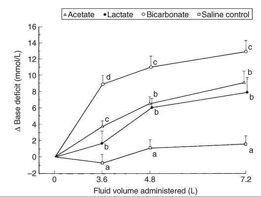
FIG. 20.8 Comparison of the alkalizing effect of various bases in diarrheic calves with severe dehydration and acidosis. At the start of the trial, all calves were at least 8% dehydrated and had a mean blood pH of 7.032 and a base deficit of 18 mmol/L. All calves received a total of 7.2 L of fluid containing 102 mmol/L of saline plus 50 mmol/L of the sodium salt of the respective alkalizing agent. Letters (a, b, c, d) are statistically different (P resulting in potential hypokalemia, which must be anticipated during fluid therapy.
Oral Electrolyte Therapy
Once the calf is able to nurse or drink, therapy is usually switched to oral electrolytes, which should continue for as long as the calf has diarrhea. This is also the route of choice for treatment of mildly affected calves on the farm. Calves with weak suck reflexes and calves that are unused to hand feeding can be tubed.
Oral electrolyte solutions need to supply sufficient sodium to facilitate normalization of extracellular fluid deficits, nutrients (glucose, citrate, acetate, propionate, or glycine) to facilitate absorption of sodium from the intestine, alkalizing agent to treat metabolic acidosis, and supplemental energy because most calves that have diarrhea are in a state of negative energy balance.614,615 The first two requirements depend on the coupled active transport of glucose and sodium ions across the brush border membranes of enterocytes, which results in passive absorption of water and other electrolytes.616 This function remains largely intact in calves with enterotoxigenic E. coli diarrhea, but where there is endothelial damage it may be impaired. Much of the research into oral electrolyte solutions has been conducted in healthy calves. Although the current understanding of the advantages and disadvantages of the different electrolytes is presented here, it is important to recognize that milk clotting, abomasal luminal pH, and abomasal emptying rates could differ between diarrheic and nondiarrheic calves.
The ideal sodium concentration for oral rehydration therapy in calves is thought to be between 90 and 130 mmol/L.615 Lower concentrations of sodium will not adequately correct dehydration. Products with a sodium concentration greater than 130 mmol/L should be avoided, as they may predispose to hypernatremia and can also delay abomasal emptying rates because of increased osmolality.617 The osmolality of the electrolyte solution is primarily determined by the concentrations of sodium and glucose. Commercially available oral electrolyte products in North America can range from approximately isotonic (280 to 300 mOsm/L) to extremely hypertonic (700 to 800 mOsm/L), with the greatest variation being in the amount of glucose added. The osmolality at the tip of the villi is approximately 600 mOsm/L, and use of solutions up to this osmolality will provide additional nutritional support.602 However, extremely hyperosmotic electrolyte solutions (700 to 717 mOsm/L) have the potential to increase dehydration and have been shown to have a slower abomasal emptying rate than calves fed iso-osmotic solutions (300 to 360 mOsm/L), increasing the risk of bloat or abomasitis and resulting in a slower rate of plasma expansion.602,617-619 Calves that are deprived of milk will lose more weight when fed isotonic low-glucose solutions.620
Oral electrolyte solutions should contain between 40 and 80 mEq/L chloride and 10 and 30 mmol/L potassium. Potassium is lost in the feces of calves with diarrhea, and with dehydration there can also be increased potassium losses from the kidney. Consequently calves with chronic diarrhea may have profound hypokalemia resulting in severe muscular weakness.602
Severely acidemic calves are unable to correct their metabolic acidosis when rehydrated with nonalkalinizing solutions, and consequently fluid therapy must be designed to address the strong ion (metabolic) acidosis and hyper-D-lactatemia.602,607,615 The ability to counteract acidosis varies greatly between oral electrolyte products. These differences are therapeutically important and are responsible for differences in survival rates between products. It is important that an oral electrolyte solution contains greater than 50 mmol/L of an alkalizing agent and has a strong ion difference ([Na+]+[K+]-[Cl-]) of at least 60 to 80.602 Commercial oral electrolyte solutions use acetate, propionate, bicarbonate, and citrate as alkalizing agents.
Understanding of the attributes of the alkalizing agents has evolved over the past few years, but there is little consensus on which is most suitable. Bicarbonate combines with hydrogen ions directly, whereas the other agents remove hydrogen ions during their metabolism within cells.604 Several studies have shown that oral electrolyte solutions containing bicarbonate (≤62 mmol/L) and citrate (≤12 mmol/L) do not interfere with milk clotting in vivo.619,621,622 Many authors still prefer bicarbonate precursors such as acetate or propionate for their energy value once metabolized, their water absorption capabilities, and the fact that they do not alkalinize the abomasum.602 It is important that solutions containing acetate and propionate also have a high strong ion difference to ensure a rapid restoration of base excess. Solutions containing bicarbonate alkalize the GI tract of milk-fed calves, and several authors have expressed concern that this may result in bacterial overgrowth in the small intestine as well as ETEC attachment and toxin production.602,623 In reality, concerns about the effects on milk clotting have been alleviated for most electrolyte solutions; it is likely that feeding a sufficient volume of an oral electrolyte solution with a recommended strong ion difference has the most significant impact on the rapid correction of dehydration 594621
and acidosis.594,621
There are a wide variety of oral electrolyte preparations on the market (Table 20.13), and many do not meet the requirements stated above, so care should be taken to select an appropriate solution. Beware of products that are designed for medicating hundreds of liters of water, as the final solution is often very dilute (support (Fig. 20.9). Maintenance metabolizable energy requirements for a 50-kg calf are about 2000 kcal, and 3500 kcal are required to support a weight gain of 0.5 kg/day. These requirements can be met by 3.3 and 5.7 L of whole cow's milk, respectively. Comparative studies indicate that weight loss in calves fed oral electrolytes are inversely proportional to the energy content of the solu- tions.620 Assuming a 4-L daily intake and 100% digestibility of oral electrolyte nutrients, regular electrolyte solutions supply between approximately 15% and 25% of energy needs. As a result, diarrheic calves that are held off milk for prolonged periods lose weight631 and can become emaciated. When maintaining body condition is a concern and little milk or solid food is being ingested, a high-energy oral electrolyte should be fed. Products such as Biolyte, Lifeguard/Enterolyte HE and RESTART H.E. provide about 50% of energy
ro
As-Fed Composition of Some Available Oral Electrolyte Solutions for Calves
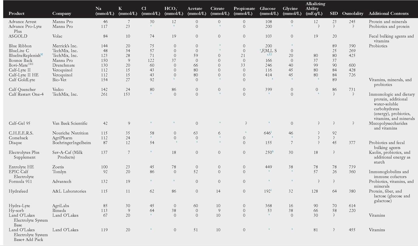
PART 3 Disorders and Management of the Neonate
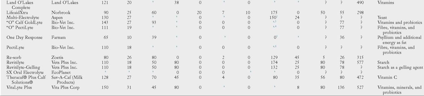
This table does not include every available product. Information updated based on TTable 20-12 in Large Animal Internal Medicine, Fifth edition. Calculated from information Hsted in the Compendium of Veterinary Products, U.S. edition, 2018, available online at https://pbs.cvpsemce.com, and on product labels. Additional information obtained from Smith GW, BerchtoldJF: Fluid therapy in calves, Veterinary Clinics of North America Food Animal Practice, 29(2):409-427, 2014; Jones C, HeinrichsJ: Comparison of commercial electrolyte products, Pennsylvania State University, PennState Extension, College of Agricultural Sciences, 2017, available at https://extension.psu.edu/table-comparison-of-commercial-electrolyte-products; and Smith G: Analysis of oral calf electrolytes, 2017, available at https://v^^v.techmixglobal.com/sites/default/files/doc/reference/Oral%20 Electrolyte % 2 OAnalysisJDr % 2OSmith~2017_0.pdf
Note: Concentrations of calcium, magnesium, phosphate, and sulfate are not included. Alolarities assume that all compounds dissociate completely in solution. Where a maximum and minimum value for a compound was provided, the mean value has been used.
aNutrient is provided by the product, but label contains insufficient information to calculate concentration.
FML,s,Ajn addition to glucose, product contains another carbohydrate source: F = fructose, M = maltodextrin, L = lactose, S = sucrose, A = amylose.
1Instead of glucose, product contains 96 mmol/L of lactose (glucose and galactose).
2Contains maltodextrins instead of or as well as glucose; value Hsted for glucose is glucose equivalents.
3Energy source is organic sugar; label does not specify the form.
4Energy source is not specified; contains reed-sedge peat and organic dried kelp.
5Instead of glucose, contains sucrose.
6Energy source is not specified; contains roughage products, reed-sedge peat, and dried kelp.
?, Published information insufficient to provide reasonable estimate; SID, strong ion difference.
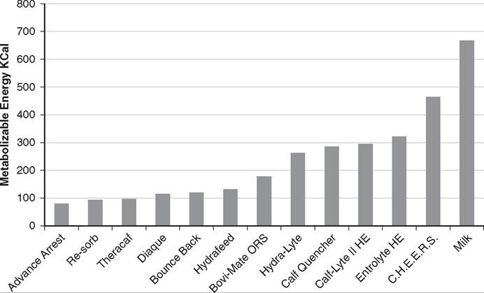
FIG. 20.9 Comparison of the energy content of various electrolyte solutions. Milk is shown for comparative purposes. Metabolizable energy content for products other than milk is estimated from glucose and other water-soluble carbohydrates of the as-fed electrolyge solution. Calculations are made with the assumption that 1 g of carbohydrate equals approximately 4 Kcal of metabolizable energy. This figure applies to electrolyte solutions fed to neonatal calves prior to rumen development. See text for manufacturers.
requirements if fed twice a day (total intake 4 L) and about 75% if fed three times a day (total intake approximately 6 L).
Milk withdrawal can reduce the severity of diarrhea and depression in severe scours. This is because malabsorption exacerbates diarrhea through the osmotic effect of unabsorbed milk nutrients and also promotes bacterial overgrowth and D-lactic acidosis. Milk also has a trophic effect on epithelial cells and maintains higher GI tract enzyme activities as well as providing protein for repair of damaged intestinal epithelium.632 In experimental trials, continued feeding of milk maintained weight gain; however, when calves were fed enough milk to fully meet their requirements and the undrunk milk was tube fed, calves initially had greater inappetence.633,634 Withdrawal of milk without a high-energy alternative can rapidly result in cachexia and malnourishment.633 In many calves, particularly the less severely affected, there is often a considerable degree of residual absorptive capacity, enough to support body weight gain if limited amounts of milk are fed. Milk withdrawal for up to 12 hours is recommended if the calf is depressed and not interested in sucking. In most cases electrolyte therapy will restore a calf's vigor and sucking drive within 1 to 2 days. Milk can then be reintroduced in small amounts (e.g., 1 L given two to four times daily). However, forced feeding (by tubing or drenching), dysfunction of the reticular groove reflex, or reflux of abomasal contents may result in ongoing acidosis due to the production of D-lactate from fermentation of carbohydrates entering the reticulorumen.600 If the calf is not interested in drinking or becomes depressed when reintroduced to milk, a high-energy oral electrolyte preparation can be tried instead. Studies indicate that diarrheic calves have a generalized malabsorption rather than specific lactose intolerance.165 Thus it may be more important to manage calves with milk intolerance by giving smaller amounts of milk in each feed rather than by changing carbohydrate source. There is little point in withdrawing milk from calves that remain alert and interested in nursing, as it is unlikely to result in clinical improvement. This is particularly likely to be the case when the calf receives whole cow's milk in frequent small quantities (i.e., by natural sucking of the dam).
Role of Antimicrobials
The majority of calves with diarrhea do not require treatment with antimicrobials, and since use of antimicrobials in neonatal calves is a driver of antimicrobial resistance (AMR) on farms, they should be used judiciously.635 Antimicrobial use in calves with diarrhea should be based on a protocol that (1) details the clinical signs indicating the use of antimicrobials; (2) specifies an antimicrobial product, dose, and duration of treatment that is appropriate for the likely pathogen and is likely to maintain a therapeutic concentration at the site of infection (small intestine and blood); (3) avoids the use of “medically important” antibiotics, unless indicated by culture results or when treatment with other antimicrobials has not been effective; (4) advocates antimicrobials registered for treating diarrhea in calves and as directed on the label or by a veterinarian whenever possible; and (5) avoids adverse local or systemic effects and violative residues. The use of antibiotics should always be accompanied by an infection control plan. The practice of continually feeding antibiotics to calves has been prohibited in many countries, but prophylactic antibiotics are still commonly used in some countries to prevent calf diarrhea.636,637 Although older studies generally demonstrated a reduction in the duration and severity of the diarrhea, there was little or no effect on diarrhea incidence or mortality rates.638 More recent studies have demonstrated benefits from stopping prophylactic and routine treatment with antimicrobials. A large study in the United States found a decrease in the prevalence of diarrhea in calves given targeted treatment for diarrhea with specific symptoms, compared with calves given both prophylactic antibiotics in the milk and routine antibiotics for all clinical cases.639 Another study demonstrated that there was no increase in mortality when a protocol for selective treatment of systemically ill calves was introduced on two large farms that had previously been using antibiotics in more than 75% of their calves with diarrhea.640 Moreover, a change in the fecal microbiota was observed in calves on both farms 12 months after the introduction of the new protocols. Although the relative proportions of the different bacteria were not always consistent across the two farms, both farms had a significant reduction in the percentage of Entero- bacteriaceae isolated in healthy calves 12 months after the protocols were introduced.
THERAPEUTIC TARGETING. The bacterial pathogens commonly associated with neonatal calf diarrhea are Salmonella and E. coli. Coliforms are normal inhabitants of the GI tract and one of the most common representatives of the aerobic gram-negative microbiota. Calves with diarrhea often have significantly increased coliform bacterial numbers in the small intestine, regardless of etiology,641-643 and this colonization is associated with altered small intestinal function, morphologic damage, and increased susceptibility to bacteremia.643 Bacterial overgrowth and subsequent fermentation in the GI tract results in the production of D-lactic acid. D-lactic acid is a major component of acidemia in diarrheic calves and is accompanied by systemic signs of weakness and ataxia. Calves with diarrhea are more likely to have failure or partial failure of passive transfer, and this group of calves, in turn, is more likely to be bacteremic.24,644 Blood cultures indicate that gram-negative bacteria account for approximately 80% of bacterial isolates, and E. coli is the most common bacteria isolated.18,19,644 In a study of 190 recumbent calves on a large calf-raising facility, 31% were determined to be bacteremic; E. coli accounted for 51% of the isolates, other gram-negatives for 25%, gramnegative anaerobes for 5.9%, gram-positive cocci for 11.8%, and gram-positive rods for 5.9%.644
Antimicrobial therapy must be targeted at a specific bacterial enteric pathogen isolated from sick calves or used in severely ill calves (as manifested by reduced suckle reflex, >6% dehydration, weakness, inability to stand, or clinical depression) to manage the risk of bacteremia. The antibiotic selected should be directed toward gram-negative organisms, particularly E. coli.
ANTIMICROBIAL SUSCEPTIBILITY. Antimicrobial susceptibility testing of fecal isolates should be specifically directed at enterotoxigenic E. coli and Salmonella spp. During a suspected outbreak of salmonellosis, fecal samples from 10 calves in the affected age-group should be cultured using an enrichment media, and antimicrobial susceptibility should be determined from the isolates obtained.645 Multiple isolates of ETEC should also be tested to determine antimicrobial susceptibility, where this is shown to be a primary etiology.
When antibiotics are used to prevent coliform septicemia, fecal samples cannot be used to determine the correct antimicrobial. The predominant strain of E. coli in the feces of a scouring calf can change several times during an episode of diarrhea, and one study has shown that 9 of 20 calves with diarrhea had different strains of E. coli isolated from upper and lower small intestines.646,647 Hence fecal E. coli strains may not be representative small intestinal E. coli strains, and multiple studies have shown that determining the appropriate antibiotic from fecal isolates is not a good predictor of clinical outcome.648-650
Antimicrobial susceptibility testing has more clinical relevance for predicting the clinical response to antimicrobial treatment when applied to bacteria isolated from blood or tissues of bacteremic calves, because the minimal inhibitory concentration break points are based on achievable antimicrobial concentrations in human plasma and MIC90 values for human E. coli isolates, which provide a reasonable approximation to achievable MIC values in calf plasma and MIC90 values for bovine E. coli isolates.647 Within a given herd there will be a diversity of bacteria isolated from bacteremic calves, so the collection of blood cultures and assessment of antimicrobial susceptibility does not necessarily provide information applicable to the next case.
When determining the correct antibiotic for prevention of septicemia in the absence of postmortem or blood cultures, antimicrobial efficacy is best evaluated by the clinical response of a number of calves to treatment, with calves randomly assigned to treatment groups, rather than the results of in vitro antimicrobial susceptibility testing performed on fecal E. coli isolates.647
MINIMIZING ANTIBIOTIC RESISTANCE. The young, preweaned calf rapidly acquires multiple-resistant commensal E. coli enteric flora within days of birth.651-654 Although colonization with E. coli-carrying AMR genes occurs regardless of antimicrobial exposure, the proportion of antimicrobial-resistant E. coli on a farm is driven by the antibiotic use on that farm.655-659 Although these coliforms are generally not pathogenic, they serve as a reservoir of AMR genes that can be shared between bacteria, including Salmonella spp., in both humans and animals.660,661
Several dietary and environmental factors influence the proportion of AMR coliforms shed. However, administration of antibiotics both fed in milk and given parenterally is a key driver, with preferential growth of antimicrobial-resistant E. coli strains starting within 3 hours of antimicrobial administration.655,659,662-666 Administration of antibiotics also converts the opportunistic pathogenic bacteria in the respiratory tract into reservoirs of AMR.659,666 As the calf gets older, changes in the commensal enteric microflora occur that result in an intestinal microbiota that is more effective at excluding these bacteria, and the proportion of resistant E. coli reduces.651,652,659,667 The prevalence of these bacteria also decreases when antibiotics are withdrawn, but a high number of resistant E. coli are sustained in the bedding for more than 1 month.668-670 Use of florfenicol and ceftiofur in one pen also results in an increase in the proportion of resistant bacteria shed by calves in adjacent pens.670 Consequently, administration of antibiotics to neonatal calves, more so than other age-groups, has been shown to be one of the key drivers of the development of AMR on a farm.635
Particularly concerning is the relationship between use of third- and fourth-generation cephalosporins on farms and the emergence and spread of broad-spectrum β-lactamase-producing E. colii.656,670-672 Broad-spectrum β-lactamases are a significant resistance mechanism that is a serious threat to the currently available antibiotic armoury, conferring resistance to some of the most commonly used antibiotics in humans, including penicillins, cephalosporins, clavulanic acid, and monobactams. In addition, these organisms are commonly multidrug resistant.673-675 It is now evident that the use of antimicrobials in calves is a major risk factor for broad-spectrum β-lactamase- producing E. coli, which is consistent with the rapid increase in antimicrobial-resistant coliforms observed following antimicrobial use in this age-group.635,667 Also of concern is the strong relationship demonstrated between the use of fluoroquinolones and the development of multidrug antimicrobial resistance, as is the co-selection of broad-spectrum β-lactamase resistance when cattle are treated with florfenicol.665,670,676
Third- and fourth-generation cephalosporins and fluoroquinolones have been identified as “highest priority critically important antimicrobials” for human medicine by national and international bodies, including the World Health Organization (WHO), Food and Agriculture Organization of the United Nations (FAO), and World Organisation for Animal Health (OIE). Hence it is imperative that veterinarians consider the impact that their selection of antimicrobial, where indicated, will have on the development of resistance. Critically important antimicrobials must not be used as the initial treatment in a disease outbreak; instead their use should be limited to those cases where culture and sensitivity has ruled out the use of other antimicrobials with likely efficacy or cases where lower priority antimicrobials result in a poor response. In some countries, the use of some of these antibiotics in food-producing animals is already banned. Logically, the range of antimicrobials used in calves on any farm should be limited, and when resistance becomes apparent, the antimicrobial type should then be changed. This strategy is likely to result in a slow decrease in resistance to that antimicrobial. However, the epidemiology of antimicrobial resistance is extremely complex, and experience in cattle and other species has shown that a reduction in resistance at a farm level can be slow.677-679 The development of AMR in Salmonella is also of concern, with cattle acting as an important reservoir of multidrug resistance.680 Administration of antibiotics or feeding of medicated milk to young calves may result in the transfer of resistance to Salmo- nella,664 but feeding medicated milk to young calves tends to reduce fecal Salmonella shedding and counterintuitively was not associated with increased shedding of antimicrobial-resistant Salmonella in epidemiologic studies.652 The presence of β-lactamase-secreting E. coli can protect other Enterobacte- riaceae, such as S. enterica, without evidence of gene transfer.681 Asymptomatic Salmonella shedding is common. Because Salmonella spp. can be recovered from dairy cattle for up to 3.5 years after clinical infection, the prevalence of AMR in Salmonella creates a risk for people in direct contact with the animal, its feces, or milk.682-685 Although several studies in the United States have failed to show a significant increase in the prevalence of AMR in Salmonella in the last 10 to 15 years, this has been attributed to variation in strain prevalence, with common cattle strains showing an increase in multidrug resistance.677,686
ANTIMICROBIAL SAFETY. A number of antimicrobials have been demonstrated to produce deleterious effects when administered orally to healthy milk-fed dairy calves. Administration of neomycin sulfate (300 mg PO q24h for the first 4 days of life) tended (P =.060) to increase the proportion of calves developing diarrhea (99 of 233 = 43%) compared with the proportion in an untreated control group (58 of 174 = 33%).687 Administration of neomycin sulfate (25 mg/kg PO q6h, n = 10), ampicillin trihydrate (12 mg/kg PO q8h, n = 6), or tetracycline hydrochloride (11 mg/kg PO q12h, n = 6) for 5 days increased the occurrence of diarrhea and decreased glucose absorption through unknown mechanisms compared with untreated controls (n = 6).688 Two other studies did not observe adverse side effects in calves administered tetracycline hydrochloride (40 mg PO q12h; 11 mg/kg PO q12h).689,690 Parenteral administration of aminoglycosides is prohibited in many countries due to prolonged slaughter withdrawal times (18 months), but there is also a potential for nephrotoxicity in dehydrated calves.
EFFICACY OF ORAL ANTIMICROBIAL THERAPY. The response to oral antimicrobial therapy is variable, with many formulations failing to demonstrate a beneficial effect.647 Oral antimicrobials may be used to treat E. coli overgrowth of the small intestine in mildly sick calves. Calves with diarrhea and moderate to severe systemic illness should be treated with parenteral antimicrobials. Efficacy of oral antimicrobials will vary depending on whether they are a liquid that will bypass the rumen or a capsule that will be deposited into the rumen. Due to reduced transit times in diarrheic calves, the antimicrobial concentration in the small intestinal lumen is lower than in healthy calves, and the rate of antimicrobial elimination is faster.662
Efficacy in either reducing mortality or reducing the duration of naturally acquired diarrhea has been demonstrated using either apramycin administered orally at either 20 or 40 mg/ kg for 5 days or marbofloxacin (a fluoroquinolone) at 1 mg/ kg/day for 3 days.691,692 The calves treated in the marbofloxacin study were younger than 5 days of age, and ETEC was isolated from more than 50% of the cases. Similarly, aminoglycosides and aminocyclitols are very poorly absorbed from the GI tract and are therefore only useful for the treatment of a confirmed enterotoxigenic E. coli infection and not for treatment of septicemia. Cephamycin C (0.75 to 20 mg/kg bid for 3 to 6 days), a broad-spectrum β-lactam antimicrobial, and enrofloxacin (5 mg/kg PO q24h for 3 days) have been shown to significantly reduce mortality in experimentally induced enterotoxigenic E. coli infections.693,694 However, it is illegal to use both fluoroquinolones and third-generation cephalosporins in some countries, as they are critically important antimicrobials for human medicine; therefore other antimicrobials should be used when indicated.
Trials with orally administered neomycin reduced the duration of diarrhea but did not reduce mortality.695,696 Similarly, trials with orally administered ampicillin failed to demonstrate a significant reduction in mortality, although the lack of success in this study was later attributed to a delay in commencement of antibiotic treatment.697 Conversely, orally administered amoxicillin trihydrate has been demonstrated to reduce mortality and the duration of diarrhea when administered at a dose of 10 mg/kg PO q12h.154,698 Amoxicillin is absorbed from the GI tract more effectively than ampicillin and can be used in combination with clavulanate potassium, providing potent irreversible inhibition of β-lactamase and increasing the antimicrobial spectrum of activity.
The results of trials with orally administered trimethoprim have been variable, with no significant improvement in outcome observed in a large field trial699 and a significant reduction in mortality observed in an experimental Salmonella challenge study where calves were administered 5 mg/kg of trimethoprim and 25 mg/kg of sulfadiazine PO daily for 5 days.700
EFFICACY OF PARENTERAL ANTIMICROBIAL THERAPY. The frequency of bacteremia is sufficiently high in calves with diarrhea that are severely ill (as manifested by reduced suckle reflex, >6% dehydration, weakness, inability to stand, or clinical depression) that routine treatment should include antimicrobials, with an emphasis on treating potential E. coli bacteremia.647
Antimicrobial drugs with an appropriate gram-negative spectrum of activity include potentiated penicillins (amoxicillin), trimethoprim-sulfonamide (TMS) combinations, aminoglycosides, sulfonamides, florphenicol, fluoroquinolones, third- generation cephalosporins (ceftiofur), and tetracyclines. There is a paucity of efficacy data to support the use of aminoglycosides, tetracycline, and nonpotentiated sulfonamides.
Potentiated sulfonamides have been evaluated in enterotoxigenic E. coli and Salmonella challenge experiments. Mortality was reduced in 2- to 3-week-old calves medicated with trimethoprim/sulfadiazine (in a 1 : 5 ratio) for 5 days 24 hours after an S. dublin oral challenge.700 Administration of either sulfadiazine or trimethoprim alone did not reduce mortality.700 TMS may be used to treat sepsis in neonatal calves, but its halflife rapidly declines as rumen function develops. In ruminating (6- to 8-week-old) calves, subcutaneous or oral administration of TMS leads to high serum levels of sulfadiazine but little or 701
no serum trimethoprim.701
Intramuscular administration of amoxicillin reduced mortality in S. dublin-challenged calves.702 In a comparative trial of amoxicillin and trimethoprim/sulfadiazine, both drugs were found to have equal efficacy in reducing adverse clinical signs of disease when dosage regimens were based on the MIC of the pathogen.703 A large study of calves with naturally acquired diarrhea demonstrated a decrease in mortality after treatment with ampicillin, and the efficacy was increased further with β-lactamase inhibition.704 Parenteral apramycin (20 mg/kg q24h, unstated route, for 5 days) significantly decreased the mortality rate of calves in a similar study.691 Fluoroquinolones are efficacious against both experimental challenge and natural ETEC infections.705,706 Florfenicol (20 mg/kg) given IM every 48 hours for two doses has been shown to be efficacious in treating salmonella.707 There are concerns about its use in treating a coliform septicemia because the MIC90 for E. coli is very high and can be achieved for only a short period when administered at 20 mg/kg IV.647,708 In most countries florfenicol is only labeled for intramuscular or subcutaneous use.
Ceftiofur has an appropriate antimicrobial spectrum, and therapeutic drug concentrations can be maintained with once- daily dosing. In a S. typhimurium challenge experiment, intramuscular administration of ceftiofur hydrochloride (5 mg/ kg q24h for 5 days) reduced the severity of clinical signs and reduced fecal shedding of Salmonella. The MIC of the challenge strain in this experiment was 1 μg7mL, and the therapeutic protocol maintained plasma concentrations above this concentration for the duration of therapy.375 Ceftiofur is a third- generation cephalosporin and is listed by the WHO in the highest priority of critically important antibiotic. Its use should be limited to appropriate cases.
A national survey in the United States found bacterial isolates to frequently be resistant to tetracycline, streptomycin, ampicillin, and ceftiofur. Less AMR was observed with aminoglycosides, fluoroquinolones, and trimethoprim-sulfas.9 The therapeutic options are subsequently limited, as the U.S. Animal Medicinal Drug Use Clarification Act prohibits extralabel use of fluoroquinolones, ceftiofur, and certain sulfonamides. In addition, there is a voluntary ban on the use of aminoglycosides, such as gentamicin and amikacin, in food-producing animals because of long-term tissue residues. 10
In summary, parenteral administration of a broad-spectrum β-lactam antimicrobial—amoxicillin or ampicillin (10 mg/kg IM q12h) or trimethoprim/sulfadiazine (20 mg/kg sulfadiazine with 5 mg/kg trimethoprim IV or IM q24h for 5 days)—is recommended for treating calves with diarrhea and systemic illness (note that these are off-label doses and require an extended meat withholding period). Antimicrobial therapy is not recommended for calves with diarrhea and no systemic illness (i.e., normal appetite for milk or milk replacer, no fever).647
ALTERNATIVES TO ANTIBIOTICS. Prebiotics and probiotics have been investigated as alternatives to antibiotics, but to date there is no evidence of clinical efficacy. The use of prebiotics and probiotics to prevent diarrhea is discussed in the Nonspecific Immunity section earlier. Lactoferrin is an iron-binding milk glycoprotein naturally found in colostrum that has antimicrobial, antiviral, antiinflammatory, and immunomodulatory effects. It has some efficacy as a preventive but has also been used in a large trial to treat calves with diarrhea on an organic farm. There was no difference in clinical scores compared with controls post treatment, but there was a significantly reduced risk of mortality and culling by 60 days in the subset of calves with severe diarrhea at enrollment.711
Antiprotozoal Drugs
Drugs reported to have some efficacy against cryptosporidia in calves include halofuginone,712-719 paromomycin,720,721 azithromycin,722 decoquinate,723,724 and β-cyclodextrin.725 Halofuginone appears to be the most efficacious, but it is not licensed in all countries. A meta-analysis of 15 cohort and clinical studies showed that prophylactic use of halofuginone delayed the shedding of Cryptosporidium, with a lower prevalence of shedding at days 4 and 7 but a higher prevalence at day 21 when treated calves were compared to controls.726 Prevalence of diarrhea was also lower on days 4 and 7 in treated calves. Because calves frequently become infected with cryptosporidia during the first 24 hours of life, prophylactic use of halfuginone administered to calves daily for the first week of life is effective at reducing disease when husbandry procedures are implemented
727
to minimize cross-infection between calves.'2' Ihe efficacy of decoquinate is questionable, with the only controlled clinical study failing to demonstrate a beneficial therapeutic effect with treatment at 2 mg/kg/day.724 Lasalocid has been trialed for treatment of cryptosporidia. Using a toxic dose of 8 mg/ kg, it was found to reduce the shedding of cryptosporidia, but the calves suffered adverse side effects. At a dose of 0.8 mg/kg, lasalocid was not effective.728 The registered dose for preventing coccidiosis in calves is 1 mg/kg per head per day.
Coccidiosis is uncommon in calves younger than 6 weeks of age. In hand-reared calves, coccidiostats (lasalocid, amprolium, or decoquinate) may be added to milk replacer. Prophylactic options for beef calves are restricted to coccidiostat-medicated pellets (monensin, lasalocid, amprolium, or decoquinate) or water (amprolium or sulfonamides). Therapeutic options include amprolium or sulfonamides such as sulfadimidine.
Both fenbendazole (5 mg/kg PO once daily for 3 days) and albendazole (20 mg/kg PO once daily for 3 days) have been shown as effective treatments for Giardia.331,414,729 Due to the high level of subclinically affected animals, all cows and their dams need to be treated, and reinfection is likely to occur unless calves are removed from environmental sources of infection.
Antiinflammatory Drugs
The routine administration of an NSAID is recommended for a calf with diarrhea that is systemically ill. It is important that administration of NSAIDs occurs after fluid therapy has commenced due to the risk of reduced renal perfusion. To avoid damaging the abomasal mucosa, no more than three doses should be given, particularly in intensive calf-rearing facilities with a history of calf deaths due to perforated abomasal ulcers.730 Although the benefits of NSAIDs have been demonstrated clinically, the underlying pathophysiology has yet to be determined and could be due to their analgesic, antiinflammatory, antisecretory, or antipyretic properties. Clinical trials have been performed with meloxicam and flunixin. Meloxicam 0.5 mg/kg SC given at the onset of diarrhea increased feed intake, hydration score, and fecal consistency and decreased signs of visceral pain compared 731732
to administering a placebo.'31,'32 A nonstatistical trend toward decreased morbidity has been reported in a study evaluating the benefits of a single or double injection of flunixin meglumine in scouring calves, but only when fecal blood was present.733 The routine administration of a corticosteroid to calves with diarrhea is not recommended, as corticosteroids suppress the immune system and diarrheic calves have been shown to have high levels of plasma cortisol when compared to healthy calves.734
Intestinal Protectants
A number of products that include intestinal protectants are marketed for treatment of calves with scours. Intestinal protectants include bismuth subsalicylate, kaolin or pectin, and activated charcoal. No efficacy data are available regarding the use of kaolin in scouring calves. Suggested advantages of bismuth subsalicylate are its neutralization of bacterial toxins and antisecretory effect through its local antiprostaglandin activity.735,736 The results of one prospective study indicated that the aforementioned nonantibiotic treatments for calf diarrhea resulted in a longer duration of treatment and increased morbidity and mortality when compared to medicated milk 737
replacer and parental antibiotic treatment.
Recent studies demonstrated that a commercially available standardized crofelemer extract, a natural product with antisecretory properties, increased fecal dry matter and reduced the severity of dehydration in both an experimental and a natural outbreak of calf scours.738,739
Prognosis
The prognosis for recovery decreases with the severity of depression. Severe hypothermia and the presence of intercurrent disease are grounds for a guarded prognosis.740-742 An initial examination should be performed. Calves with a primary problem of septicemia are not usually worth treating because of the poor prognosis. The severity of dehydration, hypothermia, and acidosis should be estimated. Recumbent calves are usually treated intravenously with a saline-based rehydrating fluid (0.9% saline, Ringer’s, lactated Ringer’s, etc.) and isotonic sodium bicarbonate (especially for older and comatose calves). Calves that can suck are treated with oral electrolytes that contain 50 to 80 mmol/L of alkalizing agent. Products that use mainly acetate as the alkalizing agent are best for calves that are still drinking cow’s milk (small quantities, frequently). Any alkalizing agent is likely effective in calves held off milk.
Summary
enterotoxigenic E. coli and Salmonella infections has been established; these diseases can be controlled by antibiotics and prevented by vaccination.743 There are public health implications to the diagnosis of cryptosporidiosis and salmonellosis. New vaccines may facilitate controlling rotavirus and coronavirus infections. Treatment of neonate diarrhea is primarily based on correcting dehydration and acidosis through the use of oral and intravenous electrolytes. Only in the case of bacterial infections can direct action be taken against the invading organism, but antibiotics may still be useful in preventing secondary bacteremias. Colostrum feeding will help reduce diarrhea in the first days of life. Management is very important in the control of diarrhea, and since infectious agents are almost always present at some exposure level, the underlying theme is to minimize the level of pathogen exposure and stress on the calf. In approaching a problem of neonatal death losses, the areas to examine should include calf immunoglobulin status, calf feeding, calf housing, cleanliness of environment, calving area, cow vaccinations, diagnosis of specific infectious agents, and treatment protocols.