Alterations in the small intestinal microflora (Small intestinal bacterial overgrowth)
Jan S. Suchodolski
Introduction
In humans, small intestinal bacterial overgrowth (SIBO) is defined as a clinical syndrome caused by an increased number of microorganisms in the small intestine.1 In dogs, the existence of SIBO, as defined in humans, is currently under debate.
In an early study by Batt et al.,2 bacterial counts in dogs with diarrhea were significantly higher compared to healthy dogs and the authors of this study defined SIBO as >104 anaerobic or >105 total bacterial colony-forming units (cfu)∕mL of fasting duodenal juice. However, these criteria for dogs are now controversial, because substantially higher bacterial counts have been found in duodenal juice of healthy dogs since the report by Batt et al.2 Recent studies performed in dogs with chronic enteropathies found no correlation between the number of bacterial colonies in the duodenum and clinical signs. Some dogs with suspected SIBO had counts that were substantially lower than 105 cfu∕ml.3 Since this condition usually responds to antibiotic treatment, some authors propose the term “antibiotic-responsive diarrhea” (ARD) rather than SIBO.4 Recently, the term “tylosin-responsive diarrhea” (TRD) was proposed for a subgroup of dogs with antibiotic-responsive diarrhea that is specifically responsive to tylosin.5 However, at this time, it is unclear if these terms (SIBO, ARD, and TRD) can be used synonymously as some dogs diagnosed with SIBO based on findings traditionally associated with SIBO do not respond to antibiotic therapy and some dogs that respond to antibiotic therapy do not have findings that have traditionally been associated with SIBO. Currently, there is no consensus on the definition and diagnostic criteria for SIBO. While it is generally accepted that a subgroup of dogs with small intestinal disease shows alterations of the small intestinal microflora, it is controversial if this is truly caused by an abnormal increase in bacterial numbers. In humans, there are several known risk factors for SIBO (Table 5.7). One of these risk factors is a decreased intestinal motility, which may result in an increase in bacterial numbers.1 The same mechanisms implicated in humans could potentially also lead to bacterial overgrowth in dogs. It is important to distinguish SIBO∕ARD∕TRD from GI disorders caused by potentially pathogenic bacteria such as Salmonella spp., Campylobacter spp., and enterotoxigenic Clostridium pefringens and difficile. SIBO has not been reported in cats as healthy cats appear to have much higher duodenal bacterial counts compared to healthy dogs, and these numbers do not differ from cats with enteropathies.6The small intestinal microflora
Based on bacterial culture results, bacterial counts in the small intestine range from 0 to >109 cfu/mL for aerobic bacteria and from 0 to > 108 cfu/mL for anaerobic bacteria in clinically healthy dogs, with a progressive increase in total bacterial numbers and microbial diversity from the duodenum to the ileum.7 Today, it is recognized that the majority of bacterial species are not amenable to routine bacterial culture tech-
niques.8 Using molecular biology methods that are based on identification of 16S ribosomal DNA, many previously uncharacterized bacterial species from canine small intestinal samples have now been identified.8 Additionally, the use of molecular profiling of the GI flora of dogs has recently demonstrated that the small intestinal microflora in dogs is highly variable and unique to each individual dog.9
The resident intestinal microflora offers nutritional benefits to the host by the production of short-chain fatty acids (e.g., butyrate, propionate, and acetate), which stimulate mucosal growth and epithelial cell proliferation. Lactate, produced by microbial fermentation, also serves as an energy source for the host. In addition, the normal intestinal microflora plays a major role in the protection of the host from invasion by harmful bacteria through the exclusion of potentially pathogenic organisms.
The implicated defense mechanisms against these harmful bacterial species include competition for oxygen and nutrient substrates, competition for mucosal adhesion sites, creation of a physiologically restrictive environment for nonresident bacterial species (e.g., production of substances that are toxic to other bacteria, changes in pH and redox potential, or hydrogen sulfide production), and secretion of antimicrobial substances (e.g., bacteriocins).10Table 5.7: Conditions associated with secondary SIBO
Small intestinal stasis
■ Anatomicabnormalities
- Congenital blind loops
- Small bowel diverticula, strictures, or adhesions
- Surgical resection of the ileo-colic valve
- Surgical blind loops (end-to-side anastomosis)
■ Partial obstructions of the small intestine
- Neoplasia
- Foreign bodies
- Chronic intussusception
■ Motility disorders
- Hypothyroidism
- Diabetic autonomic neuropathy
- Scleroderma
- Abnormal migrating motor complexes
Decreased gastric acid output
■ Atrophic gastritis
■ Administration of acid-suppressing drugs (H2-blockers, omeprazole)
Exocrine pancreatic insufficiency
■ Decreased output of pancreatic antimicrobial factors
Miscellaneous
■ Decreased mucosal immunity
Pathophysiology
There are several physiological mechanisms that regulate bacterial colonization in the small intestine, including secretion of gastric acid and antibacterial factors (i.e., pancreatic and biliary secretions), and most importantly, intestinal motility. Failure of one or more of these control mechanisms may lead to alterations in the small intestinal microflora resulting in clinical signs associated with SIBO.
Based on the underlying mechanism, SIBO can be divided into primary or idiopathic SIBO and secondary SIBO. Secondary SIBO occurs more frequently than primary SIBO. Table 5.7 summarizes the causes for secondary SIBO.
Most, but not all, bacteria ingested orally are destroyed by gastric acid.
Human patients with atrophic gastritis or those who undergo acid suppressant therapy (e.g., therapy with a proton pump inhibitor) show an increase in small intestinal bacterial counts.11 The pancreatic juice also contains antimicrobial substances that suppress excessive bacterial growth in the proximal small intestine. Dogs with experimentally induced exocrine pancreatic insufficiency (EPI) show significantly increased bacterial counts in the small intestine.12 The ileocolic valve serves as a natural barrier between the small and large intestine in dogs.13 This barrier, together with intestinal motility, is believed to prevent retrograde migration of bacteria from the highly populated large intestine into the less populated small intestine. Also, the formation of blind and stagnant small intestinal loops are a common site of bacterial overgrowth in humans.14There are several mechanisms that may lead to GI disease due to an alteration of the small intestinal microflora. Many bacterial species are able to deconjugate bile acids, which in severe cases may lead to fat malabsorption. Also, some bacterial species (i.e., Clostridium hiranonis and C. scindens) have 7α∕β- dehydroxylating activity, changing primary into secondary bile acids, which can be associated with increased toxic effects to epithelial cells.15 Bacterial toxins and metabolites can damage enterocytes. Bacterial metabolites may also cause destruction of the intestinal brush border and damage carrier proteins, resulting in malabsorption. Competition between bacteria and host cells for nutrients (e.g., cobalamin) may lead to malnutrition. Increased fat metabolism within the GI tract may lead to the production of toxic short-chain fatty acids, which can act as potent pro-inflammatory agents. Hydroxylation of fatty acids may further contribute to diarrhea. SIBO can lead to sufficient mucosal damage to cause PLE.
While some breeds (i.e., German Shepherd dogs and Chinese Shar Peis) have anecdotally been reported to be at an increased risk for developing SIBO, no genetic basis has yet been identified.
Also, IgA deficiency, which was commonly suspected to be associated with SIBO in German Shepherd dogs, has not been conclusively confirmed. A genetic susceptibility for a dysregulation in the cell-mediated immune response to a nor-
mal luminal microflora is suspected to occur in human beings with idiopathic SIBO and a similar mechanism may lead to chronic enteropathies in dogs.
Clinical signs
The clinical signs of an abnormal intestinal microflora may include chronic intermittent small and/or large bowel diarrhea. Affected dogs are usually active, but show a variable appetite ranging from poor to excessive (i.e., polyphagia). Weight loss or poor growth is frequently observed. Owners often report borborygmus and/or flatulence. Mild to moderate steatorrhea may be observed in some cases due to chronic fat malabsorption. Occasionally, dogs may show clinical signs related to the underlying cause of SIBO, such as vomiting secondary to chronic partial obstruction due to an intestinal tumor.
Diagnosis
A definitive diagnosis of SIBO is difficult to arrive at. A tentative diagnosis can be established based on clinical signs and altered serum cobalamin and /or folate concentrations, and by response to an antibiotic therapeutic trial. It must be noted that other conditions, such as undetected intestinal pathogens, may respond to antibiotic therapy, and a positive response to therapy does not necessarily confirm the presence of SIBO.
Other differential diagnoses, such as maldigestion due to EPI and causes for malabsorption such as IBD, intestinal lymphoma, lymphangiectasia, or food intolerance should be ruled out. It is also important to evaluate the patient for intestinal parasites such as Giardia spp. and known bacterial pathogens (i.e., enterotoxigenic Clostridium spp., Campylobacter spp., Salmonella spp., or enterotoxigenic E. coli).
The histopathology of the small intestinal mucosa is typically unremarkable as SIBO is usually not associated with morphological changes of the small intestinal mucosa.16 Occasionally, villous blunting or shortening may be observed.17 Diagnostic imaging may reveal causes of secondary SIBO such as anatomical abnormalities.
Findings on routine CBC, blood chemistry, and urinalysis are unrewarding in most patients. The measurement of serum cobalamin and folate can be useful for the diagnosis of SIBO.Serum cobalamin and folate concentrations
The assessment of serum cobalamin and folate concentrations is currently the most useful aid for the diagnosis of SIBO. Serum cobalamin may be decreased and serum folate may be increased in dogs with this condition. If both serum vitamin concentrations are altered, this is considered highly suggestive of SIBO; however, both have a rather poor sensitivity and specificity for the diagnosis of SIBO.4 The reported sensitivity of serum cobalamin concentration for a diagnosis of SIBO ranges from 25% to 55% and from 50% to 66% for serum folate con- centration.4
Aberrations in the small intestinal microflora may lead to an increased competition for cobalamin, resulting in decreased absorption of this vitamin. Bacteroides spp. are the principle organisms involved in the competitation for cobalamin since they can utilize cobalamin-intrinsic factor complexes, while other bacteria can only bind free cobalamin, which is present in lower concentrations in the intestinal lumen.16
Bacteria present in the distal small intestine and large intestine produce large quantities of folic acid. However, as folate carriers responsible for folate uptake are located exclusively in the proximal small intestine, folate produced in distal sections of the intestine will not be absorbed but will instead be excreted in the feces. If folate-producing bacteria migrate upwards into the proximal small intestine, folate of bacterial origin can be absorbed by the host resulting in increased serum folate concentrations.
However, cobalamin and folate uptake from the small intestine is highly complex and can be affected by several mechanisms (see 1.4.2.2). For example, a diet high in folate may lead to falsely increased serum folate concentrations, while inflammation of the ileum may damage cobalamin receptors and thus may lead to cobalamin malabsorption. Dogs with EPI have a decreased secretion of antibacterial substances with subsequent small intestinal bacterial overgrowth.17,18 As a consequence, dogs with EPI often have increased serum folate concentrations. Thus, in dogs with an abnormal serum concentration of cobalamin and/or folate, serum trypsin-like immunoreactivity (TLI) should be evaluated to rule out EPI as a secondary cause of SIBO.
Recently, it has been demonstrated that administration of tylosin does not lead, as would be expected, to a decrease in serum folate and an increase in serum cobalamin concentra- tions.19 Therefore, serum folate concentrations may not reflect therapeutic success and serum folate concentrations should always be evaluated together with the clinical picture.
Quantitative bacterial culture
Quantitative aerobic and anaerobic bacterial cultures have traditionally been considered the gold standard for the diagnosis of SIBO, but it is now recognized that there is no correlation of bacterial counts and disease status.4 Also, each dog harbors a very unique small intestinal microflora, making the determination of a normal or abnormal microflora difficult.
Miscellaneous tests
Many other tests have been proposed for the evaluation of patients suspected of having SIBO. However, many of these tests, such as the measurement of serum unconjugated cholic acid concentration (SUCA), 13C-xylose absorption test, 13C- bile acid absorption test, urinary indican test, or the hydrogen breath test, are associated with a high degree of variation between healthy animals, making them unrewarding for the diagnosis of SIBO.4
Treatment
General guidelines
It should be noted that while the altered bacterial population in the GI tract of the patient is not optimal, the resident bacterial populations are stable and able to resist acute changes or insults, such as a short course of antibiotics or temporary changes in diet. While such interventions may be associated with a transient alteration in the intestinal flora and some degree of clinical improvement, the aberrant microflora is likely to repopulate after the withdrawal of short-term therapy.10 Therefore, in most cases long-term therapy is required.5
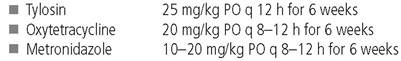
The treatment of choice for SIBO is the administration of a broad-spectrum antibiotic that is effective against both aerobic and anaerobic bacteria. Tylosin, metronidazole, and oxytetracycline are the most commonly used antibiotics for the treatment of SIBO (Table 5.8). Some patients may require several days to weeks of therapy before responding. However, if no clinical response is observed within two weeks, a therapeutic regimen with another antibiotic should be considered. The initial treatment period should last for 6 weeks. Thereafter, antibiotic treatment should be reinstated when clinical signs recur.
Underlying factors predisposing dogs to secondary SIBO (Table 5.7) should be corrected if they can be identified. For example, in most dogs with EPI and clinical signs of SIBO, the microflora returns to normal within a few weeks after initiation of enzyme replacement therapy. However, some patients may require antibiotic therapy.
Tylosin
Tylosin is often proposed as the antibiotic of choice in patients with suspected derangements of the intestinal microflora.5 Ty- losin is a macrolide antibiotic, which inhibits bacterial protein synthesis by binding to the 50S ribosomal subunit. Tylosin has antibiotic activity predominantly against gram-positive bacteria (e.g., Staphylococcus spp., Streptococcus spp., and Clostridium spp.) and also against some Mycoplasma and Chlamydia spp.
Table 5.8: Antibiotic agents used for the treatment of ARD/SIBO
While tylosin also has an effect against some gram-negative bacteria (e.g., Campylobacter spp., Helicobacter pylori, Hemophilus spp., Pasteurella spp., and Legionella spp.), it has no effect against members of the Enterobacteriaceae group (e.g., Escherichia coli and Salmonella spp.). It is speculated that tylosin also may exhibit immunomodulatory effects, however, no mode of action has yet been identified.5
Tylosin is very well tolerated and considered safe for longterm use. Several dosages of tylosin have been reported in the literature, underlining its broad therapeutic safety. Tylosin can be purchased as a powder for use in poultry and can be mixed into the food. Due to its broad dynamic and safety range, the dosage can be roughly estimated. For small patients, however, compounding of the powder may be necessary. Most commonly used dosages range between 15-25 mg/kg PO q 12 h. However, recent data would suggest that tylosin is only bactericidal at higher dosages and the author recommends a dosage of 25 mg/kg PO q 12 h for routine use. The initial treatment period should last at least for 6 weeks, unless adding tylosin into the treatment protocol does not lead to any improvement, in which case tylosin can be discontinued after 2 weeks. Some patients may have a recurrence after treatment is discontinued. Such patients should be further evaluated for any underlying cause of SIBO, but if one cannot be identified, long-term therapy can be initiated. After improvement of clinical signs, a reduction in the frequency of administration or a reduction in dosage, to the lowest frequency and/or dosage necessary to control clinical signs, may be attempted.
Oxytetracycline
Oxytetracycline is an excellent antibiotic choice for the treatment of SIBO due to its unusual metabolism. Oxytetracycline (administered at a dosage of 20 mg/kg PO q 8-12 h) is secreted in the bile and undergoes enterohepatic circulation, reaching high penetration in the small intestine and bile. Oxytetracycline can have some side-effects, and should thus not be administered in very young or pregnant animals. Also, calcium in the diet chelates oxytetracycline, rendering it ineffective. Oxytetracycline should, therefore, not be administered with food. As for tylosin, oxytetracycline should initially be given for a period of 6 weeks if the antibiotic therapy leads to an improvement in the clinical signs. It should also be noted that the oral formulation of oxytetracycline has limited availability, currently only being available in some European countries.
Metronidazole
Because of its activity against anaerobic bacteria, metronidazole at a dosage of 10-20 mg/kg PO q 8-12 h, is a commonly used antibiotic for the treatment of SIBO. In addition to its antibiotic properties, it also has been proposed that metronidazole exhibits immunomodulatory functions with beneficial effects in the treatment of intestinal inflammation. Metronidazole has, however, potential side effects and long-term therapy, as often required in patients with SIBO, may not be optimal. In vitro studies have shown the mutagenic properties of metronidazole.20 While no conclusive data about the mutagenic potential of metronidazole in vivo are available, given the fact that tylosin is a safe and effective alternative for longterm administration, it appears prudent that prolonged administration of metronidazole should only be attempted if tylosin has proven to be ineffective for controlling clinical signs.
Cobalamin supplementation
Dogs with SIBO may be cobalamin (Vitamin B12) deficient and parenteral supplementation of cobalamin (cyanocobalamin) is indicated. The typical dose in small dogs (5-15 kg) is 500 μg cobalamin SC per injection; in dogs > 15 kg, the typical dose is 500 to 1,200 μg cobalamin SC per injection depending on body size. Supplementation should be given for several weeks using the following dose regimen: one dose weekly for six weeks, one dose every other week for six weeks, and one more dose a month later. Serum cobalamin concentration should be re-evaluated a month after the last dose and if the serum cobalamin concentration is in the lower part of the reference range, supplementation should be continued.
Dietary management
Dietary management may be useful in some patients with SIBO. A highly digestible, fat restricted diet, containing a pre- biotic can be offered to these patients. A recent study showed that feeding a diet containing fructo-oligosaccharides, as a prebiotic agent, had similar effects as did antibiotic therapy.21 The use of a diet containing a prebiotic is aimed at promoting the growth of beneficial bacteria in the gut and limiting the growth of more harmful species. While the study compared two treatment groups that received either antibiotic therapy or dietary management, it seems prudent to combine antibiotic and dietary management in clinical patients. If combined antibiotic and dietary management leads to amelioration of the clinical signs, the antibiotic can be discontinued after 6 weeks. However, dietary management should be continued long-term.
Probiotics
Empirical administration of a probiotic compound (e.g., Lactobacillus spp. or Bifidobacterium spp.) may be considered as an ancillary treatment. However, it should be noted that no studies evaluating the clinical utility of probiotics in canine patients with SIBO have been published.
Prognosis
The prognosis for patients with secondary SIBO is excellent if the underlying cause can be effectively treated. However, it should be noted that an underlying cause can rarely be identified in dogs with SIBO. In dogs with primary SIBO that respond to treatment but show recurrent clinical signs after withdrawal of antibiotics, clinical signs can often be controlled with long-term administration of antibiotics.
??9 Key Facts
■ SIBO is a clinical syndrome that is difficult to definitively diagnose.
■ Patients diagnosed with SIBO need to be evaluated for an underlying cause.
■ Serum cobalamin may be decreased and serum folate may be increased in patients with SIBO. If both serum vitamin concentrations are altered, this is highly suggestive of SIBO.
■ Empirical administration of a broad-spectrum antibiotic effective against both aerobic and anaerobic bacteria in combination with dietary management is the treatment of choice for SIBO.
■ The treatment should be re-evaluated after approximately six weeks; many dogs will require repeated or long-term treatment to control clinical signs.