Anatomy
The large bowel ranges in length from 28-90 cm in dogs and 20-45 cm in cats.1,2 It begins at the ileocolic junction and terminates at the anus. Anatomically, the large bowel is divided into the cecum, colon, and rectum.
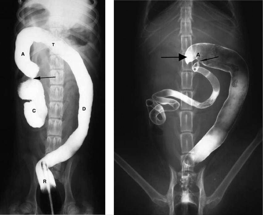
The cecum is a sigmoidshaped diverticulum of the proximal colon and joins the colon through the cecocolic orifice. This junction is in close proximity to the ileocolic orifice, also referred to as the ileocolic valve (Figure 6.1). The cecum is variable in length and measures 8-30 cm in dogs and 2-4 cm in cats (Figure 6.2).Figure 6.1 (left):
Barium enema in a dog. This figure shows a ventrodorsal abdominal radiograph after a barium enema in a dog showing the cecum (C), ascending colon (A), transverse colon (T), descending colon (D), rectum (R), and cecocolic junction (arrow).
Figure 6.2 (right): Barium enema in a cat. Ventrodorsal abdominal radiograph after a barium enema in a cat showing the ileocolic junction (thin arrowhead), the short cecum (thick arrowhead), and the short ascending colon (A).
The colon is divided into ascending, transverse, and descending portions and their connecting flexures. These subdivisions are identified on the basis of their relative position within the body. The ascending colon is a short segment that begins at the ileocolic sphincter and courses cranially to the right (hepatic) colic flexure. Spatially, the cecum and ascending colon lie to the right of the median plane and are in close association with the descending duodenum, right limb of the pancreas, and stomach. From the right colic flexure, the transverse colon runs, cranial to the root of the mesentery, to the left (splenic) colic flexure, where it joins the descending colon. The transverse colon is in close proximity to the left limb of the pancreas, stomach, and loops of small intestine.
The descending colon, the longest segment, passes caudally, following the left lateral abdominal wall into the pelvic inlet, where the rectum begins. It is usually covered by the greater omentum and lies adjacent to the ascending portion of the duodenum. The uterus or prostate and the urinary bladder lie ventral to its terminal portion.Blood is supplied to the large bowel by the cranial and caudal mesenteric arteries. Venous drainage occurs through the cranial and caudal mesenteric veins, which empty into the portal vein. Lymphatic drainage occurs through the right, middle, and left colic lymph nodes.
Histologically, the large bowel is similar to the small bowel, and contains mucosal, submucosal, muscular, and serosal layers. The anatomical modifications of the colonic mucosa, which increase its absorptive surface area, are not as distinctive as those in the small bowel. The colonic mucosa is devoid ofvilli, and the microvilli of colonic epithelial cells are less abundant than their counterparts in the small bowel. Despite the absence of villi, there are numerous crypts that extend from the absorptive surface through the entire thickness of the mucosa. These crypts of Lieberkuhn, contain epithelial, mucus, and endocrine cells. Compared to the small bowel, mucus cells are more prominent and endocrine cells are fewer in number. The deeper portion of the crypts consist mainly of undifferentiated cells, which migrate along the crypts as they proliferate and mature, ultimately giving rise to the aforementioned epithelial, mucus, and endocrine cells. At the mucosal surface, the cells undergo apoptosis, degenerate, and are sloughed into the lumen. Cell turnover in the colon is slower than in the small intestine, requiring 4-7 days. Within the lamina propria moderate numbers of diffusely distributed lymphocytes and plasma cells can be found.3,4
Both the intrinsic and extrinsic nervous systems regulate large bowel function. Intrinsic innervation occurs through the intramural network of neurons contained in the myenteric plexus, which lies between the longitudinal and circular muscle layers, and the submucosal plexus. Intrinsic control allows the large bowel to autonomously regulate functions based on intraluminal conditions, such as the degree of distension and the type and quantity of fluid and other intraluminal contents. Extrinsic neural control occurs through the autonomic nervous system. Parasympathetic innervation to the proximal portion of the large bowel is through the vagus nerve with the remainder of the large bowel supplied by the pelvic nerves. Sympathetic innervation arises from the paravertebral ganglia and follows the splanchnic nerves to the wall of the large intestine. Parasympathetic preganglionic fibers and sympathetic postganglionic fibers synapse on cell bodies and neurons of the intrinsic nervous system, respectively.
6.3