Anatomy of the Udder and Malformations
The goat's udder is composed of two glands, called halves or sides. In the male, teats are present but the glands usually remain rudimentary.
NormalAnatomy
Each gland has a single large teat.
Six to nine large milk ducts join to form the gland cistern. The gland cistern blends with the teat cistern, which ends at a single streak canal and a single teat opening (Turner 1952; Heidrich and Renk 1967), as illustrated in Figure 14.1. Teats may be funnel, cylindrical, or bottle shaped (James et al. 2009). The major blood supply is provided by the external pudendal artery. Venous return is via the external pudendal vein and the subcutaneous abdominal vein. The genitofemoral nerve, which passes through the inguinal ring with the external pudendal vessels, supplies most of the udder. Some skin innervation is also supplied by the lumbar cutaneous nerves cranially and the mammary branch of the pudendal nerve caudally.The glands are separated by a median suspensory ligament. The lateral laminae of the suspensory apparatus are lateral to the external pudendal vessels and attach to the symphysial tendon caudally and the tunica flava abdominis cranially. The mammary (superficial inguinal) lymph nodes are located deep to the lateral laminae and caudal to the external pudendal arteries (Garrett 1988; Constantinescu 2001).
The ultrasonographic anatomy of the bovine teat and udder has been well described (Trostle and O'Brien 1998) and the images supplied should be applicable to the goat
Goat Medicine, Third Edition. Mary C. Smith and David M. Sherman. © 2023 John Wiley & Sons, Inc. Published 2023 by John Wiley & Sons, Inc.
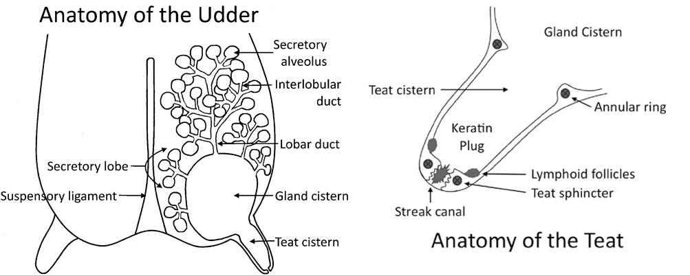
Figure 14.1 Anatomy of the udder and teat. Source: Courtesy of Dr. Paula Menzies.
udder. More recently, ultrasonography of the sheep udder (Barbagianni et al.
2017) and the goat udder (Fasulkov et al. 2010) has also been described. A 5 MHz rectal probe can be used, but a 7.5 MHz probe gives better resolution. Isopropyl alcohol or coupling gel is applied to achieve good contact with the skin, and the teat is scanned in longitudinal and transverse planes. The gain of the machine is adjusted until the normal milk appears anechoic. Using a commercial standoff device or immersing the teat in water in a flat-sided container improves the image, especially of superficial tissues. The near teat wall can be used as a convenient standoff for imaging lesions of the far teat wall.Supernumerary Teats
Small supernumerary teats, completely separate from and usually cranial to the main teats, are occasionally observed in goats. Almost 12% of Barbari goats in one study had supernumerary teats (Bhat 1988), as did 30% of West African Dwarf goats in Ghana (Oppong and Gumedze 1982), while goats with supernumerary teats predominated in a population of native goats in Japan (Nozawa 1970). These teats represent remnants of the embryologic mammary line that extends to the vulva, where ectopic milk-secreting tissue also may be located. The distinct supernumerary teats are commonly amputated with scissors while the doe kid is still young. Tetanus prophylaxis (such as 200-300 IU antitoxin) is recommended (Smith and Roguinsky 1977).
Rarely, the extra teats are of a size comparable to the normal teats, resulting in an udder divided into four equal glands (Balasurbramanian et al. 1994).
Double or Fused Teats
It is relatively common for goats, especially of breeds that have not been selected for dairy purposes, such as Pygmy and meat goats, to have double teats on one or both sides of the udder. The abnormal teats may be fused to the tip, but have two orifices (including fishtail teats). Forked teats (Figure 14.2), where both ends are of similar size, and small spurs protruding from the side of larger teats are other variations. Double teats are probably inherited in a complex way (Cunningham 1931-1932).
Kids (both does and bucks) of dairy breeds should be examined at birth, and all those with teat malformations other than simple, discrete, supernumerary teats should be marked for culling. Breed associations vary in the
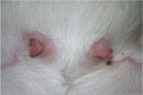
Figure 14.2 Bilateral forked teats in a Boer kid. Source: Courtesy of Dr. M.C. Smith.
importance they place on abnormal teats. The American Veterinary Medical Association has determined that removing an extra teat that would have interfered with milking is not ethical in an animal to be shown, bred, or sold (AVMA 1976). Owners who are unconcerned about the possible genetic basis for these malformations occasionally choose to cut off part of a double teat. Sometimes the wrong guess is made and the doe freshens with a small spur off a separate, larger gland that no longer has a teat orifice. Infections may also become established in the remaining gland at time of spur removal.
Weeping Teats and Teat Wall Cysts
Some goats have milk-secreting tissue in the wall of the teat, especially near its base. If this tissue communicates through one or more pores in the skin to the outside, milk oozes out. The condition is noticed when the hand of the milker becomes wet. Cauterization of the opening(s) with silver nitrate sticks after each milking may solve the problem. If the glandular tissue does not communicate with the outside or to the teat cistern, a cyst may form in the wall of the teat and interfere with milking. Real-time ultrasound clearly demonstrates a fluid-filled cavity. If the cyst is large, it may need to be drained occasionally using aseptic techniques. Weeping teats are probably hereditary (Biffani et al. 2020).
An extreme version of this condition, possibly hereditary, has been reported in Israel, where 19 of 324 lactating mixed-breed goats in one herd had multiple cysts 3-8 mm in diameter in the wall of the teat near its base.
Most of these cysts contained clear rather than milky fluid (Yeruham et al. 2005b).Blind Udder Halves
A presumably congenital abnormality has been described in a single Saanen goat in which the primary milk ducts ended blindly instead of joining the gland cistern. The mammary gland was capable of secretion, but no milk could be obtained from either teat (Turner and Berousek 1942). Absence of leukocytic infiltration in the udder tissue made a bacterial or retroviral etiology unlikely. Blind teats are common in artificially reared calves that suck on each other, and presumably scar tissue in the teat end could cause a similar problem in goats. Such acquired causes are discussed below, under the topic of teat obstruction and stenosis.
Poor Udder Suspension
The various suspensory ligaments of the udder should be strong and broad so that the udder is held tightly against the body with the floor of the udder above hock level (Considine and Trimberger 1978). Low-slung udders are prone to injury and are also bruised by alternately wrapping around one hind leg and then the other as the goat runs. Affected does and their offspring should not be allowed to contribute more to the gene pool in the herd. The heritability of the medial suspensory ligament as scored by the American Dairy Goat Association linear appraisal program has been estimated at 0.33 (Hatchette et al. 2001). An enlarged, pendulous udder may also be the result, rather than the cause, of mastitis (Addo et al. 1980). Unfortunately, poor udder attachment may correlate with higher milk production, so udder conformation should be included in selection indices to avoid unwanted changes in the structure of the udder (McLaren et al. 2016).
Neoplasia and Fibrocystic Disease
Neoplasms of the parenchyma of the udder are rarely reported in goats. In one Indian slaughterhouse study of 2000 female goats 4 years of age or older, multiple gray-white dots seen grossly in three udders were identified as intraductal carcinomas.
The tumors were multicentric, with diffuse intraluminal proliferation of cells. Metastasis to the supramammary lymph node had occurred in one animal (Singh and Iyer 1972). In another study of more than 4000 mature goats, two more multicentric intraductal carcinomas were identified (Sharma and Iyer 1974). Diffuse involvement of both glands with a mammary adenocarcinoma was observed in a 7-year-old doe that had not kidded or lactated for five years. Tumor metastases were found in the bronchial lymph node (Miller 1992). An aged Toggenburg doe with a chronic history of mastitis was found at necropsy to have a locally infiltrative adenocarcinoma in the udder and a granulosa cell tumor on one ovary, sug - gesting that high estrogen concentrations might be a risk factor for mammary cancer (Cooke and Merrall 1992). Lohr reported mammary adenocarcinomas in seven goats in a survey of 100 goats with neoplasia in the United States (2013).Slaughterhouse studies from India report a condition termed fibrocystic disease or cystic hyperplasia (Singh and Iyer 1973; Sharma and Iyer 1974; Tripathi et al. 1989; Upadhyaya and Rao 1994). Affected goat udders are enlarged and very firm/indurated, with multiple pea- to grape-sized nodules containing straw-colored fluid. Periductal and perilobular fibrosis with mononuclear cell infiltration surrounds dilated ducts filled with proteinaceous material. A predisposition of fibrocystic disease to the development of intraductal carcinomas has been proposed (Tripathi et al. 1989). A unilateral fibroepithelial hyperplasia of the udder of a nulliparous goat has also been reported and compared to mammary fibroepithelial hyperplasia in young, sexually intact cats (Andreasen et al. 1993). According to anecdotal reports, the histologic findings in biopsies from the udders of goats with inappropriate lactation syndrome (see below) are often suggestive of neoplasia. Thus, these various syndromes may be related.