Anatomy1-2
Lisa E. Moore
The esophagus is the connection between the oropharynx and the stomach; the major function of which is to carry ingested material from the oral cavity to the stomach.
The esophagus is divided into a cervical, thoracic, and abdominal portion. It begins at the upper esophageal sphincter (pharyngoesophageal sphincter), which is comprised of the cricopharyngeus and thyropharyngeus muscles. The cervical portion of the esophagus lies dorsally and to the left of the trachea. The thoracic portion of the esophagus extends from the thoracic inlet to the diaphragmatic hiatus. It lies dorsal to the trachea at the carina, where it then crosses the midline and lies to the right of the aortic arch. From here, it lies very close to the median plane as it passes between the caudal lung lobes. The short terminal portion of the esophagus lies in the abdominal cavity between the diaphragm and stomach.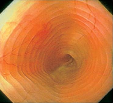
Figure 3.1:
Normal feline esophagus. Endoscopic view of a normal feline distal esophagus showing the "herringbone" pattern of the mucosal folds. (Image courtesy of Dr. David Twedt, Colorado State University, Ft. Collins, CO.)
The esophagus has several layers: adventitia, muscularis, submucosa, and mucosa. In the dog, the muscular layer is comprised of two oblique layers of striated muscle in the form of spiral fibers. These continuous oblique bundles spiral around the esophagus and cross each other at right angles, thus making up the two main muscular layers. About 5-10 cm from the cardia, the muscle fibers of the inner layer become more transverse while those of the outer layer become more longitudinal. The gastroesophageal sphincter is comprised of an outer layer of longitudinal striated muscle and an inner layer of circular smooth muscle.
These longitudinal striated fibers continue and partially blend with some of the gastric smooth muscle fibers. In the cat, the muscular layer cranial to the base of the heart is comprised of striated muscle, whereas caudal to the heart it is comprised of smooth muscle. The gastroesophageal sphincter in the cat is comprised solely of smooth muscle.The submucosal layer loosely attaches the mucosal and muscular layers, and contains mucus glands. In the dog, this loose connection allows the relatively inelastic mucosal layer to be thrown into longitudinal folds. In the cat, the mucosal layer contains longitudinal and transverse folds from the level of the heart base distally. This combination of longitudinal and transverse folds gives the mucosal layer of the esophagus of the cat its characteristic “herringbone” pattern (Figure 3.1). The mucosal layer is comprised of cornified, stratified squamous epithelium, which contains openings of the ducts belonging to the submucosal glands.
Branches of the cranial and caudal thyroid arteries serve as blood supply to the cervical portion of the esophagus. The esophageal portion of the bronchoesophageal artery supplies the cranial two-thirds of the thoracic portion of the esophagus, the remaining third being supplied by branches of the dorsal intercostal arteries. The terminal portion of the esophagus is supplied by a branch of the left gastric artery. Venous drainage occurs via satellites of the arteries that supply it. These veins mostly empty into the azygos vein. Lymphatic vessels from the esophagus drain into various lymph nodes including the retropharyngeal, mediastinal, bronchial, and portal nodes. Lymphatics and lymphatic vessels of the esophagus lie in the submucosal layer.
The striated muscle of the upper esophageal sphincter and esophageal body are innervated by branches of the vagus nerve. The vagus nerve also contains autonomic nerves to the smooth muscle and visceral afferents from sensory receptors. Sensory innervation distributes to the spinal segments C1-L2 with the cervical portion reaching segments C2-C6 and T2- T4, the thoracic portion reaching segments T2-T4 and T8- T12, and the lower esophageal sphincter reaching segment T1-L3. Sympathetic nerves also innervate the esophagus.
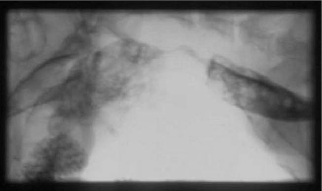
Figure 3.2:
Fluoroscopy of a dog with cricopharyngeal achalasia. Fluoroscopic image of a 6-year-old castrated male mixed-breed dog with adult-onset cricopharyngeal achalasia. The head is to the left. Note the barium remaining in the pharynx after repeated attempts at swallowing. The upper esophageal sphincter fails to relax and allow the barium to pass.
3.2