Background Information of Clinical Importance
Anatomy and Physiology
A detailed review of the musculoskeletal anatomy of the goat is beyond the scope of this text and the information is available from other sources (Chatelain 1987; Constantinescu 2001; Popesko 2008).
A representation of the caprine skeleton is given in Figure 4.1 and the topographic muscular anatomy of the goat in Figure 4.2.Normal Skeletal Variations
Goats commonly have numeric variation in vertebrae. The normal vertebral formula is 7 cervical, 13 thoracic, 6 lumbar, 5 sacral, and 7-12 coccygeal vertebrae. One survey of 185 goats revealed that 24% had numeric variation of vertebrae or the presence of structurally transitional vertebrae (Simoens et al. 1983). Common variations include 12 thoracic, 5 lumbar, and 4-6 sacral vertebrae. These variations are rarely, if ever, associated with clini - cal disease.
An extra sesamoid bone is infrequently found at the lateral head of the gastrocnemius muscle (Rajtova 1974). Floating ribs, with no connection to the costal arch, also occur infrequently (Hentschke 1980).
Bone Growth
There are several reports on epiphyseal closure times in growing goats (Dhingra and Tyagi 1970; Rajtova 1974; Ho 1975; Dhingra et al. 1978). The findings are widely disparate for numerous epiphyses; the reasons for these disparities are unclear. In the most recent study in Korean native goats, the distal humeral epiphysis fused at 8-12 months; proximal radial epiphysis and distal tibial epiphysis fused at 1 year; and proximal and distal epiphyses of ulna and femur, proximal epiphyses of
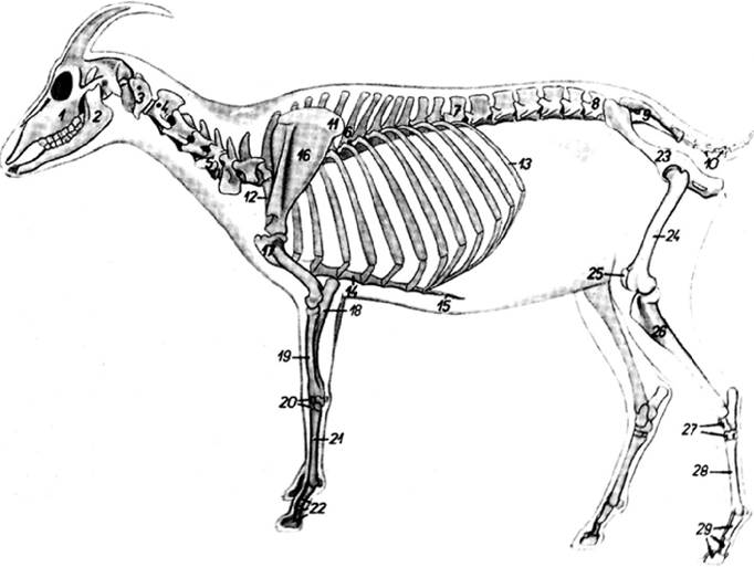
Figure 4.1 The caprine skeleton. (1) Maxilla, (2) mandible, (3) atlas, (4) axis, (5) fifth cervical vertebra, (6) sixth thoracic vertebra, (7) thirteenth thoracic vertebra, (8) sixth lumbar vertebra, (9) sacrum, (10) coccygeal vertebrae, (11) cartilage of scapula, (12) first rib, (13) thirteenth rib, (14) body of sternum, (15) xiphoid cartilage, (16) scapula, (17) humerus, (18) ulna, (19) radius, (20) carpal bones, (21) third and fourth metacarpal bones, (22) bones of digits of thoracic appendage, (23) os coxae, (24) femur, (25) patella, (26) tibia, (27) tarsal bones, (28) third and fourth metatarsal bones, (29) bones of digits of pelvic appendage.
Source: Reproduced with permission from Popesko, P. (2008). Atlas of Topographical Anatomy of Domestic Animals, new revised English edn. Bratislava: Vydavatelstvo Priroda. www.priroda.sk.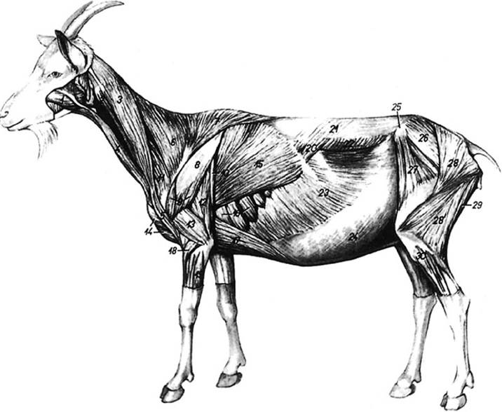
Figure 4.2 Topographic anatomy of the superficial caprine musculature. (1) Masseter muscle, (2) brachiocephalic muscle,
(3) cleido - occipital muscle, (4) sternomandibular muscle, (5) cleidobrachial muscle, (6) cervical part of trapezius muscle,
(7) thoracic part of trapezius muscle, (8) aponeurosis of deltoid muscle, (9) deltoid muscle, (10) tensor fasciae antebrachii muscle, (11) omotransverse muscle, (12) long head of triceps brachii muscle, (13) lateral head of triceps brachii muscle, (14) superficial pectoral muscles, (15) latissimus dorsi muscle, (16) thoracic ventral serratus muscle, (17) deep pectoral muscle, (18) extensor carpi radialis muscle, (19) extensor carpi ulnaris muscle, (20) caudal dorsal serratus muscle, (21) thoracolumbar fascia, (22) internal oblique abdominal muscle, (23) external oblique abdominal muscle, (24) aponeurosis of oblique muscles of abdomen, (25) tuber coxae, (26) middle gluteal muscle, (27) tensor fasciae latae muscle, (28) gluteobiceps muscle (28: superficial gluteal muscle, 28': biceps femoris muscle), (29) semitendinosus muscle, (30) peroneus longus muscle. Source: Reproduced with permission from Popesko, P. (2008). Atlas of Topographical Anatomy of Domestic Animals, new revised English edn. Bratislava: Vydavatelstvo Priroda, www.priroda.sk.
humerus and tibia, and distal epiphysis of radius fused at. 1 year or later (Choi et al. 2006). Sexual dimorphism occurs in caprine bone growth. Males have longer and wider bones and later closure of epiphyses than Iemales (Rajtova 1974).
The Parathyroid Glands
The parathyroid glands play an important role in the development and maintenance of normal bone through the action of parathyroid hormone on calcium and phosphorus metabolism (Hove 1981; Care and Hove 1982).
Histologic evaluation of the glands can be a valuable aid in the diagnosis of metabolic bone disease. These glands may be difficult to locate in the goat because of small size and variable relationship to other tissues. There are usually two pairs, though accessory parathyroid tissue can occur.The anterior pair are usually located deep in the anterior portion of the neck at the bifurcation of the common carotid artery. In the young goat, thymus is present at this site and the parathyroid gland may be recognized as a small, reddish-brown mass about 5 mm in diameter at the cranial pole of the thymus. Thymic tissue is atrophied in older goats, and the external parathyroid gland may be found caudal to the bifurcation of the common carotid artery. The posterior pair are usually located within the paired thyroid glands, most often in the middle portion on the medial aspect of the thyroid. The parathyroid tissue may be isolated in a distinct connective tissue capsule or mingled with thyroid tissue.
Conformation
There is increasing application of linear appraisal systems to generate sire summary information for the genetic improvement of dairy goats in breeding programs. The linear appraisal system is modeled after that used in dairy cattle and attempts to define a structural conformation consistent with functional durability as well as reproductive and productive efficiency. Type traits related to conformation that are included in linear appraisals are stature, rump angle, rump width, and rear legs (Wiggans and Hubbard 2001).
Skeletal conformation is also a significant component of dairy goat judging. Feet and legs are distinct structural categories subject to scoring. Recognized defects that reduce an animal's score include excessive spreading of the toes, shallow heels, turning out of the front feet or legs, rolled or turned-over claws, weak pasterns, winged-out shoulders or elbows, bowed or crooked forelimbs, straight stifles, rear limbs too close together (impinging on udder), turned-in hocks, and enlarged joints (Considine and Trimberger 1978).
The selection and judging of meat goats also include skeletal and conformational criteria (Martinez et al. 1991).Clinical Pathology
The most commonly used clinical chemical parameters for assessment of bone in health and disease are serum alkaline phosphatase (AP), calcium, and inorganic phosphorus. Variations in these parameters associated with different metabolic and nutritional bone diseases of goats are summarized in Table 4.1. Serum vitamin D and magnesium levels are less frequently evaluated. For evidence of muscle damage, serum activities of creatine kinase (CK), aspartate aminotransferase (AST), and lactate dehydrogenase (LDH) are most commonly measured. Reported values for these parameters in goats are given in Table 4.2. As discussed later, variation may occur with regard to age, breed, sex, and stage of production or preg - nancy status.
Alkaline Phosphatase
In goats, as in other species, serum AP is higher in young animals compared with adults, due to AP activity associated with the increased osteoblast function of growing bone (Castro et al. 1977a; Sugano et al. 1980; Bogin et al. 1981). In healthy adults, most of the serum AP present is derived from liver, particularly bile duct epithelium. Tissue enzyme profiles in goats demonstrate that AP activity is 50-100 times greater in kidney than in liver (Kramer and Carthew 1985). However, in renal disease, AP from tubular epithelium is released into the urine rather than the blood. There are now immunoassays available for measurement of bone-specific AP that were developed for use in humans, validated in sheep, and successfully applied to goats (Liesegang et al. 2006).
There is considerable variation in serum AP activity between individual goats even within the same age group, resulting in a very wide range of reported normal values. In any individual healthy goat, however, serum AP levels usually remain constant (Kramer and Carthew 1985). The measurement of bone-specific AP in Saanen does showed that serum concentrations dropped progressively through pregnancy and reached their lowest point in the week following parturition.
This reflects remodeling and calcium release from bone in response to the skeletal demands of the fetus and early lactation (Liesegang et al. 2006, 2007). There is evidence that high and low AP enzyme levels in goats are heritable, with the high level being genetically dominant (Lode 1970). Therefore, detection of a single high serum AP value in an individual goat may be difficult to interpret clinically. Increases in serum AP on serial samples within the same goat are a more reliable indication of bone or liver disease.Table 4.1 Clinical pathologic changes associated with metabolic bone and muscle diseases in goats.
| Disease | Serum alkaline phosphatase | Serum calcium | Serum phosphorus | Other changes |
| Rickets | Increased | Decreased | Decreased | Decreased serum vitamin D levels may also occur |
| Epiphysitis | Normal | bgcolor=white>NormalNormal | Excessive calcium in ration likely | |
| Fibrous osteodystrophy | Increased | Normal or decreased | Normal or increased | Excessive phosphorus in ration likely |
| Chronic fluorosis | Normal or increased | Normal or decreased | Normal or increased | Increased fluoride levels in bone |
| Enzootic calcinosis | Increased | Increased | Increased | Calcification of soft tissues |
| Hypervitaminosis D | Highly variable | Increased | Increased | Increased serum vitamin D3 levels |
Table 4.2 Some normal values for bone- and muscle-related enzymes and electrolytes in goat serum.
| Parameter | Unit | Goat description | Mean | ±SD | Range | Reference |
| Alkaline phosphatase | IU/L | F, pygmy, bgcolor=white>M, F, Israeli, 2-5 years of age | 190 | 34 | Bogin et al. (1981) | |
| IU/L | F, Alpine, adult | 217-586 | Garnier et al. (1984) | |||
| IU/L | Adult goats from multiple farms | 176 | 50.6 | Stevens et al. (1994) | ||
| Magnesium | mEq/L | M, F, Pygmy, all ages | 2.1 | 0.3 | Castro et al. (1977b) | |
| mg/dL | General values | 2.8-3.6 | Brooks et al. (1984) | |||
| mg/dL | Saanen kids | 2.23-2.49 | Boss and Wanner (1977) | |||
| Phosphorus | mEq/L | M, F, Pygmy, all ages | 4.8 | 0.9 | Castro et al. (1977b) | |
| mg/L | Lactating Saanen, adult | 43.5 | 23.9 | Ridoux et al. (1981) | ||
| mg/L | Lactating Alpine, adult | 59.9 | 20.2 | Ridoux et al. (1981) | ||
| mg/dL | M, F, Israeli, 3-4 months of age | 9.3 | 0.5 | Bogin et al. (1981) | ||
| mg/dL | M, F, Israeli, 2-5 years of age | 7 | 1.4 | Bogin et al. (1981) | ||
| mEq/L | General values | 1.7-4.3 | Brooks et al. (1984) | |||
| mg/dL | Adult goats from multiple farms | 5.5 | 1.7 | Stevens et al. (1994) |
F, female; M, male; SD, standard deviation.
Calcium
The reported range of normal serum calcium in goats is fairly narrow. Nevertheless, within that range, significant differences have been noted with regard to age, breed, pregnancy status, and lactation status. Young goats tend to have higher serum calcium than mature goats (Bogin et al. 1981; Ridoux et al. 1981). In a French study, Saanen goats had higher mean serum calcium levels than Alpine goats (Ridoux et al. 1981). Mean serum calcium was significantly higher in black Bengal goats during pregnancy than during lactation. Levels were higher in early pregnancy than in late pregnancy, but higher in late lactation than in early lactation (Uddin and Ahmed 1984).
Phosphorus
The range of inorganic phosphorus concentrations found in the serum of normal goats is often wider than calcium. Serum phosphorus levels are significantly higher in young animals than in mature animals (Boss and Wanner 1977; Bogin et al. 1981; Ridoux et al. 1981). The calcium-to- phosphorus ratio also is altered with age, reported as 1.1 in goats 3-4 months of age and 1.37 in goats 2-5 years of age (Bogin et al. 1981).
Breed differences have also been noted between Saanens and Alpines, with a lower mean serum phosphorus in the Saanen breed. Serum phosphorus concentration may be significantly higher during pregnancy than during lactation, but shows less variation during different stages of pregnancy or lactation than does serum calcium (Uddin and Ahmed 1984).
Magnesium
Serum magnesium levels have received less scrutiny in the goat than in other species. No variations of serum magnesium levels were noted as a function of age in goats 3-4 months of age compared with goats 2-5 years of age (Bogin et al. 1981). Similarly, no differences were noted in Saanen kids whose magnesium levels were measured every 20 days up to 8 months of age. The mean serum magnesium ranged from 2.23 to 2.49 mg/dL (Boss and Wanner 1977).
Vitamin D
There is little published information on normal caprine serum or plasma levels of vitamin D and its metabolites in health or disease. Studies of vitamin D metabolism suggest that the conversion of vitamin D3 to 25-(OH)D3 is limited in goats compared with other farm animal species (Hines et al. 1986).
One comparative study of sheep, camels, and Sinai desert goats kept in direct sunlight in summer showed the goats to have lower mean plasma levels of 25-(OH)D3 than either of the other species: 23.9 ± 5.7 ng/mL versus 40.7 ± 9.1 ng/ mL for sheep and 443 ± 96 ng/mL for camels (Shany et al. 1978). Studies on the effects of pregnancy and lactation on bone metabolism in goats indicate that serum 1,25 dihydroxy vitamin D levels peak in the first week of lactation under the influence of parathyroid hormone, to stimulate active calcium transport in the intestine to help restore the serum calcium pool in the face of lactational demand (Liesegang et al. 2006). Serum vitamin D levels return to prepartum levels by one month post partum.
Creatine Kinase
Enzyme activity of CK in tissues other than cardiac and skeletal muscle is minimal in the goat, making CK the most reliable indicator of muscle damage (Kramer and Carthew 1985). Age differences have been observed in serum CK levels, with young, growing goats having higher concentrations than older goats (Sugano et al. 1980). Enzyme activity within muscle itself also decreases with age (Braun et al. 1987). No significant differences were observed in serum CK levels of adult female goats measured two weeks pre partum, three weeks post partum, and two months into lactation (Garnier et al. 1984).
The reported range of normal serum CK in goats is fairly narrow, yet the potential for elevation is enormous in the face of widespread muscle necrosis. Mild, non-pathologic increases in serum CK may be noted due to low-level muscle damage after transport, fighting, poor venipuncture technique, or intramuscular (IM) injections, while in acute nutritional muscular dystrophy (NMD) serum CK levels may easily increase a hundredfold or more.
Aspartate Aminotransferase
AST is found in muscle and is increased in the blood when myodystrophy occurs. It is less specific for muscle damage than CK, however, because it also occurs in high concentration in the livers of goats (Kramer and Carthew 1985). When evaluated alone, AST cannot be used to discriminate between muscle and liver disease.
Lactate Dehydrogenase
Serum LDH is a collection of LDH isoenzymes derived from a variety of tissues including liver and muscle. It is also present in high concentration in red blood cells, so hemolysis during handling of blood samples can artificially elevate serum LDH. It is not as reliable an indicator of muscle damage as is CK. Evaluation of isoenzymes of LDH would enhance the specificity for muscle, liver, and other tissue forms, but this analysis is not routinely performed. The relative proportions of LDH isoenzymes in the serum of normal goats are LDH-1, 29-51%; LDH-2, 0-5%;
LDH-3, 24-40%; LDH-4, 0-6%; and LDH-5, 14-36% (Brooks et al. 1984). Proportional increases in serum levels of LDH-3, LDH-4, and especially LDH-5 are indicative of skeletal muscle damage. An increase in LDH-5 was also noted in normal, healthy goat kids between birth and 3 weeks of age (Sobiech et al. 2005).
There is conflicting data on whether significant differences in serum LDH levels occur in relation to age (Castro et al. 1977a; Bogin et al. 1981; Varshney et al. 1982). In adult females, the range of serum LDH levels may be higher during lactation than during pregnancy (Garnier et al. 1984). Females have significantly higher serum LDH levels than males (Castro et al. 1977a). Normal serum LDH levels are generally lower in goats than in cattle or sheep. The LDH activity in muscle itself is significantly decreased in adult goats compared with kids (Braun et al. 1987).
Diagnostic Procedures
Imaging Techniques
Plain radiography can be extremely helpful in the diagnosis of skeletal disorders. The convenient size and relative cooperativeness of goats make radiography a realistic option in caprine medicine. When necessary, goats can be blindfolded or sedated with xylazine or other sedatives to facilitate radiographic procedures. If sedated goats are made recumbent for radiographic procedures, care must be taken to avoid bloat and/or regurgitation. When possible, the animal should be fasted for 12 hours before sedation, the head placed lower than the body to facilitate drainage of ingesta out the mouth, and the left side should be dorsal to facilitate access to the rumen in case of bloat. Procedures for sedation are discussed in Chapter 17.
Reports of special radiographic procedures related to the caprine locomotor system are limited. Angiography of the caprine digit has been described (Burns and Cornell 1981). Bone scans using nuclear scintigraphy can help to identify localized areas of inflammation in the skeleton. Radiopharmaceuticals, however, are not approved for use in animals intended for the human food supply, so application may be limited to pet goats. The procedure has been done in goats using intravenous injection of 99m-technetium pyrophosphate, followed by scanning three hours later with a rectilinear scanner (Milhaud et al. 1980b). Good definition of ribs, joints, kidneys, and cervical vertebrae was achieved, while excessive accumulation of technetium in the urinary bladder interfered with visualization of the bony pelvis. Scintigraphy has been used in goats to quantitate the severity of joint inflammation in caprine arthritis encephalitis (CAE) and has been found to correlate well with histopathologic grading (Papageorges et al. 1991). Radiolabeled 99mTc-ciprofloxacin scintigraphy was also applied in the workup of a case of acute hindlimb paresis in a 3-week-old goat that demonstrated suppurative vertebral osteomyelitis and diskospon- dylitis at necropsy (Alexander et al. 2005).
Computed tomography (CT) has been used to confirm vertebral osteomyelitis in a 1-month-old male goat kid (Szafranski et al. 2020). Reports of the use of magnetic resonance imaging (MRI) in clinical caprine medicine are rare, but there are reports of its application in research studies of osteochondrosis (Toth et al. 2015), osteochondritis dessicans (Toth et al. 2017), and osteoarthritis (Schrauth et al. 2016) using goats as a model.
Electromyography
Normal electromyographic (EMG) responses of calves, sheep, and goats have been compared. Average action potentials of the superficial digital flexor muscle were 8.1 ms in the goat, 8.2 ms in the sheep, and 10.5 ms in the calf. The average peak potentials were 6.3 ms in the goat, 5.7 ms in the sheep, and 4.8 ms in the calf. Average amplitudes of the potentials were 322 μV in the goat, 253 μV in the sheep, and 337 μV in the calf. The proportion of biphasic and triphasic potentials was 93% in the goat, 92% in the sheep, and 85% in the calf (Mielke et al. 1981).
The EMG response can be used in the diagnosis of goats with myotonia congenita. Other applications of the EMG reported in goats include the diagnosis of ruminal adhesions to the abdominal wall (Cheong et al. 1987) and the identification of skeletal muscle denervation in swayback (Wouda et al. 1986).
Arthrocentesis and Synovial Fluid Analysis
Arthrocentesis and synovial fluid analysis can aid in the differential diagnosis of arthropathy. Strict asepsis should be observed during arthrocentesis. A 1.5 in. (3.8 cm) 18- or 20-gauge needle can be used to obtain fluid. Sedation facilitates the procedure.
The carpal joints are most often enlarged in goats with joint problems. The radiocarpal joint is distinct, while the midcarpal and carpometacarpal joints communicate. Access to these joints is improved when the carpus is flexed to a 90° angle. The extensor carpi radialis tendon running centrally over the anterior aspect of the carpus should be identified as a landmark. This may be complicated by the common occurrence of thick skin, callus, or hygroma on the carpi of goats. The radiocarpal joint is entered just lateral to the lateral edge of the extensor carpi radialis tendon, while the more distal midcarpal joint is entered medial to the medial edge of this tendon. The carpometacarpal joint is not accessible directly (Sack and Cottrell 1984).
The shoulder joint is approached laterally. The acromion and the notch in the proximal border of the greater tubercle of the humerus are used as landmarks. The needle is inserted into the joint distal to the midpoint between the two landmarks, with the tip of the needle directed medial to the greater tubercle (Sack and Cottrell 1984).
The elbow joint is approached from the lateral aspect. The two landmarks are the lateral epicondylar crest of the humerus and the cranial border of the olecranon, which together form an angle that points distally. The needle is inserted into the angle and directed mediodistally and slightly cranially into the olecranon fossa (Sack and Cottrell 1984). For aspiration of joint fluid from the stifle, the joint can be approached from the proximolateral side between the patella and the lateral patellar trochlea (Rorvik 1995). Approaches to other joints have not been described specifically in goats, but techniques described in cattle are generally suitable (Greenough et al. 1981).
Normal caprine synovial fluid should be clear and colorless to slightly yellow, free of particulate debris, and should not clot. Mean total protein has been reported as 1.84 ± 0.22 g/dL (Nayak and Bhowmik 1990). Total cell counts should be less than 500/mm3 (Crawford and Adams 1981). Monocytes and lymphocytes should predominate in normal synovial fluid and neutrophils should not exceed 10% of the total cell count. Abnormalities in synovial fluid reported in various caprine diseases are discussed throughout the chapter.
Muscle and Bone Biopsy
No special concerns have been identified for performance of muscle or bone biopsies in goats. The rib is commonly used for biopsy in the diagnosis of metabolic and nutritional bone disease. The costochondral junction is especially useful when rickets is suspected.
Regional Anesthesia
Intravenous regional anesthesia can be used successfully in goats to perform minor or major surgical operations on the distal forelimbs or hindlimbs, including amputations (Babalola and Oke 1983). This procedure is described in Chapter 17.
Foot Trimming
Therapeutic and prophylactic foot trimming are important procedures in goats. Careful paring of the foot may promote healing in cases of foot rot, foot abscesses, and puncture wounds by allowing aeration and drainage of affected tissues. Corrective trimming also promotes normal comfort and conformation in cases of chronic laminitis.
Prophylactic trimming is required to maintain normal hoof structure and provide a strong base for support of the
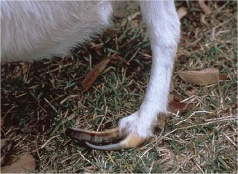
Figure4.3 Severely neglected, overgrown foot of a goat. Such excessive growth is caused by continuous confinement and lack of exercise. Source: Reproduced by permission of Dr. C.S.F. Williams.
limbs. Overgrowth of the feet is particularly a problem in intensively managed goats, where normal wearing of the hoof associated with exercise is limited (Figure 4.3). Overgrown feet cause abnormal gait and undue stress on joints, tendons, and ligaments. Goats in confinement should have their feet trimmed a minimum of twice a year. Goats that have a history of laminitis or clinical CAE will need more frequent trimming.
A pointed hoof shears or a hoof knife is appropriate equipment. Unlike sheep, goats resist being tipped on their rumps for foot trimming. The feet are simply lifted for trimming with the animal in a standing position. Large bucks and squirmy kids are often easier to trim if restrained in lateral recumbency by a knee placed on the neck. The basic steps of foot trimming are illustrated in Figure 4.4. They include removal of hoof wall that has overgrown the sole, shortening of the toe, and leveling of the sole and heel.
In the properly trimmed foot, the coronary band should be parallel to the weight-bearing surfaces of the claws. Excessive shortening of the toe by trimming will cause the animal to “break forward” at the fetlock, while inadequate trimming of the toe causes the animal to rock backward on the foot, reducing contact between the anterior sole and the ground, thus causing undue stretching of the flexor tendons.