Diagnosis of Musculoskeletal
Disease by Presenting Signs
This section is a guide to the differential diagnosis of musculoskeletal problems and attempts to include all possible diagnoses independent of geographic occurrence or relative incidence.
Such information is included in subsequent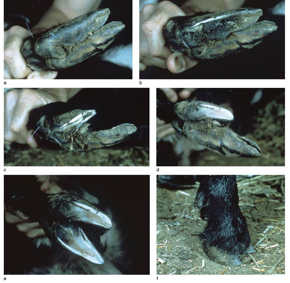
Figure 4.4 Procedure for proper foot trimming in the goat. (a) Plantar view of untrimmed goat foot prior to beginning. (b) Overgrown wall covering sole is trimmed away, revealing debris packed between wall and sole. (c) Excessive toe growth is trimmed back. (d) Sole is paired down to healthy fresh tissue. (e) The same steps are repeated on the other claw of the foot. (f) The completed, properly trimmed foot. Source: Reproduced by permission of Dr. C.S.F. Williams.
discussions of the specific diseases later in the chapter or elsewhere in the text.
Abnormally Appearing or Sore Feet
Infectious causes of abnormally appearing or sore feet include foot scald, foot rot, foot abscess, foot and mouth disease (FMD), bluetongue, and dermatophilosis. In FMD and bluetongue, which are most often subclinical in goats, oral lesions may be seen in conjunction with hoof lesions. Dermatophilosis, or mycotic dermatitis, can affect the feet and distal limbs as well as other areas of skin. This is also true of mange mite infestations, particularly chorioptic mange.
Metabolic and nutritional causes include zinc deficiency and Iaminitis. In zinc deficiency, hoof lesions are seen along with dermatitis. Acute and chronic forms of lamini- tis occur. In acute laminitis feet are predominantly sore and hot, while in chronic laminitis they are malformed and overgrown.
Chronic selenosis is the only reported toxic cause of abnormally appearing feet. Traumatic causes include overgrown hooves secondary to inadequate trimming, foreign bodies such as stones or wood chips lodged between the claws, bruising of the sole, and puncture wounds of the foot, especially those that lead to foot abscesses.
Swelling Around the Joints
Swollen joints usually imply arthritis. It is important to differentiate in the goat between true arthritides and periarticular or superficial swellings. Goats maintained in confinement, particularly on hard surfaces, develop a marked thickening of the skin, particularly over the carpi, hocks, and sternum, which can make the joint itself appear swollen. Goats are also prone to carpal hygromas. These are soft, fluctuant, periarticular swellings on the anterior aspect of the carpi, referred to in lay terms as “water on the knee.” However, filling of the carpal bursa is also an early sign of arthritis caused by CAE virus infection, and this must be differentiated from an uncomplicated hygroma.
Infectious causes of arthritis include the CAE virus, various Mycoplasma spp., and a variety of bacterial agents. Polyarthritis in neonates or young goats is usually bacterial or mycoplasmal in origin. Chlamydial arthritis is a well- known cause of arthritis in sheep, but documentation of chlamydial arthritis in goats is lacking (Nietfeld 2001), despite the fact that it is sometimes cited as a cause of arthritis in the species. Similarly, erysipelothritic polyarthritis caused by Erysipelothrix rhusiopathiae is a well- known endemic problem in young lambs, but its occurrence in goats is not well documented.
Nutritional and metabolic causes of swollen joints include rickets and so-called nutritional arthritis or osteopetrosis. Rickets is actually an enlargement or distortion of the improperly calcified epiphyses adjacent to a seemingly swollen joint. Nutritional arthritis, secondary to excessive calcium feeding, is reported primarily in bucks of dairy breeds.
Traumatic injury to the joints is common in goats because of their propensity to fight and to climb and explore. Swellings may occur secondary to avulsions of tendons and ligaments, dislocations, and hemarthrosis. Does in advanced pregnancy sometimes develop edematous swellings around and above the coronary bands, particularly on the hindlimbs.
This is presumably because of circulatory compromise and usually resolves with parturition. However, it may also be associated with pregnancy toxemia, as discussed in Chapter 19.Degenerative osteoarthritis may be seen, especially in very old goats or in goats with poor conformational characteristics, such as excessive straight-leggedness in the hind limb.
Stiff, Painful, or Abnormal Gait
Abnormalities of gait can be caused by neurologic dysfunction or musculoskeletal diseases. The differential diagnoses for neurologic causes of gait abnormality are given in Chapter 5. While most neurologic diseases cause some degree of paresis or ataxia, tetanus is noteworthy in producing a stilted, stiff gait, which suggests musculoskeletal pain. This is also true in the case of spastic paresis, a neurogenic disease that causes intermittent stiffening of the hindlimbs. Similarly, peritonitis, pleuritis, or mastitis may cause sufficient pain on motion that affected animals show a guarded, stilted gait.
Regarding primary musculoskeletal diseases, any of the causes of painful feet or swollen joints identified above can contribute to development of a stiff, abnormal, or painful gait. Additional causes of muscle origin include NMD in its early or mild stages, parasitic myositis involving tapeworm cysts of the muscle, and myotonia congenita, in which a stiff gait is intermittent and may be preceded by a generalized muscular contraction, causing the goat to fall down, as seen in “fainting goats.” Enzootic calcinosis caused by ingestion of Trisetum flavescens or yellow oat grass can cause calcification of tendons and ligaments, with a resulting painful gait.
Additional causes of skeletal origin include fibrous osteodystrophy associated with excessive phosphorus in the diet; rickets in young, growing goats or osteoporosis in adult goats, both associated with dietary imbalances of calcium, phosphorus, and vitamin D; osteopetrosis in bucks or epiphysitis in young, growing goats associated with excess calcium intake.
Chronic fluorosis can lead to abnormal bone growth and apparent bone pain. Copper deficiency in kids causes a primarily neurologic disease known as swayback or enzootic ataxia. However, affected kids may also have bone pain associated with abnormal osteogenesis and subsequent bone fragility.Failure to Extend a Limb or Limbs
The differential diagnosis for failure to extend a limb or limbs varies markedly with the age of the affected animal. In newborn goats, arthrogryposis or persistent flexure of joints may be caused by congenital Akabane disease or Cache Valley or Schmallenberg virus infections, inherited beta mannosidosis in goats of the Nubian breed, possible congenital lupinosis, or contracted tendons associated with positional constraints in utero during fetal growth. An inherited tendon shortening also occurs in Australian Angora goats. In older kids, enzootic ataxia (nutritional copper deficiency) sometimes is accompanied by flexor contracture of the forelimbs.
In mature animals, ankylosis of joints in a flexed position is a common outcome of chronic CAE virus arthritis. The carpi are most often affected. However, any traumatic or infectious cause of arthritis may result in a reduced range of motion when chronic in nature. Elbow joints are frequently involved. Dislocations or luxations may also cause pain on extension of joints, with animals favoring a flexed position.
When goats are recumbent from any cause, tendon contracture occurs rapidly. Initiation of regular physical therapy to manually straighten the front limbs is advisable if goats are anticipated to be recumbent more than 24 hours.
Non-weightbearing on a Single Limb
This refers to animals that are able to extend the affected limb but are unwilling to bear weight on it. Differential diagnoses include fractures, dislocations, severe arthritis involving a single joint, puncture wounds of the foot, severe foot rot, foot scald, or foot abscesses. Fractures may be primarily traumatic in origin or may be predisposed by increased bone fragility, such as occurs in rickets, fibrous osteodystrophy, copper deficiency, and chronic fluorosis.
Osteomyelitis may also predispose to fracture or cause sufficient pain to result in non-weightbearing.Bowed Limbs
Bowing of the forelimbs is seen primarily in metabolic bone diseases. Known causes include epiphysitis, rickets, and a condition known as bowie, or bentleg, associated with phosphorus deficiency. Bowing of the hindlimbs may occur in zinc-deficient goats. Poor conformation in individual goats with weak attachments of the shoulder assembly may lead to winged-out elbows and the appearance of bowed limbs.
Conditions Restricted to a Forelimb
Luxation of the scapulohumeral joint occurs with some frequency in goats. One veterinary referral center reported on five cases seen over a 26-month period. The affected limb is carried in a semi-flexed position and abducted and rotated outward, with apparent pain on attempts at extension. The condition is successfully managed by surgical intervention to stabilize the joint (Purohit et al. 1985).
Goats, similar to sheep (Scott 2001), may suffer a traumatic injury to the elbow joint, probably initially involving a collateral ligament. Due to new bone formation around the joint, flexion is decreased and the leg is carried stiffly. Radiographic evaluation can assist in the diagnosis of this condition, as bony proliferation may be seen around the elbow joint though the joint itself is not distended.
Carpal hygromas are common in goats maintained on rough, hard flooring. They must be differentiated from other causes of carpal swelling as discussed above in the section on swollen joints.
Conditions Restricted to a Hindlimb
Lateral luxation of the patella occurs in goats and is believed to be congenital, associated with hypoplasia of the lateral trochlear ridge. Progressive lameness occurs and the laterally displaced patella is palpable along with crepitus. Affected goats keep the stifle and hock flexed, because extension of the limb is difficult and painful when the patella is displaced. Successful surgical correction may be accomplished via trochlear sulcoplasty and desmotomy (Baron 1987).
Rupture of the gastrocnemius muscle belly above the tendon can occur in goats as in cattle, producing a palpable swelling on the caudal aspect of the hindlimb above the hock and a characteristic hindlimb position where the hock remains on the ground while the animal is standing (Figure 4.5). A similar stance was reported in a goat with a ruptured calcaneal tendon secondary to dog bite (Hunt
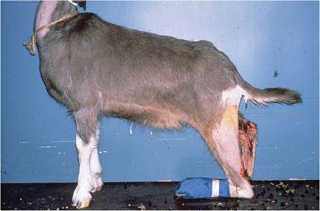
Figure 4.5 Typical stance of goat with ruptured gastrocnemius muscle. Note that the hock is resting on the ground while the animal is standing. Source: Courtesy of Educational Media, Cummings School of Veterinary Medicine at Tufts University, Mr. David Wilman photographer.
et al. 1991). That case was successfully treated by transposition of the tendon of the peroneus longus muscle. Careless shearing technique could also sever the calcaneal tendon.
Spastic paresis, a progressive neurologic disorder, manifests as intermittent episodes of muscular rigidity in the gastrocnemius muscle and hyperextension of one or both hindlimbs. Affected goats may actually lift their hind ends off the ground during active episodes and walk only on the forelimbs if both hindlimbs are affected simultaneously. The condition occurs in several breeds of cattle and is generally considered to be a genetic disorder with a complex manner of inheritance (Scarratt 2004). The condition is uncommon in goats and it is unclear if it also represents a genetic disorder in this species. In cattle, surgical treatments include tibial neurectomy or gastrocnemius tenotomy, but there are no reports of treatment in goats. The condition in goats is discussed further in Chapter 5.
Weakness and Recumbency
This clinical presentation encompasses a broad range of differential diagnoses, as weakness and recumbency can occur in a variety of neurologic and systemic diseases. Only primary muscle and skeletal problems are listed here.
Muscular diseases associated with weakness and recumbency include clostridial myositis, NMD, ingestion of myo- degenerative plants such as Cassia roemeriana or Karwinskia humboldtiana, and milk fever or hypocalcemia, a metabolic disease discussed in Chapter 19. Myotonia congenita can cause affected goats to be recumbent, but the effect lasts only approximately one minute.
Myasthenia gravis, a cause of profound muscle weakness in humans and dogs, has been reported in one veterinary text as occurring in the goat, but no primary reference is cited (Fraser 1986). A published review of this disease does not identify myasthenia gravis as a naturally occurring disease in goats (Lindstrom 1979) and a search of the literature through 2020 produced no case reports in goats. However, myasthenia gravis has been produced experimentally using goats as an animal model by the induction of autoantibodies to acetylcholine receptors. These antibodies block neuromuscular transmission (Lindstrom 1976).
Arthritis can be sufficiently painful to inhibit ambulation. Acute mycoplasma arthritis is especially associated with recumbency in goats. Severe hoof diseases, such as foot rot, chronic selenosis, and laminitis, can also keep goats recumbent.
Bone abnormalities can produce recumbency through either bone pain or secondary fracture. Metabolic bone diseases associated with recumbency include rickets and fibrous osteodystrophy. Traumatic fractures, especially those of the vertebral column, must also be considered in goats unwilling to rise. Osteomyelitis can also predispose to fracture and recumbency, especially if involving the vertebrae.