Background Information of Clinical Importance
Anatomy and Physiology
Liver
The caprine liver comprises four distinct lobes: right, left, quadrate, and caudate. It is located in the dorsal two-thirds of the right anterior abdomen, in contact with the diaphragm and the abdominal wall from the seventh to last rib (Figure 11.1).
Dorsally, the liver is covered by the lung as far caudally as the ninth rib space, but the liver is percuss- ible or accessible for biopsy between the ventral lung border and the costochondral junctions in the seventh through ninth rib spaces. The gallbladder extends below the ventral border of the liver.Structural variation has been reported in the biliary apparatus of the goat. The cystic duct from the gallbladder enters the right hepatic duct in about half of goats examined, while in the other half it enters at the junction of the left and right hepatic ducts, so that the common bile duct is not preceded by a common hepatic duct (Robinson and Dunphy 1962). The distribution of the various branches of the hepatic veins of the goat has also been identified as distinct from the arrangement traditionally described for all ruminants (Brikas and Tsiamitas 1980).
Goat Medicine, Third Edition. Mary C. Smith and David M. Sherman. © 2023 John Wiley & Sons, Inc. Published 2023 by John Wiley & Sons, Inc.
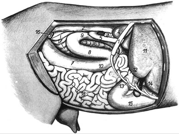
Figure 11.1 Location of the liver in the abdomen of the goat and relationship to other visceral structures. 1, thirteenth rib; 2, descending colon; 3, pancreas, right lobe; 4, right kidney; 5, descending part of duodenum; 6, distal loop of ascending colon; 7, cecum; 8-10, proximal loop of ascending colon (8, middle gyrus; 9, dorsal gyrus; 10, ventral gyrus); 11, liver; 12, cranial part of duodenum; 13, costal arch; 14, gallbladder; 15, abomasum; 16, ovary and uterine tube; 17, spiral loop of colon; 18, jejunum.
Source: Reproduced with permission from Popesko P. (2008). Atlas of Topographical Anatomy of Domestic Animals, new revised English edn, Bratislava: Vydavatelstvo Priroda, www.priroda.sk.Pancreas
The pancreas is located in close association with the portal vein in the craniodorsal abdomen to the right of the midline. It is composed of a larger right lobe, extending along the descending duodenum, and a smaller left lobe (Lukens 1938; Naranjo et al. 1986). The pancreatic duct enters the common bile duct (Robinson and Dunphy 1961). Histologically, the structure of the goat pancreas is similar to that of other domestic ruminants (Reddy and Elliott 1985; Lone et al. 1989).
Clinical Pathology
Enzymology
Hepatic parenchymal disease that causes a disruption of hepatocytes can lead to measurably increased serum levels of a number of intracellular hepatic enzymes in goats. Information about normal serum levels of these enzymes and their variation in goats due to age, breed, and sex is given in Table 11.1.
Increases in aspartate aminotransferase (AST) and lactate dehydrogenase (LDH) are not liver specific, and also result from muscle cell damage, as discussed in Chapter 4. Basal levels of sorbitol dehydrogenase (SDH) in goat liver and serum are much higher than those reported for cattle and sheep.
It is advised that alanine aminotransferase (ALT), formerly serum glutamic pyruvic transaminase (SGPT), commonly used as an indicator of hepatic disease in dogs and cats, should not be used in the evaluation of liver disease in ruminants because the livers of cattle, sheep, and goats contain low levels of ALT (Kramer 1989). Changes in serum ALT concentrations are highly variable and unpredictable in response to hepatic injury (Hanifa Moursi et al. 1979; Abu Damir et al. 1982; Jones and Shah 1982; Clark et al. 1984; Shimizu et al. 1986; El Dirdiri et al. 1987).
Concentrations of liver-specific enzymes are generally higher in acute liver disease than chronic liver disease and may be within normal limits in the later stages of prolonged hepatic disease, so careful interpretation of laboratory values in conjunction with clinical findings is essential.
Biliary stasis or obstruction can occur concurrently or independently of parenchymal damage, and different enzymes are used to evaluate cholestasis, namely alkaline phosphatase (AP) and gamma-glutamyl transferase (GGT). Increases in serum AP or GGT are associated with irritation or destruction of biliary epithelium and can occur in obstructive conditions of the biliary system, such as fas- cioliasis. While serum elevations of GGT are essentially liver specific, AP occurs in other tissues, especially bone, and serum concentrations may be elevated in young, growing animals because of normal osteoblastic activity or
Table 11.1 Some normal values reported for liver- associated enzymes and other blood parameters associated with liver disease.
Goat description (number
| Enzyme | Unit | of animals) | Mean ± SD | Range | Reference | ||
| Alanine aminotransferase | IU/L | General values | 24.0-83.0 | Kaneko (1980) | |||
| (ALT, SGPT) | IU/L | Adult, lactating F, Saanen and Alpine (79) | 18.7 ± 3.1 | 12.5-24.9 | Ridoux et al. (1981) | ||
| IU/L | Adult, lactating F, Saanen only (25) | 20 ± 3.6 | Ridoux et al. (1981) | ||||
| IU/L | Adult, lactating F, Alpine only (54) | 18.1 ± 2.7 | Ridoux et al. (1981) | ||||
| Albumin | g/dL | bgcolor=white>General values2.7-3.9 | Kaneko (1980) | ||||
| Alkaline phosphatase (AP) | IU/L | General values | 219 ± 76 | 93-387 | Kaneko (1980) | ||
| IU/L | Adult, lactating F, Saanen and Alpine (63) | 184±120 | 0.0-424 | Ridoux et al. (1981) | |||
| Ammonia | mmol/L | Adult Nubian goats (11) | 40.2-43.7 | Ali and Abu Samra (1987) | |||
| Arginase | IU/L | 0.5-2.5 years, F, breed not stated (bgcolor=white> | IU/L | Adult, non-lactating F, Alpine (35) | 217-417 | Garnier et al. (1984) | |
| IU/L | Adult, lactating F, Alpine (70) | 300-586 | Garnier et al. (1984) | ||||
| Ornithine carbamyl transferase | IU/L | Adult, lactating F, Saanen and Alpine (69) | 13.4 ± 5.5 | 2.4-23.4 | Ridoux et al. (1981) | ||
| IU/L | 1 year, lactating F, Saanen and Alpine (42) | 12.2 ± 5.3 | Ridoux et al. (1981) | ||||
| IU/L | Adult, lactating F, Saanen only (23) | 15.8 ± 4.3 | Ridoux et al. (1981) | ||||
| IU/L | Adult, lactating F, Alpine only (46) | 11.9 ± 5.1 | Ridoux et al. (1981) | ||||
| Sorbitol dehydrogenase | IU/L | Not specified | 19.4 ± 3.6 | 14.0-23.6 | Kaneko (1980) | ||
| IU/L | 4-month-old adult, MC and F, Saanen - feral (5) | 37 ± 7.2 | Kramer and Carthew (1985) | ||||
| IU/L | Adult, lactating and non-lactating F, Alpine (105) | 2.6-11.6 | Garnier et al. (1984) |
F, female; IU, international units; M, male; MC, castrated male; SD, standard deviation.
secondary to pathologic conditions of bone unrelated to biliary disease, as discussed in Chapter 4.
Thus, serum GGT is the preferred test for assessment of biliary integrity in goats. While the goat kidney contains high levels of GGT, the enzyme is passed in the urine in association with renal damage and does not enter the blood. High levels of GGT in the serum of newborn kids can serve as an indirect indicator of colostrum intake, as discussed further in Chapter 7 in the section on failure of transfer of passive immunity.Total bilirubin concentration in the serum of normal goats is generally reported to be in the range of 0.0-0.1mg∕dL (Kaneko 1980). Higher bilirubin concentrations in the range of 0.2-0.8, however, have been reported in normal Pygmy goats, with values varying with both age and sex (Castro et al. 1977). As in cattle and sheep, circulating bilirubin Iev- els increase only modestly in the face of severe, generalized hepatic disease (Sen et al. 1976). The most dramatic increases in serum or plasma bilirubin in the goat are due to hemolytic crises rather than liver dysfunction (Wasfi and Adam 1976).
Serum bile acids can be a useful indicator of liver dysfunction and may be a more reliable indicator of liver disease than either circulating bilirubin levels or transaminase activities (Kaneko 1980). Because the liver is responsible for the uptake, conjugation, and secretion of bile acids, there is a decreased secretion of bile acids into bile with liver dysfunction and an associated increase in blood concentrations. However, to date there has been little reported use of bile acids for diagnostic purposes in goats. In one case of hepatoencephalopathy in a 16-week-old female Nubian kid due to a congenital portosystemic shunt, an elevated serum bile acid concentration of 95 μmol∕L was noted, with a reference interval cited as 0-50 μmol∕L (Wilkerson et al. 2008). Serum bile acid concentrations have been reported for normal goats before, during, and after fasting, with no fasting effect on serum bile acid concentrations noted (Rudolph et al.
2000).Serum cholesterol levels have been used as an indicator of liver dysfunction in various species, but there are few reports in goats on variation of serum cholesterol levels in disease. Serum cholesterol levels in goats, and in some cases lipid profiles including high-density lipoprotein, low- density lipoprotein, and triglycerides, have been reported in relation to various physiologic and nutritional states, including pregnancy, lactation, fasting, and diet, but the results of similar studies are sometimes contradictory, so it appears that more assessment is needed before serum cholesterol levels and lipid profiles will be a useful diagnostic tool in goat medicine.
Liver Function Tests
Galactose elimination, a liver function test used in humans, is not appropriate in goats due to a high percentage of galactose loss in the urine (Treacher 1972). Sulfobromophthalein (Bromsulphalein, BSP) clearance is used to evaluate liver function in ruminants and other species. A delay in the clearance of this dye from the blood by the liver suggests functional impairment or decreased flow of bile. Focal liver diseases, such as abscesses, do not interfere with BSP clearance. Prolongation of clearance time in the goat is most often seen with generalized hepatic lipidosis (fatty liver) secondary to pregnancy toxemia, with chronic toxic hepatitis, or with biliary obstruction. The T1∕2 for BSP clearance in normal goats was reported as 2.13 ± 0.19 minutes (Sen et al. 1976). It increased to 4.04 ± 0.24 minutes subsequent to a single dose of carbon tetrachloride, to 16.5 minutes after repeated doses of carbon tetrachloride, and to more than 34 minutes after bile duct ligation.
In another study, Lal et al. (1991) used percent retention of BSP rather than BSP clearance to assess liver function. Normal goats had a mean percent retention time of 4.2 ± 0.34 at 10 minutes after intravenous injection of BSP dye at a dose of 5 mg/kg bodyweight (bw). Twelve hours after the induction of ruminal acidosis by feeding of wheat, percent retention of BSP in those goats increased to 15 ± 1.08.
Other Laboratory Tests
The liver is the primary site of albumin production, and hepatic insufficiency leads to hypoalbuminemia. Normal serum albumin levels in the goat are reported in the range of 2.7-3.9 g/dL (Kaneko 1980). Hepatic dysfunction can also cause hypoglycemia. Normal blood glucose levels in the goat are reported in the range of 50-75 mg/dL (Kaneko 1980). When goats are in negative energy balance, such as occurs in pregnancy toxemia or lactational ketosis, the liver produces increased ketone bodies that are detectable in serum, milk, or urine. Blood ammonia levels may be elevated in goats with generalized liver disease, particularly when neurologic signs are present, but it is not a consistently reported finding. In a case of hepatic encephalopathy associated with paratuberculosis in a goat, blood ammonia concentration was reported at 230 μmol∕L compared to a value of 27 μmol∕L in a healthy control goat (Rubin et al. 1999), while in a case of hepatoencephalopa- thy associated with portocaval shunt in a goat, plasma ammonia concentration reached 599 μg∕dL (352 μmol∕L), with a normal reference value range given as 25.5-109 μg∕ dL (15-64 μmol∕L) (Humann-Ziehank et al. 2001).
Beyond the identification of ketone bodies in urine, interpretation of urinalysis in goats for diagnosing liver disease has received little attention. Normal goat urine does not contain bilirubin, so presumably evidence of conjugated bilirubin in the urine may indicate obstructive liver disease or severe hemolytic disease. The absence of urobilinogen would suggest complete biliary obstruction, while increases in urobilinogen could represent active hepatocellular disease.
Diagnostic Procedures
Biopsy
Liver biopsy can be an aid in the diagnosis of suspected hepatic disease, particularly in cases of chronic liver disease when clinical findings suggest hepatic involvement but clinical chemistry results are equivocal. A prolonged BSP clearance time may also be justification for liver biopsy to determine the basis of impaired function. In focal hepatic disease, the liver biopsy may be misleading because a circumscribed lesion may be missed altogether.
Access to the liver for obtaining a biopsy sample is limited in the goat, and when possible ultrasound needle guidance is desirable. Unguided liver biopsies should be performed cautiously, using a transthoracic approach. Tranquilization of excitable individuals may be helpful. With the animal standing, the preferred site for introduction of the biopsy instrument is the right ninth intercostal space (Fetcher 1983). Entry should be dorsal to a line drawn from the point of the elbow to the craniodorsal angle of the paralumbar fossa. Entering at the eighth intercostal space can cause a puncture of the caudal lung lobe, while use of the tenth space may put the biopsy instrument behind the caudal edge of the liver. The correct site should be surgically prepared and a local anesthetic applied. The biopsy instrument must traverse the caudal thorax and penetrate the diaphragm before entering the liver. The instrument should be directed anteriorly after passing through the intercostal muscle to avoid puncture of the gallbladder, especially if the animal has been anorexic and the gallbladder is distended. It should not be necessary to introduce the biopsy instrument to a depth more than 3-4 cm, because the transthoracic space in this area is less than 2 cm and the liver thickness is approximately 4-5 cm.
Ultrasound
Ultrasonography can be helpful in identifying uncommon mass lesions in the liver such as abscesses, hydatid cysts, and rarely neoplasms. It has also been reported to be useful in the diagnosis of fatty liver in goats, with affected livers showing multiple hyperechoic foci of fatty infiltration distributed throughout an isoechoic liver parenchyma (Gonenci et al. 2003). When available, ultrasonography can be particularly helpful in performing accurate liver biopsies.
Cholecystography
Intravenous cholecystography has been performed successfully in goats using 20 mL of intravenous contrast material (biligrafin). In normal goats, the gallbladder can be visualized 30-60 minutes after injecting contrast media. It is located between the caudal border of the eighth rib and the cranial border of the twelfth rib, 6-8 cm below the vertebral column. In goats with liver disease, visualization may be delayed and the position of the gallbladder may shift subject to hepatomegaly (Singh et al. 1990).