Bacterial Diseases
Secondary bacteria, especially staphylococci, commonly invade almost any skin lesion on a goat. Thus, other etiologies should be ruled out before assuming that bacteria isolated from the surface of a lesion are causative.
Other organisms, such as Corynebacterium pseudotuberculosis and Dermatophilus congolensis, are usually significant if present. Foot rot, an interdigital dermatitis caused by Dichelobacter and Fusobacterium spp., is discussed in Chapter 4.Staphylococcal Dermatitis
Staphylococcal skin infections are common in goats and may be primary or secondary. The bacteria are also normal skin flora. In one Spanish study, 346 strains of staphylococci were isolated from axillary skin or udder of 133 healthy goats, and 21% of these isolates were coagulase positive (S. aureus and Staphylococcus hyicus; Valle et al. 1991).
Etiology
Impetigo is a superficial pustular dermatitis that does not involve hair follicles. Staphylococcal folliculitis is an infection and inflammation of hair follicles (Scott 1988). The species involved often has not been reported. Staphylococcal species isolated from goats with skin disease include S. intermedius, S. aureus, S. chromogenes, and S. hyicus (Scott 1988; Andrews and Lamport 1997; Mahanta et al. 1997). Species identification is not a good predictor of antibiotic sensitivity (Biberstein et al. 1984).
Clinical Signs and Diagnosis
The primary lesion is a non-follicular or follicular papule that develops into a pustule. Lesions may enlarge or coalesce, discharge purulent or serosanguinous exudate, and become encrusted (furunculosis; Scott 2018). Alopecia and scaling are prominent in the chronic or healing stage. Multiple small pustules of impetigo frequently appear on the teats and udder (see Figure 14.5) or perineum and underside of the tail. Because these pustules may be preceded by vesicles and followed by scabs, they may be confused with lesions of contagious ecthyma (Smith 1981).
Direct smears show degenerate neutrophils with phagocy- tosed cocci (Scott 2018). When lesions remain localized they are relatively benign and self-limiting, except that lesions on the teats predispose to staphylococcal mastitis. Fly bites on the udder (fly worry) are said to resemble a staphylococcal infection, but to be more pruritic (Matthews 2016).In some goats the infection becomes general, involving the skin of the abdomen, inner thighs, and even the neck and back. Other distributions are possible, especially if the staphylococci are secondary to another condition, such as chorioptic mange. Periocular alopecia and crusts comprise yet another possible manifestation (Scott 1988). Confusion with mycotic infections or nutritional deficiencies is possible.
A presumptive diagnosis is often based on inspection alone. Gram stains and cultures document the presence of staphylococci, while evaluation of skin biopsy specimens should help to rule out other diseases that might have been primary and still require therapy.
Treatment and Prevention
Localized lesions on the udder may be washed with an iodophor or chlorhexidine shampoo, dried, and then coated with an antiseptic or antibiotic ointment. Affected does should be milked last. Single-service paper towels and attention to hand washing by the milker decrease the risk of spread to other does. Rubber gloves protect against transmission of infection to humans and goats. Similar treatment of lesions around the tail is possible, but these seem to be less important and frequently heal spontaneously.
If a generalized infection is suspected, culture of the organism and determination of antibiotic sensitivity are recommended. Systemic antibiotic treatment (one to two weeks of therapy) may start with penicillin (20 000 IU/kg/d intramuscularly), pending results of sensitivity testing. As the concern over methicillin-resistant staphylococci increases in both veterinary and human medicine, it is very important for the practitioner to remember that many of the antibiotics used to control these infections in other species are forbidden by law in all sheep and goats in the United States, because of their status as food animals.
Thus, no matter what the laboratory reports for a sensitivity pattern, chloramphenicol, fluoroquinolones such as enrofloxacin, and glycopeptides such as vancomycin are absolutely forbidden in all goats.Autogenous bacterins may be tried for control of chronic or epizootic infections (Scott et al. 1984a), but bacterins have not received scientific evaluation in caprine dermatology.
Dermatophilosis
Dermatophilosis, also known as streptothricosis, is a common skin infection in goats worldwide. Cattle, sheep, horses, various wildlife species, and humans are also affected (Stewart 1972; Hyslop 1980).
Etiology
Dermatophilus congolensis is a Gram-positive, pleomorphic, facultative anaerobic actinomycete. It produces motile zoospores that invade the skin.
Pathogenesis
D. congolensis may survive in soil or in dust on an animal's hair coat during dry weather. It is introduced into the epidermis following injuries of any sort, including those caused by tick bites and thorny vegetation. Its life cycle is activated by moisture (Bida and Dennis 1976). Outbreaks often occur during periods of heavy rain or high humidity (Memery and Thiery 1960; Yeruham et al. 2003).
Clinical-Signs
Several localizations of the disease have been reported. Ears are commonly involved, especially in young kids (Larsen 1987). Tiny wart-like scabs first appear on the inner hairless surface of the ear pinna. They are easily rubbed off, exposing dry, circular, light-colored areas beneath. Raised scabs with matted hairs form on the external portions of the ears and are more tightly attached (Figure 2.8). The lesions are non-pruritic and benign and last two to three months in kids if not treated (Munro 1978).
Other affected areas of the body include the nose, muzzle, feet, scrotum, and underside of the tail (Memery 1960; Yeruham et al. 2003; Loria et al. 2005; Dalis et al. 2009: Scott 2018; Blick et al. 2019). These areas of skin frequently are exposed to moisture or mild abrasion from vegetation.
Thick proliferative crusts may be mistaken for lesions of contagious ecthyma (sore mouth; Tiddy and Hemi 1986). In fact, the simultaneous presence of both diseases has been reported in splenectomized kids (Munz 1969, 1976); in Yaez goats, a cross between domestic goats and wild ibex (Yeruham et al. 1991); and in a herd outbreak in Boer goats (Blick et al. 2019). The dry crusts, scaling, and alopecia of healing or chronic lesions resemble ringworm. Secondary bacterial infections (e.g., staphylococci, corynebacteria, Fusobacterium necrophorum) are to be expected and may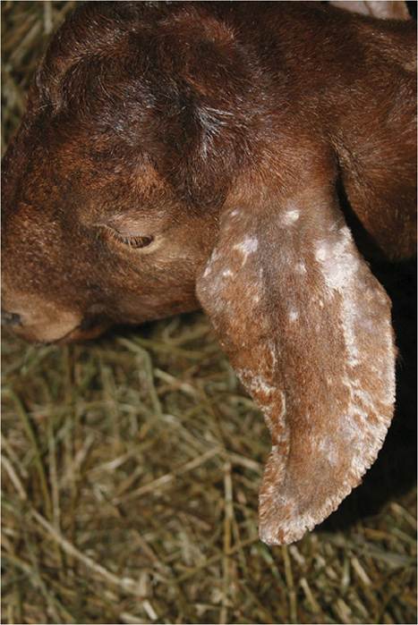
Figure 2.8 Dry scabs typical of dermatophilosis on the external surface and margin of the ear. Source: Courtesy of Dr. M.C. Smith.
lead to pruritus or pain. Sometimes the entire dorsum of the goat is involved, with lesions clinically resembling rain scald in horses; continuous exposure to wet weather presumably is an important etiologic factor (Bida and Dennis 1976; Scott et al. 1984a). Damage to hides can be extensive. Suppurative lymphadenitis has been reported in Beetal goats from which D. congolensis was demonstrated by smear and culture (Singh and Murty 1978).
Diagnosis
The diagnosis can be confirmed in several ways. When lesions are moist, an impression smear of the underside of a scab reveals Gram-positive branching filaments, either unsegmented or in railroad track arrangements of two to eight parallel rows of cocci (Scott 2018). Giemsa or Diff Quik stain may also be used. The organisms in smears also fluoresce under ultraviolet light after stain - ing with acridine orange (Mathieson 1991). Fluorescent antibody techniques have been used for rapid identification of the organism in smears of exudate (Pier et al. 1964). In dry lesions, skin biopsy is necessary to demonstrate the organism. In addition to superficial exu - date, there is hyperkeratosis and infiltration of the epi - dermis and hair follicles with neutrophils.
Bacterial filaments in the biopsy sample are periodic acid-Schiff (PAS) positive (Loria et al. 2005).Culture of the organism by a diagnostic laboratory will also confirm the diagnosis, but this is best done under microaerophilic conditions with increased carbon dioxide (Scott 1988). Scabs are ground up in saline and cultured at once and also after 24 hours at room temperature. A medium that selects for Gram-positive organisms (such as colistin-nalidixic acid medium) is helpful. Tiny gray adherent colonies composed of branched mycelia may be visible after 48 hours and should then be subcultured; it is common for the original plate to be rapidly overgrown by contaminants.
Several serologic tests have been used to identify antibodies against D. congolensis. The purpose was to potentially monitor the prevalence of exposed animals. The tests have included passive hemagglutination (which showed 23% of slaughtered goats in a Nigerian study to be seropositive; Oyejide et al. 1984) and radial immunodiffusion (Makinde 1980). Where available, a PCR test can be used to confirm presence of the organism (Chitra et al. 2017).
Therapy and Prevention
Penicillin-streptomycin was commonly recommended for dermatophilosis in individual animals in the past, but this product is no longer available in the Unitisd Stateis. Tetracycline is also effective at a dose of 20 mg/kg Subcutaneously or intramuscularly once. Where feasible, shelter from the rain and bathing (iodophors, 2-5% lime sulfur) and grooming to remove crusts should be recommended. Improved nutrition and control of external parasites (especially ticks) are also desirable for treatment and prevention. Cleansing thick crusts with hydrogen peroxide will help to control secondary anaerobic infections. Brushes should be disinfected before being used on other animals. Goat handlers should also be warned that people are occasionally infected with this organism. Carrier animals appear to be the reservoir for the agent, but the organism can also survive for many months in the environment.
Vaccination against der- matophilosis has not been successful (Bida and Dennis 1976), and recovery does not appear to provide immunity.Corynebacterium pseudotuberculosis
C. pseudotuberculosis is usually associated with lymph node enlargement (caseous lymphadenitis) and is discussed in detail in Chapter 3. However, small nodules and draining tracts in the skin sometimes occur in goats (Scott 2018) and may be a source of infection to others. Diagnosis is made by culture and by skin biopsy, which reveals a tuberculoid granulomatous reaction (Scott 1988). Affected animals should be isolated or culled.
ActinobaciLLosislActinomycosis, Mycobacteriosis, and Protothecosis
Actinobacillosis, a suppurative to granulomatous disease of sheep caused by Actinobacillus Iignieresii, has not been well documented in goats, but the organism is occasionally isolated from abscesses of the face and mouth of goats (Matthews 2016). Diagnosis is based on demonstration of cheese-like granules less than 1 mm in diameter in pus or by aerobic or anaerobic culture of the organism. In direct smears, club-like bodies radiate from the center of the granules and crushing reveals small Gram-negative bacilli. Rinsing and culturing the granules, rather than simply swabbing a fistulous tract, are recommended to avoid overgrowth with secondary bacteria (Scott 1988). Treatment, at least for sheep, typically involves sodium iodide (20 mg/kg of sodium iodide as a 10% solution intravenously or subcutaneously) weekly for four to five weeks and streptomycin (20 mg/kg/day) for five to seven days.
A case of pyogranulomatous dermatitis has been reported on the udder of an aged dairy goat. A diagnosis of Actinomyces sp. was based on the appearance of the organism in Gram-stained smears and the presence of sulfur granules. Raised knot-like lesions were yellowish brown or reddish black, and abscesses extended into the parenchyma of the udder. Udder amputation was the proposed therapy, but the goat died first (Hotter and Buchner 1995).
A tuberculoid granulomatous dermatitis with dark crusted nodular lesions and ulceration as well as Langhans-type multinucleated cells has been reported on the teats and udder skin of goats in France. A mycobacterial pathogen closely related to Mycobacterium leprae was identified by PCR techniques (Chartier et al. 2016).
A case of pyogranulomatous dermatitis around the nares caused by a Prototheca sp. has been reported from a mature goat in Brazil (Macedo et al. 2008). Ulcerated nodules contributed to inspiratory dyspnea and weight loss. Oval to spherical, non-budded, walled sporangia typical of this algal-like organism that lacks chlorophyll were demonstrated in histologic section. A similar case of Prototheca Wickerhamii infection involving the nostrils and skin of the face of an immunocompetent adult goat in Brazil was treated unsuccessfully with long- term fluconazole (Camboim et al. 2011). Various antifungal agents are used to treat human and canine cutaneous protothecosis, but euthanasia is recommended for affected goats.