Etiologic Diagnoses
Readers who desire a more exhaustive reference list for any of the conditions described below should consult D.W. Scott's Large Animal Dermatology textbook (1988). Several review papers also discuss dermatologic diseases of goats (Smith 1981, 1983; Mullowney and Baldwin 1984; Scott et al.
1984a, b; Manning et al. 1985; Jackson 1986; Corke and Matthews 2018) and many are illustrated in a recent text (Scott 2018).Viral Diseases
Contagious ecthyma and capripox viruses cause prominent skin lesions in goats. Virus infections involving other body systems also may have cutaneous manifestations. Warts (cutaneous papillomas) in goats have not been conclusively proven to be of viral origin and are discussed under neoplastic conditions.
Contagious Ecthyma
Contagious ecthyma is a contagious, zoonotic disease of goats and sheep (and camelids) that has several alternative names, including orf, soremouth, scabby mouth, and contagious pustular dermatitis. It has worldwide distribution.
Etiology and Epidemiology
The cause is an epitheliotropic parapoxvirus that enters the goat through skin abrasions (Mayr and Buttner 1990). The virus replicates in proliferating keratinocytes in the damaged epidermis (McKeever et al. 1988) and then causes a primary viremia to lymph nodes, bone marrow, and liver. In some cases, the virus then becomes generalized, with a second viremic phase, and spreads to the head, extremities, udder, genitals, lungs, and liver (Mayr and Buttner 1990). The morbidity in young kids often approaches 100%, while mortality from starvation and secondary infections may be as high as 20% (Van Tonder 1975), but is usually much lower. In one outbreak that followed introduction of kids from an infected herd to a naive herd without quarantine, 93% of 38 suckling kids died because of feeding difficulties (Mazur and Machado 1989).
Scabs that fall to the ground during resolution of lesions have long been incriminated as the source of infection to other animals months or even years later (McKeever and Reid 1986), and this is indeed possible if the environment remains dry. More recently, persistently infected carrier sheep, some of which are asymptomatic, have been demonstrated to be an important source of contagion (Lewis 1996). Presumably carrier goats also occur, and infection can be activated by stress (Mayr and Buttner 1990).
Clinical Signs
The incubation period is three to eight days (Mayr and Buttner 1990). Papules progress rapidly to vesicles, pustules, and scabs. Crusty, proliferative lesions typically form on the lips, but can also affect the face, ears, coronary band, scrotum, teats, or vulva. In one outbreak, where exposure presumably occurred in contaminated pens at a show, in seven goats lesions occurred on the neck, chest, and flanks rather than on the lips or teats (Coates and Hoff 1990). In another case report, lesions were most common on haired skin of adult goats and the first crusty scabs noticed were on the caudal aspect of the hind legs (Moriello and Cooley 2001). The scabs frequently harbor secondary bacteria (such as staphylococci) or even screwworm maggots (Boughton and Hardy 1934). Sometimes large masses of granulation tissue develop under the scabs. Lesions regress in three or four weeks.
Most adult goats with lesions on the lips (Figure 2.4) continue to eat and milk well. Occasional goats, especially young kids exposed to other diseases or management deficiencies, will develop generalized lesions or severe secondary bacterial infections. Lesions on the teats of milking animals may compromise the health of the sphincter and predispose to bacterial mastitis. Associated pain may cause the doe to reject nursing efforts by its kid.
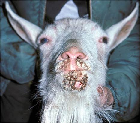
Figure 2.4 Healing crusts from contagious ecthyma on the muzzle of a mature doe.
One large scab has fallen off, leaving healthy skin beneath. Source: Courtesy of Dr. M.C. Smith.
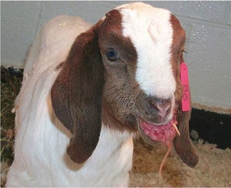
Figure 2.5 Severe contagious ecthyma lesions on the gums of a Boer kid. Source: Courtesy of Dr. M.C. Smith.
Severe generalized and persistent proliferative lesions have been seen in Boer goats and their crosses in the United States (Figure 2.5). Draining lymph nodes are markedly enlarged in these animals, and thymic atrophy is often present. Preliminary research has not proven whether this variation represents a viral strain difference or a difference in immune response of the affected Boer goats (de la Concha-Bermejillo et al. 2003; Guo et al. 2003; Avra et al. 2018). Close examination of many sheep and goats with a more typical presentation of contagious ecthyma often reveals a mild lymphadenopathy.
Diagnosis
Diagnosis is usually based on clinical signs alone, although electron microscopy or immunologic techniques to demonstrate antigen in scabs or serology could be used for confirmation or to rule out capripox infection (Robinson and Balassu 1981). More recently, polymerase chain reaction (PCR) tests have been developed to identify the virus (Mondal et al. 2006; Nandi et al. 2011). In lambs experiencing contagious ecthyma, serum antibodies often do not appear until after reexposure (Mayr and Buttner 1990). Similarly, rural physicians usually make the diagnosis in their human patients on clinical signs alone, but urban dermatologists lacking experience with the disease may insist on biopsy for histologic or electron microscopic examination (Gill et al. 1990; Centers for Disease Control and Prevention 2006). Skin biopsies of ruminants reveal ballooning degeneration of keratinocytes and eosinophilic intracytoplasmic inclusions (Robinson and Balassu 1981; Scott 2018), although the inclusions are not always detectable (Housawi et al. 1993). The crusts consist of multiple layers of necrotic cellular debris and neutrophils.
Histopathology helps to distinguish contagious ecthyma from the convalescent stages of peste des petits ruminants (PPR). Capripox, foot and mouth disease (FMD), dermat- ophilosis, and staphylococcal dermatitis are other differentials to consider (Nandi et al. 2011). Diagnostic tests for contagious ecthyma, including differentiation from other diseases, have been reviewed by Spyrou and Valiakos (2015).Therapy
The possible beneficial effects of treatment must be weighed against the danger of zoonotic infection (Figure 2.6). Any person handling an affected goat should wear gloves. Numerous products have been used topically,
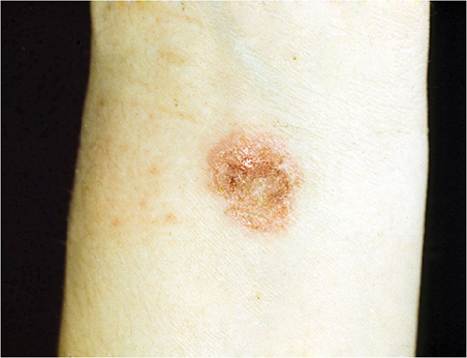
Figure 2.6 Orf (contagious ecthyma) lesion on the author's wrist. Source: Courtesy of Dr. M.C. Smith.
with anecdotal reports of faster healing. However, these products have been used with minimal consideration of meat and milk residues. These include kerosene mixed with lard, penetrating oil spray (WD-40, WD-40 Company, San Diego, CA, USA), and bismuth subsalicylate (Pepto- Bismol, Procter & Gamble, Cincinnati, OH, USA). Parapoxviruses are said to be sensitive to ether (Avra et al. 2018). Systemic antibiotics are indicated if secondary bacterial infections are severe. An udder salve is indicated to keep scabs on the teats pliable. If painful, proliferative lesions within a kid's mouth cause feeding to decrease, the kid could be anesthetized and subjected to debridement (electrocautery after spray cryotherapy). This approach has been used in lambs, with good results (Meynink et al. 1987).
Vaccination
Commercially available vaccines often are unattenuated live virus preparations (basically ground-up scabs) or tissue culture strains, although the level of protection afforded by the latter appears to vary with the strain (Pye 1990). In one study (Musser et al. 2008), a vaccine prepared from goat strains of the virus seemed to protect the goats better from wild-type virus than did commercial sheep-strain vaccines.
Caution is advised, however, before introducing a particularly virulent vaccine strain to a farm experiencing mild disease. An autogenous vaccine can be made by crushing, in saline, a few grams of scabs between two spoons or with a mortar and pestle. The suspension is filtered through cheesecloth and a few drops of antibiotic solution such as penicillin/streptomycin are added to control bacteria (Bath et al. 2005). The skin in a hairless, protected area is lightly scarified and the virus suspension is rubbed in. Sites for vaccination include the inside of the ear pinna, the underside of the tail, or the axilla. Avoid the medial aspect of the thigh, because the infection can be spread to the lips by chewing and to the udder and teats by direct contact (Lewis 1996). Scabs appearing at the vaccination site in one to three days indicate a “take.” Monitoring this reaction as evidence of continued vaccine viability permits owners to economize by freezing leftover vaccine for later use. If some animals in the herd develop vaccination scabs but others do not, a preexisting immunity is probably responsible for absence of a take.Where a newer, parenteral vaccine is available, subcutaneous vaccination with a live cell culture vaccine avoids postvaccinal disease or excretion of the virus. Use of this vaccine every 6-12 months has been recommended in noninfected herds, and in the face of an outbreak (Mayr and Buttner 1990).
In countries where capripox virus exists, vaccinating goats for capripox sometimes provides solid immunity against contagious ecthyma, whereas vaccination or natural infection with the contagious ecthyma virus provides no protection against capripox (Sharma and Bhatia 1958).
There are several controversies associated with vaccination. The first is whether to recommend vaccination in a herd that is not endemically infected. The vaccine, because it is unattenuated, will introduce the disease to such a herd. In herds in which buying or showing of goats occurs regularly, vaccination prevents the occurrence of an outbreak during the show season or in milking animals.
It is important to vaccinate at least six weeks before the show season so that vaccine scabs will be gone before the first show. (Presumably this procedure would increase the prevalence of subclinical carriers at shows and thereby increase the risk to unvaccinated animals in attendance.) When soremouth has appeared on the premises, it may be desirable to vaccinate all as yet unaffected goats to limit the duration of the outbreak. A program of vaccination for all young kids, often in conjunction with annual revaccination of late pregnant adults, is then established. Disinfection of the pens after all lesions have cleared is recommended if the owner chooses not to follow a routine vaccination program. Suitable disinfectants include 5% creolin solution, formalin, detergents, and commercially available virucidal disinfectants (Mayr and Buttner 1990).The occurrence of colostral immunity in kids from vaccinated animals is disputed (Robinson and Balassu 1981). French enterprises that assemble kids from many sources, however, have found it advisable to pay a premium for kids from vaccinated dams. This is because vaccination of the dam seems to be more effective than vaccination of kids at birth in preventing adverse effects of the disease on the quality of kid skins (Faure 1988). In an experimental study in Mexico, kids born to dams vaccinated (virulent vaccine) in late pregnancy were challenged by skin scarification with virulent virus. Kids younger than 45 days old resisted challenge, whereas kids older than 45 days developed characteristic lesions (Tortora Perez 1989).
Work with sheep has suggested that vaccinating at the time of drying off is preferable to vaccinating later in pregnancy when lambs are to be raised by the ewes. Lymphocytes migrating to the udder at the end of lactation produce antibodies in the milk that may protect the lips and mouth of nursing lambs (Le Jan et al. 1978).
Capripox
The malignant pox diseases of sheep, goats, and cattle are not host specific, although they show host preferences. Strains can be distinguished by restriction endonucleases, but not by several serologic tests (Black 1986). Currently all strains are included in the Capripox genus of poxvirus.
Etiology and Pathogenesis
Capripoxviruses are distinct from parapoxviruses. They are acid labile and sensitive to lipid solvents. Malignant sheep and goat pox infections occur in the Middle East, Turkey and Greece, Far East, and Africa north of the equator (Davies 1981; Babiuk et al. 2008). A benign form of goat pox has been reported from California (Renshaw and Dodd 1978) and Scandinavia (Bakos and Brag 1957), but the agents involved were not confirmed to be capripox viruses (Committee on Foreign and Emerging Diseases 2008). Skin lesions and scabs are major sources of virus. The virus resists desiccation and may survive in scabs for at least three months. Transmission is often through skin abrasions or by inhalation, with an incubation period of three to eight days. Viremia occurs, and the virus is carried to other sites in the skin, regional nodes, spleen, kidney, and lungs. The virus is excreted from skin lesions and in nasal and ocular exudates and milk. Night herding (congregating herds at night for protection) and stabling favor spread of the disease. Wild ungulates are not thought to serve as reservoirs.
Clinical Signs
The severity of signs varies with the strain of capripox. Young animals are most severely affected. Early signs include rhinitis, conjunctivitis, and pyrexia 104-107.6 °F (40-42 °C). The animals stand with arched back and are anorectic. Cutaneous lesions (reddish macules and papules, 0.5-1.5 cm diameter, Figure 2.7) and lesions on the external nares and lips and within the mouth appear one or two days later. Skin lesions persist for four to six weeks. In some outbreaks, vesicular lesions of the skin coalesce. Oral lesions on the tongue and gums tend to ulcerate. Regional
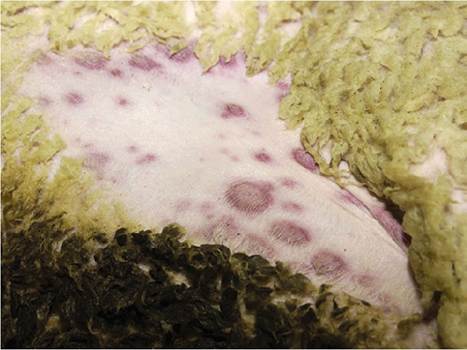
Figure 2.7 Early macules of capripox infection on the skin of an experimentally infected sheep. Source: Courtesy of National Veterinary Services Laboratories, Ames, IA, USA.
lymph nodes may be enlarged up to eight times their normal size (Committee on Foreign and Emerging Diseases 2008). Animals that die frequently have lesions in the lungs and alimentary tract.
The hair is erect over skin lesions, the skin is thickened, and crusts of exuded serum form on the surface. Healing may leave an ulcer and then a permanent scar after the full skin thickness sloughs. Damage to hides causes important economic losses.
When the disease first enters a susceptible flock, morbidity may be more than 75%, and 50% of affected animals die. Mortality may increase to 100% in kids or when superimposed on other virus infections such as PPR. The morbidity rate is lower in endemic flocks. Some animals convert serologically without development of clinical signs. Imported breeds, notably European breeds, are generally more severely affected than indigenous breeds in endemic areas (Karim 1983; Kitching 1986). Animals that recover from the disease are immune for life (Babiuk et al. 2008).
There is also a nodular form (“stonepox”) in sheep and goats that resembles lumpy skin disease of cattle (also caused by a capripox; Patnaik 1986). Vesicles and pustules are absent and there is no cross-immunity with more typical strains of goat pox or with contagious ecthyma. The virus is present in blood and skin throughout the course of the disease, which is often fatal (Haddow and Idnani 1948). This viral goat dermatitis, apparently restricted to the Indian subcontinent, reappeared briefly in Pakistan in 2004 (Muhammad et al. 2008).
In the benign goat pox form, vesicles and pustules develop from papules on the lips and udder (and sometimes on the perineum and inside of the thigh). Pock lesions heal in five to eight weeks, leaving behind permanent scars.
Goat pox, like contagious ecthyma, has been considered to be a zoonotic disease (Bakos and Brag 1957; Sawhney et al. 1972), but more recent authors dispute this (Committee on Foreign and Emerging Diseases 2008).
Diagnosis
Capripox is most likely to be confused with contagious ecthyma because lesions may be limited to the lips, oral mucous membranes, or udder. Electron microscopy (Hajer et al. 1988) and serologic tests (such as immunodiffusion and serum neutralization) readily differentiate capripox from the parapoxvirus of contagious ecthyma. Histopathology reveals large eosinophilic intracytoplasmic inclusions, vasculitis, thrombosis, and necrosis (Davies 1981).
Control
Import restrictions covering animals and animal products from endemic areas are required to avoid introduction of this disease to non-infected regions. Quarantine and slaughter of diseased and contact animals would be recommended if introduction occurred (Babiuk et al. 2008). A carrier state has not been documented to occur.
In endemic countries, vaccination is the only effective method of disease prevention (Bhanuprakash et al. 2011). Prophylactic vaccination reduces morbidity in these (often nomadic pastoral) regions. Most trials have shown excellent cross-protection with various strains of sheep and goat pox (Davies 1981). Live, attenuated vaccines (using a mild strain) are preferred, but difficult to distribute (Kitching 1986). An experimental subunit vaccine reduced the severity of signs without risking introduction of the disease (Carn et al. 1994). A recombinant capripoxvirus vaccine has been produced that protects goats against PPR as well as against capripox (Romero et al. 1995). Use of autogenous vaccines may increase the incidence of disease (Das et al. 1978). Contagious ecthyma vaccines do not protect against goat pox.
Miscellaneous Virus Infections
As already discussed under the heading of pruritus, goats with rabies or pseudorabies may show skin lesions that result from severe pruritus. These conditions are discussed in Chapter 5. Scrapie in goats (also discussed under neurologic diseases in Chapter 5) may be pruritic, as demonstrated by biting and rubbing at the legs, flanks, lumbar region, and neck, and by alopecia in these areas (usually without scab formation). The clinical course of scrapie may last three to four months (Hadlow 1961; Brotherston et al. 1968; Harcourt and Anderson 1974).
Peste des Petits Ruminants
PPR is a morbillivirus infection that causes serious losses in sheep and goats throughout its range (Committee on Foreign and Emerging Diseases 2008). The major clinical signs of stomatitis, enteritis, and pneumonia are described in the appropriate chapters. During early stages of the disease, the lips are edematous and brown scabs cover eroded and ulcerated epithelium. Goats that survive the acute phase of the disease may develop labial scabs that persist for up to 14 days; histologically, acanthosis and hyperkeratosis are evident. Necrotic epithelium is infiltrated with degenerating neutrophils. There is no papilliform proliferation or ballooning degeneration typical of contagious ecthyma, although lesions are grossly similar (Whitney et al. 1967; Abraham et al. 2005). Syncytial multinucleated giant cells and eosinophilic cytoplasmic inclusions may be seen in the epithelium (Cam et al. 2005). Goats that are vaccinated with inactivated vaccine (Nduaka and Ihemelandu 1975) or that are reexposed to PPR after recovery from the virus also develop labial scabs that heal in about 10 days (Ihemelandu et al. 1985). In these animals, histology reveals proliferation of macrophages and lymphocytes, suggesting an immune response.
Bluetongue
Bluetongue is a disease of sheep and cattle caused by an orbivirus that has at least 27 serotypes and is spread by Culicoides insects. Signs in sheep include fever, stomatitis, coronitis, and birth of lambs with congenital brain anomalies. Goats are susceptible to bluetongue in that viremia and fever occur and antibodies develop (Luedke and Anakwenze 1972; Backx et al. 2007), but overt clinical signs are rarely seen or described in goats in the United States. During an outbreak in cattle in Israel, two Saanen goats were found with swollen lips and marked salivation (Komarov and Goldsmit 1951). During the recent outbreak of bluetongue in northwestern Europe, a small number of goats developed edema of the lips and head, small scabs on the nose and lips suggestive of mild contagious ecthyma, and erythema of the udder skin (Dercksen et al. 2007). Goats may serve as a natural reservoir for the bluetongue virus (Erasmus 1975). Virus isolation and serology help to distinguish bluetongue from FMD and PPR, and historically from rinderpest, which was globally eradicated in 2011. Bluetongue is discussed in detail in Chapter 10.
Caprine Herpesvirus
Experimental inoculation of kids with a herpesvirus isolate has produced vesicles, ulcers, and crusts on the muzzle and feet (Waldvogel et al. 1981). Ulcers also occurred in the mouth, esophagus, rumen, and intestines. In one naturally occurring outbreak, numerous foci of necrosis and hemorrhage were found in the skin of a single kid (Mettler et al. 1979). Any histologic finding of acidophilic intranuclear inclusion bodies in epithelial cells suggests the possibility of herpesvirus infection. The disease is discussed in Chapters 12 and 13.
Foot and Mouth Disease
The picornavirus that causes FMD is a very important and exceedingly contagious disease of cattle in South America, Europe, Africa, and Asia, but is currently absent from the United States, Canada, and Australia. Signs in cattle include fever, stomatitis with vesicles and bullae, anorexia, agalactia, and a very prolonged convalescence. The disease in sheep and goats is usually mild, and only important in that these animals and meat from them may transmit the disease to cattle. However, during outbreaks of FMD, other (often idiopathic) oral lesions can cause great concern to regulatory authorities, at least in sheep (Watson 2004). Lameness is often the most pronounced clinical sign in goats; vesicles or bleeding ulcers may be found in the interdigital space or at the coronary band (McVicar and Sutmoller 1968; Mishra and Ghei 1983). Goats are routinely vaccinated in endemic regions (Mansoor et al. 2018). The disease is discussed in detail in Chapter 4.
Vesicular Stomatitis
Vesicular stomatitis is a rhabdovirus disease primarily affecting horses, cattle, and swine, and limited to the Western Hemisphere. Typical signs in these species are oral vesicles and ulcers, salivation, coronitis, and teat lesions. Regulatory officials should be notified so that the disease may be differentiated from FMD. The epidemiology is poorly understood, but may include insect vectors such as sand flies (Lutzomyia), midges (Culicoides), and black flies (Simulidae) (Committee on Foreign and Emerging Diseases 2008). Goats are considered to be resistant, but, according to unpublished reports, vesicular stomatitis in goats has been accompanied by vesicles at the commissures of the lips, which must be differentiated from early lesions of contagious ecthyma.
Malignant-Catarrhal-Fever
Infection of goats with the gammaherpesvirus ovine herpesvirus-2, the causative agent of sheep-associated malignant catarrhal fever in cattle, has rarely been reported to cause cutaneous lesions in goats. Erythematous papules on the distal limbs progressed to generalized erythema, scaling, and alopecia without pruritus (Foster et al. 2010). The skin was infiltrated with macrophages and multinucleated giant cells; diagnosis was by PCR testing for the sheep virus. Contact with sheep, which commonly carry the virus subclinically, is the most likely source.