Bacterial Diseases
Mycoplasma Arthritis
Mycoplasma infections account for serious morbidity and mortality in goats throughout the world. Several distinct disease entities are recognized, notably contagious caprine pleuropneumonia (CCPP), contagious agalactia, and infectious keratoconjunctivitis.
However, mycoplasma infections are usually septicemic, and polyarthritis frequently occurs in these and other Mycoplasma spp. infections of goats. The multisystemic effects of mycoplasma infection occur concurrently with sufficient frequency that a specific term, MAKePS syndrome, has been proposed for the constellation of signs that may be observed, namely mastitis, arthritis, keratitis, pneumonia, and septicemia (Thiaucourt and Bolkse 1996). The discussion here will focus mainly on manifestations of arthritis.Etiology
The study of caprine mycoplasmosis remains an active and dynamic field, with new species and strains being identified and old species and strains being renamed and reclassified, due in large part to the application of genetic sequencing techniques (Fischer et al. 2012). The frequent identification of new isolates, coupled with historical problems of erroneous taxonomic identification of old isolates, has produced some considerable confusion about the various causes of caprine mycoplasmosis. The status of important mycoplasma infections in goats has been reviewed (Ruffin 2001; Al-Momani and Nicholas 2006; Yatoo et al. 2018).
Genera of the prokaryotic class Mollicutes that infect goats include Mycoplasma, Ureaplasma, and Acholeplasma. Species associated with goats are identified in Table 4.4, with the main pathogens being Mycoplasma spp. Several of these belong to the so-called Mycoplasma mycoides cluster, a group of ruminant pathogens with shared biochemical,
Table 4.4 Mycoplasma spp. isolated from goats.
Infected tissues
Mycoplasma species Other hosts Primary Other
Associated diseases in goats Pathogenicity
Geographic distribution
| Mycoplasma capricolum subsp. Capripneumoniae (formerly strain F38) | Goats only | Respiratory tract | Potential for septicemia | Contagious caprine pleuropneumonia (CCPP) | High | Currently Near East, Africa |
| M. capricolum subsp. capricolum | Sheep | Joints | Udder, lungs, ears, urogenital tract, eyes; septicemia possible | Polyarthritis, mastitis, pneumonia, neonatal death, keratoconjunctivitis | High | Australia, India, Europe, United States, Egypt |
| Mycoplasma mycoides subsp. capri | Goats only | Respiratory tract | Joints, ears | Pleuropneumonia, arthritis | Moderate | Africa, Middle East, western Asia, southern and eastern Europe, North America |
| M. mycoides subsp. mycoides (large-colony type)a | Rare in sheep; very rare in calves | Respiratory tract | Udder, joints, eyes, ears, potential for septicemia | Pleuropneumonia, mastitis, arthritis, keratoconjunctivitis, neonatal death, abortion | Moderate | Europe, Africa, Asia, Australia, North America |
| Mycoplasma agalactiae | Sheep | Udder | Joints, eyes, urogenital tract, ears, rarely lungs | Contagious agalactia (CA), arthritis, pneumonia, keratoconjunctivitis, vulvovaginitis | High | Europe, United States, former USSR, Asia, North Africa |
| Mycoplasma arginini | Sheep, chamois, many others | Respiratory tract | Urogenital tract, joints, eyes | Pneumonia, arthritis, keratoconjunctivitis, vulvovaginitis | Very low or non-pathogenic | Worldwide |
| Mycoplasma conjunctivae | Sheep, chamois | Eyes | Respiratory tract, rarely joints | Keratoconjunctivitis, pneumonia, arthritis | Moderate | Worldwide |
| Mycoplasma ovipneumoniae | Sheep | Respiratory tract | Eyes, urogenital tract | Initiator of pneumonia | Low | Worldwide |
| Mycoplasma putrefaciens | Goats only | Udder | Joints, ears | bgcolor=white>Mastitis, arthritisVariable | United States, France, Australia | |
| M. mycoides subsp. mycoides (small-colony type) | Primarily cattle; very rare in goats | Respiratory tract | Joints | Pleuropneumonia, polyarthritis | Unknown | Africa |
| Mycoplasma bovigenitalium (same as ovine/ caprine M. serogroup 11)b | Primarily cattle and buffaloes; also sheep and goats | Reproductive tract | Udder, joints | Vulvovaginitis, cervicitis, endometritis, epididymitis, oophoritis, mastitis, arthritis | Variable with strain | Worldwide |
Table 4.4 (Continued)
| Mycoplasma species | Other hosts | Infected tissues | Associated diseases in goats | Pathogenicity | Geographic distribution | |
| Primary | Other | |||||
| Mycoplasma yeatsii, Mycoplasma auris, and Mycoplasma cottewii | Commensals found in the ear canal of goats | Non-pathogenic, though one report of M. auris as a cause of clinical mastitis | Australia, North America, Europe, possibly elsewhere | |||
| Acholeplasma laidlawi | Many other hosts | Urogenital tract | Respiratory tract | No clinical disease produced | Non-pathogenic | Worldwide |
| Acholeplasma oculi | Sheep, cattle, horses, pigs | Eyes | Urogenital tract, lungs | Keratoconjunctivitis | Not well established | United States, United Kingdom, Japan, India |
| Ureaplasmas | Goat-specific strains exist | Urogenital tract | Respiratory tract | Vulvovaginitis | Not well established | Worldwide |
aIn 2009, M.
mycoides subsp. mycoides large-colony type was reclassified as a serovar of M. mycoides subsp. capri (Pettersson et al. 1996; Manso-Silvan et al. 2009), but is retained separately in the table as older literature refers to M. mycoides subsp. mycoides large-colony type as a distinct species.b It has been proposed that M. bovigenitalium and ovine/caprine Mycoplasma serogroup 11 be combined under the designation M. bovigenitalium because of serologic, structural, and genetic similarities (Nicholas et al. 2008).
serologic, antigenic, and genomic characteristics (Fischer et al. 2012). Species in the M. mycoides cluster that are pathogenic for goats include the cause of CCPP, Mycoplasma capricolum subsp. capripneumoniae, as well as M. capricolum subsp. capricolum and M. mycoides subsp. capri.
Historically, there have been two forms of the species called M. mycoides subsp. mycoides in the M. mycoides cluster that shared similar immunologic and serologic characteristics, but differed on the basis of colony morphology and specific biochemical tests (Cottew and Yeats 1978). The small-colony (SC) type is the cause of contagious bovine pleuropneumonia (CBPP) and has been isolated from goats only rarely. The large-colony (LC) type is principally a pathogen of goats and has a much wider geographic distribution. It only rarely infects cattle.
Reports of M. mycoides subsp. mycoides from diagnostic laboratories should clearly distinguish between these two organisms based on colony size and other discriminating factors, to avoid confusion about the potential spread of CBPP to areas free from the disease, as CBPP is a highly regulated disease globally. This problem is less likely in the future as more sophisticated techniques such as PCR are applied for organism identification, and because M. mycoides subsp. mycoides LC has been reclassified as a serovar of M. mycoides subsp. capri (Pettersson et al. 1996; Manso-Silvan et al. 2009).
In addition to the established mycoplasmas identified in Table 4.4 as caprine pathogens, other mycoplasmas are reported from goats and new strains are continually being recognized.
M. bovis, ordinarily a cattle pathogen, was isolated from a goat with pneumonia and found to be capable of causing caprine mastitis (Ojo and Ikede 1976). Mycoplasma bovigenitalium has been isolated from the genital tracts of does with repeat breeding problems in India (Pathak et al. 1989), and it has been proposed that strains of the Mycoplasma ovine/caprine serogroup 11 be reclassified as M. bovigenitalium (Nicholas et al. 2008).Several Mycoplasma spp., generally considered as non- pathogenic, including Mycoplasma cottewii, Mycoplasma yeatsii, and Mycoplasma auris, have been isolated from swabs of the ear canal of goats in Australia (Cottew and Yeats 1982), the United States (DaMassa 1983a; DaMassa et al. 1994), and Mexico (Otero Negrete et al. 2009) and from a goat milk bulk tank sample in France (Chazel et al. 2010). In the ear canal they are often found in association with ear mites of the genus Raillietia and sometimes in conjunction with known pathogenic species such as Mycoplasma agalactiae and M. mycoides subsp. capri. It has been suggested that the ear canal may offer an ecologic niche in which horizontal gene transfer can occur between pathogenic and non-pathogenic Mycoplasma species (Dordet-Frisoni et al. 2013). M. auris has been identified as the cause of clinical mastitis in goats in Spain (Garcia- Galan et al. 2020).
Mycoplasmas are distinguished from other bacteria in that they do not have a rigid cell wall, and they possess only a cell membrane. They have the smallest genome of any free-living organism, but are capable of considerable phenotypic variation, producing variable surface proteins through complex genetic mechanisms that assist in evasion of the host immune system and adherence to host cells (Ruffin 2001). Mycoplasmas can be grown in vitro on culture media, but they are very fastidious and have numerous special growth requirements, including serum factors and yeast extracts. Many species produce characteristic spreading colonies with a “fried-egg” appearance.
In general, these organisms are fragile away from the host and are rapidly inactivated by sunlight, dehydration, and heat, but are more resistant to cold. Effective disinfectants include formalin, phenols, cresols, peracetic acid, and iodophores. Procedures for the cultivation and differentiation of important caprine mycoplasmas have been reviewed (Nicholas and Baker 1998; Nicholas 2002).Epidemiology
Arthritis caused by Mycoplasma in goats has been associated with numerous species. Major causes of importance include M. agalactiae, M. capricolum, M. mycoides subsp. capri, M. mycoides subsp. mycoides LC type (now recognized as a serovar of M. mycoides subsp. capri), and Mycoplasma putrefaciens. Minor causes include Mycoplasma arginini, Mycoplasma conjunctivae, and M. mycoides subsp. mycoides SC type.
M. agalactiae is one of the mycoplasma species identified as a cause of contagious agalactia in goats and sheep and is discussed further in Chapter 14. Currently the disease is reported from Africa, Europe, Asia, and the Middle East, and is enzootic in much of the Mediterranean region. Sporadic reports of M. agalactiae associated with arthritis and mastitis in individual goats have come from California (Jasper and Dellinger 1979; DaMassa 1983b). Goats are considered more susceptible than sheep. The highest incidence of disease is recorded in late spring and summer, when does have kidded and are lactating. The morbidity can approach 100% and mortality ranges from 10% to 30%. The disease is characterized by the concurrent appearance of mastitis, arthritis, and keratoconjunctivitis, which are unlikely to be found together in a single animal but are likely to be found throughout the herd. Respiratory involvement is minimal or absent. Transmission is by direct contact with infected animals or their discharges, including milk, urine, and lacrimal and nasal secretions. Introduction into the udder during milking is important in the spread of infection. Chronically ill or convalescent goats can sporadically shed the organism (Blaha 1989).
Contagious agalactia is considered by the OIE to be a transmissible disease of socio-economic importance within affected countries and also significant in the international trade of animals and animal products. As such, it is regulated by governments in the international trade of livestock. Historically, M. agalactiae was recognized as the definitive etiologic agent of contagious agalactia. However, in recent years it has become clearer that M. capricolum subsp. capricolum, M. putrefaciens, and M. mycoides subsp. capri also cause contagious agalactia syndromes in goats that are largely indistinguishable from classic contagious agalactia. Therefore, the OIE now recognizes all four organisms as causes of contagious agalactia for regulatory purposes (OIE 2018c). In France, which is the fifth largest goat milk-producing country in the world, contagious agalactia is endemic, but national surveillance data over a six- year period indicated that M. agalactiae is infrequently the cause in affected herds. M. mycoides subsp. capri was the most common isolate, followed by M. capricolum subsp. capricolum and then M. putrefaciens (Chazel et al. 2010). All four organisms frequently produce arthritis in goats in addition to mastitis, and they may also produce keratoconjunctivitis and/or pneumonia.
M. capricolum subsp. capricolum has a worldwide distribution and is considered to be highly pathogenic, yet disease due to this organism has been reported only sporadically from diverse geographic locations, including California (Cordy et al. 1955), Spain (Talavera Boto 1980), Australia (Littlejohns and Cottew 1977), Egypt (El- Zeftawi 1979), India (Banerjee et al. 1979; Sikdar and Uppal 1983), and Morocco (Taoudi et al. 1988). Morbidity and mortality in these reports are quite variable. Arthritis is the predominant sign in M. capricolum subsp. capri- colum infection, but neonatal septicemia, pneumonia, keratoconjunctivitis, mastitis, and agalactia can also be seen. Transmission of M. capricolum subsp. capricolum occurs by direct contact with infected goats; inhalation of the organism from feces, urine, or respiratory discharges; and especially by ingestion of infected milk by sucking kids (DaMassa et al. 1983a).
M. mycoides subsp. mycoides LC type, now recognized as a serovar of M. mycoides subsp. capri, has a worldwide distribution. It is a frequently reported caprine mycoplasma infection in the United States, particularly from eastern and western coastal states (DaMassa et al. 1983b; Kinde et al. 1994) and also from the midwest (Johnson et al. 2019). Kids are more frequently and severely affected than adults and most likely to show signs of arthritis. Though variable, mastitis may affect 25-33% of does, while morbidity and mortality rates may exceed 90% in kids on the same premises.
The most common clinical findings in adults are fever, mastitis, pleuropneumonia, and arthritis. In kids, arthritis, septicemia, and meningitis are more common. In addition, polyserositis, osteomyelitis, keratoconjunctivitis, abscesses, and abortion are also reported. Reports from France, California, and Israel suggest that morbidity and mortality are highest in intensive commercial goat dairy operations (Bar Moshe and Rapapport 1981; Perreau et al. 1981; DaMassa et al. 1983b; East et al. 1983). In an outbreak in a goat dairy in North Carolina in 1996, 47 of 65 kids (72.3%) either died or had to be euthanized due to polyarthritis and/or septicemia (Butler et al. 1998). Economic losses caused by decreased production, diagnosis and treatment costs, and death of replacement stock can be devastating in severe outbreaks.
Infected, lactating does can be asymptomatic carriers, with high numbers of organisms being shed in the milk. These does may become clinically ill themselves as a result of management, nutritional, or climatic stresses. The most explosive disease outbreaks occur among kids after the onset of kidding season. The principal mode of transmission to kids is the oral route via daily ingestion of infected colostrum and milk (DaMassa et al. 1983b). The mode of transmission among adults is not as clear. Transmission by direct contact is possible but not very efficient (Rosendal 1983). Transfer of infection during the milking process by introduction into the teat seems to play a larger role, particularly because improvement of sanitary procedures slows infection rates in known infected herds (East et al. 1983). Infection may be introduced into a herd by sub- clinically infected milking does (East et al. 1983). In New Zealand, an outbreak of polyarthritis in dairy calves due to M. mycoides subsp. mycoides LC type was linked to consumption of unpasteurized bulk goat milk from an affected dairy goat herd (Jackson and King 2002).
From a historical perspective, it is worth noting that M. mycoides subsp. capri was long considered to be the primary cause of CCPP. Though the organism can indeed produce a pleuropneumonia, the causative agent of the classically described disease CCPP, which occurs exclusively in goats, is now known to be M. capricolum subsp. capripneumoniae, and was formerly known as the Mycoplasma F38 strain. While M. capricolum subsp. capripneumoniae infects only goats, M. mycoides subsp. capri can also infect sheep and cattle experimentally.
Mycoplasma putrefaciens infects goats in the United States, Europe, and the Middle East. It was first isolated from mastitic goats in California in 1955 and identified as a distinct species in 1974. It has been identified with arthritis in kids in Spain (Rodriguez et al. 1994), mastitis and arthritis in goats in France (Gaillard-Perrin et al. 1986), and mastitis and arthritis in goats in California. The potential for economic catastrophe with M. putrefaciens infection is pronounced. An outbreak in California in 1987 resulted in the destruction of an entire herd of 700 goats because of widespread mastitis, arthritis, and abortions (DaMassa et al. 1987).
The major mode of transmission of M. putrefaciens among adults is by introduction through the teat due to poor hygiene during milking. For kids, the disease is spread by feeding of infected colostrum or milk. Stresses such as crowding, poor nutrition, and lack of shelter from wind and rain may predispose kids to clinical disease. Intensive dairy operations are likely to be hardest hit. A serologic survey in California indicated that Angora goats recently imported from Texas had a higher prevalence of infection than indigenous dairy breeds, but clinical disease caused by M. putrefaciens has not been recorded in Angoras (Abegunde et al. 1981). In France, M. putrefaciens was identified as a cause of mastitis in a dairy goat herd and was treated with apparent success. However, at the beginning of the next lactation, M. putrefaciens could still be cultured from the udders, even though udders and milk were normal (Mercier et al. 2000).
All of the major mycoplasma species discussed above have been found in the ear canals and ear mites of normal goats. This represents an effective means for maintaining a carrier state and transmitting new infections to susceptible goats (Cottew and Yeats 1982; DaMassa 1983a, 1990). In addition, all the species except M. mycoides subsp. capri have been isolated from the nasal and oral cavities and tonsils of clinically normal goats at necropsy (Cottew and Yeats 1981).
Among the minor causes of caprine mycoplasma arthritis, M. arginini is probably the least consequential. This ubiquitous organism causes minimal disease alone (Goltz et al. 1986) and is often isolated from clinically normal goats. It is frequently isolated in conjunction with other bacterial pathogens such as Pasteurella spp. from pneumonic lungs, and may serve as an initiator of bacterial pneumonia. There are occasional reports of M. arginini being isolated from the joints of arthritic goats (Barile et al. 1968; Al-Aubaidi et al. 1972), but the etiologic significance in arthritis is doubtful.
Mycoplasma conjunctivae is found worldwide and associated primarily with keratoconjunctivitis in sheep and goats. However, it has the potential to become septicemic, and concurrent pneumonia and arthritis have been observed in at least one outbreak of keratoconjunctivitis caused by M. conjunctivae in a goat herd (Baas et al. 1977). The ocular disease is discussed in more detail in Chapter 6.
M. mycoides subsp. mycoides SC type is the cause of CBPP. Although the disease is enzootic in cattle in many parts of Africa, and occurs sporadically in southern Europe, with the last outbreak occurring in Portugal in 1999, the organism rarely infects goats. There are four published reports of caprine infection. The organism was isolated from joints of polyarthritic goats in New Guinea in 1955 and again from goats in Sudan and Nigeria in the 1960s. These represent, respectively, the O, P, and Vom strains (Cottew 1979). It also has been isolated in Portugal from a ewe with mastitis and two goats with pneumonia (Brandao 1995). CBPP is a highly contagious disease and there is considerable effort to control its spread, particularly in Africa. There are concerns that goats may serve as a reservoir for M. mycoides subsp. mycoides SC type and thereby confound efforts to eradicate the disease in cattle (Sharew et al. 2005).
Mixed or concurrent mycoplasma infections causing arthritis and mastitis in goat herds can occur. An outbreak in a Saanen herd in Spain involved M. agalactiae and M. putrefaciens (Gil et al. 1999), while a large goat herd in California experienced arthritis, mastitis, and sudden death due to M. agalactiae and M. mycoides subsp. mycoides LC type (Kinde et al. 1994). Both organisms were producing disease in the herd, but no affected individual goat was infected with both agents.
Pathogenesis
Mycoplasmal arthritis in goats always occurs as a result of septicemia. Therefore, it frequently involves multiple joints and is accompanied by generalized malaise, fever, and evidence of infection at other organ sites. Septicemic goats may die without manifestation of localizing signs.
Experimental infections with M. mycoides subsp. mycoides (LC type) in goats suggest that vasculitis, coagulopathy, and thrombosis play significant roles in the pathogenesis of mycoplasma septicemia, leading to infarction and necrosis in numerous organs (Rosendal 1981; Bolske et al. 1989). Complement activation by M. mycoides subsp. mycoides LC type may also be an important initiator of inflammation (Rosendal 1984). The pathogenic mechanisms of M. mycoides subsp. mycoides SC type have been reviewed (Pilo et al. 2007) and may serve as a framework for understanding mycoplasma pathogenicity in general.
Unlike other bacterial species whose virulence is determined by factors such as toxins and invasins, the virulence factors of Mycoplasma species seem to be determined by intrinsic metabolic or catabolic pathway functions or by constituents of the mycoplasmal outer surface. In M. mycoides subsp. mycoides SC type, a few virulence determinants have been identified, including capsular polysaccharide, which may give the organism the capacity to persist and disseminate in the host. In addition, there are adhesion factors, immunomodulating factors, and toxic metabolic pathway products, which exert cytotoxic effects. One example of the latter is the production of large amounts of cytotoxic H2O2, facilitated by the organism’s capacity to import and metabolize glycerol (Schumacher et al. 2019).
Clinical Findings
Mycoplasma arthritis may be seen in all ages and breeds of goats, but is most common in young kids and yearlings in dairy goat breeds. In many instances, there is a history of purchased goats introduced into the herd some months before the onset of disease. Arthritis rarely occurs alone in an outbreak of mycoplasmosis. Mastitis, abortion, agalactia, pneumonia, or keratoconjunctivitis may occur concurrently or precede the onset of arthritis. The pres - entation may vary with age or production group. For example, mastitis may occur in lactating does several months before arthritis is noted in kids. This underscores the importance of obtaining a thorough history. There is a tendency for disease incidence to increase around the kidding season.
Affected animals usually first show evidence of septicemia, including fever of 40-42.5 °C (104-108.5 °F), anorexia, depression, weakness, and rough haircoat. One exception is in M. putrefaciens infection, where fever does not regularly occur (DaMassa et al. 1987). Some animals may die of septicemia without developing additional localizing signs. In most cases involving arthritis, lameness develops concurrently or within several days of the onset of fever, but may precede the onset of joint swelling. Though a single joint may be affected, polyarthritis is the rule. Any diarthrodial joint may be involved, but the carpi, tarsi, and stifle joints are most frequently affected, followed by the elbow and fetlock joints. The joints become swollen, hot, and painful. The pain is frequently so severe that affected goats refuse to bear weight on a single affected leg, or become persistently recumbent when multiple joints are involved. The clinical course is usually 4-10 days and commonly ends in death if not treated. Occasional spontaneous recoveries occur, sometimes with complete resolution of joint swelling and lameness noted over several weeks. Morbidity and mortality are almost always higher in kids and yearlings than in adults.
Goats with hot swollen joints should be carefully examined for indications of other organ system involvement, especially nasal discharges, increased lung sounds, corneal opacity or conjunctivitis, and mastitis or agalactia, all of which can occur in mycoplasmosis. Diarrhea is not usually associated with mycoplasmosis, but has been reported in affected kids concurrently experiencing coccidiosis (East et al. 1983).
Clinical Pathology and Necropsy
Leukopenia with neutropenia is likely in the early stage of septicemia, and neutrophilic leukocytosis with hyperfibrinogenemia in the later stages. Synovial fluid analysis can be useful in diagnosing arthritic mycoplasmosis. While the volume of fluid may not be increased, the color is frequently yellow to red-brown, and cytologic examination reveals increased cellularity, particularly with increased numbers of neutrophils.
Diagnosis of mycoplasmosis requires isolation or identification of the organism or confirmation of serologic evidence of infection. Culture techniques are available for isolation of the organism, and have been improved in recent years, making culture a more reliable diagnostic method than in the past. Nevertheless, two species - M. capricolum subsp. Capripneumoniae, the cause of CCPP, and M. conjunctivae - are highly fastidious and remain more difficult to culture reliably.
The OIE recommendations for preferred samples from living animals include nasal swabs and secretions, milk from mastitic females or from apparently healthy females where there is a high rate of mortality/morbidity in kids, joint fluid from arthritic cases, and eye swabs from cases of ocular disease. Blood may yield mycoplasmas during the acute stage of the disease when there is septicemia, but blood may also be taken for antibody detection from affected and non-affected animals. The ear canal can be another productive source of pathogenic mycoplasmas, although the presence of non-pathogenic mycoplasmas in the ear canal may make confirmation difficult. At necropsy, samples should include udder and associated lymph nodes, joint fluid, lung tissue taken at the interface of healthy and diseased lung, and pleural/pericardial fluid. Samples should be dispatched quickly to a diagnostic laboratory in a moist and cool condition.
Biochemical tests historically played an important role in differentiating isolates from culture, but identification of isolates can also be achieved using specific antisera in growth inhibition tests, film inhibition tests, the indirect fluorescent antibody (IFA) test, or a rapid dot immunobinding test. PCR assays have emerged as important diagnostic tools for identification of mycoplasmas, directly from field specimens or from culture isolates. For instance, a set of PCR tests was developed that could detect all members of the M. mycoides cluster and distinguish M. mycoides subsp. mycoides LC type and M. mycoides subsp. capri from M. capricolum subsp. capripneumoniae and M. capricolum subsp. capricolum (Bashiruddin et al. 1994). Another PCR can specifically identify M. capricolum subsp. capripneumoniae (Woubit et al. 2004) and another specifically M. agalactiae (Tola et al. 1997). A newer diagnostic test based on PCR of the 16S rRNA gene with Mycoplasma-specific primers and separation of the PCR product according to primary sequence using denaturing gradient gel electrophoresis (DGGE) allowed for the differentiation of 67 Mycoplasma spp. of human and veterinary origin (McAuliffe et al. 2005). Immunomagnetic capture-PCR methods exist for detecting M. agalactiae from milk samples (Sanna et al. 2014). A LAMP test for the diagnosis of contagious agalactia in goats has also been developed, and was reported to be considerably more sensitive than PCR for detection of M. agalactiae in milk samples (Rekha et al. 2015).
CF, ELISA, and immunoblotting tests are used for sero - logic testing. ELISAs currently tend to be favored over CF tests because of their greater sensitivity and ease of use for large-scale testing (Nicholas 2002). Serologic tests are not generally used for the diagnosis of individual cases of mycoplasmosis, but can be valuable on a herd basis when acute and convalescent samples are obtained three to eight weeks apart. The application of diagnostic techniques for small ruminant mycoplasmosis has been reviewed (Nicholas and Baker 1998) and details of test methods recommended by the OIE for contagious agalactia syndrome and CCPP are available on the internet (OIE 2018c, d).
A variety of gross necropsy findings may occur in goats with septicemic mycoplasmosis. These include patchy or diffuse pneumonia; pulmonary congestion or edema; a watery, yellow, or red-tinged hydrothorax; fibrinous pleuri- tis with adhesions to the chest wall; fibrinous pericarditis; serous or fibrinous peritonitis; meningitis; mastitis with firm, hyperemic glandular tissue; swollen, edematous lymph nodes; and fibrinopurulent arthritis.
In acute or peracute arthritis, synovial fluid is as described above. In advanced or severe cases, the affected joint is filled with a white to yellow fibrinopurulent exudate. There may be erosions of the articular cartilage; the joint capsule and periarticular tissues are hyperemic, thickened, and edematous; and fibrin tags may be noted extending up tendon sheaths.
Histologically, the joint lesion is characterized by necrosis of the synovium and joint capsule, with fibrin covering the joint surfaces. There is congestion and edema of the subsynovial tissue with invasion of neutrophils into necrotic tissue and the joint spaces. Vasculitis and thrombosis may be observed with perivascular infiltrates of macrophages. Neutrophilic infiltration of adjacent bone marrow and foci of osteomyelitis may be present. Pneumonic and mammary lesions are described, respectively, in Chapters 9 and 14. Other possible microscopic lesions observable in septicemic myco - plasmosis include myocardial and adrenal cortical necrosis, renal infarction, glomerulitis, enteritis, focal hepatic necrosis, focal splenic necrosis with depletion of white pulp, and lymphadenitis.
Diagnosis
Definitive diagnosis of mycoplasmosis of goats depends on isolation or identification of a known pathogenic Mycoplasma spp. from affected tissues, either directly or following culture. This is increasingly being accomplished with the aid of PCR assays. Numerous PCR assays exist and selection of the appropriate assay may affect diagnosis, as illustrated in a recurring outbreak of polyarthritis in goat kids due to M. mycoides subsp. capri reported from Missouri, where the infection remained unconfirmed for two annual kidding seasons when a general mycoplasma PCR was used, and was confirmed only in the third year when a technique using universal 16S primers and amplicon sequencing was employed (Johnson et al. 2019).
Veterinarians should suspect mycoplasma infection when arthritis occurs in conjunction with fever and generalized malaise, especially in young kids, and when other localized infections such as pneumonia, mastitis, abortion, and keratoconjunctivitis occur in the herd or flock, either concurrently or historically. Mycoplasma infection should be considered even when a single case of arthritis is observed in a herd or flock, as morbidity is not always high and sporadic cases may precede a generalized outbreak. The differential diagnosis for caprine arthritis is given earlier in the chapter. The differential diagnoses for pneumonia, mastitis, abortion, and keratoconjunctivitis are discussed elsewhere in this text.
Treatment
The prognosis for recovery is generally poor in clinical mycoplasmosis involving arthritis, pneumonia, and mastitis, even with aggressive therapy. An extended course of parenteral antibiotic therapy is required, usually ranging from 5 to 14 days. Complications of lameness from repeated injections are common. Because mycoplasmosis can spread rapidly through a herd or flock, all contact animals should be treated at the same time when the dis - ease is identified in individual animals. Treatment, no matter how successful in controlling clinical disease, may not eliminate the carrier state. In Iact, under some regulatory control programs, treated animals are regarded as infected.
In general, Mycoplasma spp. are most often sensitive to tetracyclines, the macrolide antibiotics (tylosin, erythromycin, oleandomycin, spiramycin, tilmicosin), and tiam- ulin. The aminoglycosides, including spectinomycin, are sometimes effective, as are nitrofurans, chloramphenicol, and florfenicol. Lincomycin has also been used successfully. Susceptibility of the various Mycoplasma spp. to these different antibiotics is variable, even within the same drug class, so that in vitro culture and sensitivity should be carried out whenever possible (Adler and Brooks 1982; Al-Momani et al. 2006). Antibiotics that inhibit cell wall peptidoglycan synthesis are not effective, and it is not unusual for owners to have treated myco - plasma infections unsuccessfully with penicillin before calling the veterinarian. There is also evidence that Mycoplasma spp. develop resistance to antibiotics. The in vitro susceptibility of various caprine mycoplasmas to a wide range of antibiotics has been reported (Al-Momani et al. 2006; Antunes et al. 2007).
A wide range of doses has been suggested for treatment of caprine mycoplasmosis, and antibiotic therapy is often met with less than satisfactory results. All the doses given here are for once-daily IM injection administered for at least five days, except for oxytetracycline, which should be given subcutaneously (SC): oxytetracycline at 15 mg/kg bodyweight (bw); streptomycin at 30 mg/kg bw; tiamulin at 20 mg/kg; and tylosin at doses of 5-44 mg/kg bw, but most commonly at 20 mg/kg. Spiramycin has been given at a loading dose of 50 mg/kg followed by daily doses of 25 mg/ kg (Perreau 1979). The fluoroquinolone danofloxacin was reported to be highly effective in the treatment of CCPP when given at a dose of 6 mg/kg bw subcutaneously (SC) once and repeated at 48 hours (Ozdemir et al. 2006). Florfenicol has been used at a dose of 40 mg/kg (route not specified) every four days, with moderate success in a herd with M. mycoides subsp. capri infection (Johnson et al. 2019).
Tiamulin was reported to be severely irritating at the injection site, producing general excitation in young goats (Ojo 1984). In poultry and swine, tiamulin given in conjunction with monensin produces a toxic myopathy (Pott and Skov 1981). Although not reported in goats, caution is advised when monensin is used as a coccidiostat at the time of tiamulin therapy. A combination of lincomycin at a dose of 5 mg/kg and spectinomycin at 10 mg/kg given IM once daily for three days produced recovery in 55-80% of treated goats and sheep with contagious agalactia in a field trial in Greece (Spais et al. 1981). Lincomycin used orally, however, has been associated with severe toxic reactions and high mortality in a large sheep flock in the United States (Bulgin 1988).
Control
Specific mycoplasmal diseases such as contagious agalactia and CCPP are of great regulatory concern to countries free of disease, and importation of goats from known infected countries is prohibited. Outbreaks are usually controlled by stamping out procedures. In enzootic regions, restrictions on animal movement, testing and culling of infected animals, and in some cases vaccination are carried out. In France, vaccination for contagious agalactia is prohibited and control is based on test and slaughter.
Vaccines are used in various countries against M. agalac- tiae, and in some cases also M. mycoides subsp. capri and M. capricolum subsp. capricolum. They may be commercially available or in some cases autogenously produced, and may be either live or inactivated. Live vaccines may be more immunogenic, but they also have been associated with transient infection and shedding of the organism, placing other animals at risk if they are not also vaccinated. In Europe, formalin inactivated vaccines are used. Unfortunately, inactivated vaccines remain suboptimal, as they will reduce clinical severity but may not prevent new infections or milk excretion. The strain included in the vaccine as well as the inactivation method and the adjuvant used are all factors that influence vaccine efficacy (Jay and Tardy 2019).
There are several recommendations for controlling the introduction and spread of mycoplasmas in individual goat herds. Closed herds should be maintained where there is no history of mycoplasmosis, especially in commercial dairy operations. No animals should be purchased, and animals from the herd should not be brought to shows because of the risk of exposure to carrier animals.
When mycoplasmosis has occurred on the premises, management procedures should be evaluated, particularly in the areas of milking and kid rearing, and aggressive efforts made to reduce the risk of transmission (East et al. 1983; Rowe 2006). When mastitis has occurred, milk samples should be cultured from all lactating does, and infected carrier does should be culled or at least managed as a separate string, and housed and milked separately.
In the milking parlor, strict hygiene should be observed; a number of specific recommendations have been made (Rowe 2006). Does should be spray prewashed or predipped and individual paper towels used to dry the udder. Postmilking teat dipping is also essential. Milkers should wear gloves and disinfect them between does. Teat cups should be back-flushed or dipped in disinfectant between does, and thorough cleanup of the pipeline and milking equipment must be done after each milking. Does with elevated California mastitis test (CMT) reactions, elevated somatic cell counts (SCC), or clinical mastitis should be removed immediately from the milking string and a milk sample frozen for culture. Dairies should have weekly samples frozen from the tank for routine monitoring, and increase in SCC or increase in CMT on the dairy should be aggressively pursued. See Chapter 14 for further discussion on interpreting CMT reactions in goats.
Infected groups of kids should be culled to slaughter. Then new kids should be separated from dams at birth and fed heat-treated colostrum, prepared as described earlier in this chapter for control of CAE virus infection. Kids should then be housed separately from adults and fed pasteurized milk or milk replacer. Strict hygiene should be observed, with regular disinfection of feeding utensils after use. When these kids mature and reach their first lactation, the milk should be cultured and the doelings hand-milked until a negative culture allows them to enter the milk string. Long-term surveillance by milk cultures should be continued in herds that have previously experienced mycoplasmosis. Regular herd-wide treatment of ear mite infestations is also advisable because of the potential role of these parasites in mycoplasma transmission. Systemic ivermectin is effective against Psoroptes cuniculi, but may not be effective against Raillietia capra. It should not be used in lactating does.
Bacterial Polyarthritis
Bacterial polyarthritis, also known as joint ill, occurs predominantly in neonates as a sequela to omphalophlebitis or bacteremia.
Etiology and Pathogenesis
A variety of bacteria have been isolated from joints of goats with arthritis. The most commonly reported isolates from kids include Trueperella (formerly Arcanobacterium, formerly Actinomyces, formerly Corynebacterium) pyogenes, Escherichia coli, Streptococcus spp., and Staphylococcus spp., all common environmental contaminants that can gain entry into kids via the umbilicus (Guss 1977; Adams 1983; Nayak and Bhowmik 1988). Streptococcus dysgalactiae (Blanchard and Fiser 1994), Pasteurella multo- cida (Bhomik and Dalapati 1995), and Klebsiella pneumoniae (Bernabe et al. 1998) also have been reported as causes of caprine arthritis. Polyarthritis occurs secondary to bacteremia, when circulating bacteria localize in joints and produce a destructive inflammatory response. Underlying immunodeficiencies can predispose to the development of bacterial polyarthritis.
Historically E. rhusiopathiae, a soil-borne pathogen, has been responsible for a high incidence of polyarthritis in young lambs after castration and tail docking, or as a result of navel infection. Improved hygiene and disinfection in lamb management have reduced the incidence of this disease and it is now considered to be of minor importance (Kimberling 1988). Some sources refer to E. rhusiopathiae polyarthritis as also occurring in kids (Guss 1977; Vaissaire et al. 1985). However, documented cases of infectious arthritis due to this organism in goats are scarce (Eamens et al. 1985), with one report of putative E. rhusiopathiae polyarthritis in 4-month-old goat kids from the United Kingdom in 2001 (Wessels 2003). The organism was not isolated from affected joints and the diagnosis was based on high serum agglutination titers in affected goats, which also showed evidence of bronchopneumonia and systemic amyloidosis.
Similarly, texts or review articles sometimes refer to chlamydial arthritis in goats. One such article suggests that chlamydial arthritis in goats might be seen in conjunction with signs of abortions, pneumonia, and keratoconjunctivitis in the herd or flock (Rodolakis 2001). Nevertheless, while chlamydial polyarthritis is well established in lambs, documentation of the condition in goats remains lacking (Nietfeld 2001). Chlamydial infections in goats are primarily associated with abortion, as discussed in Chapter 13.
Epidemiology
Bacterial polyarthritis is most often seen in newborns and young kids after bacteremia. Factors that predispose these goats to bacteremia include kidding in contaminated environments, poor sanitation in housing, the development of omphalophlebitis after failure to disinfect the navel at birth, and failure of transfer of passive immunity via colostrum from the dam. Navel infection is not a prerequisite to bacteremia and polyarthritis. An outbreak due to K. pneumoniae occurred on a farm where navel disinfection was routine. It was postulated that infection occurred via the intestinal route in association with a recent switch to feeding artificial colostrum (Bernabe et al. 1998). Polyarthritis in kids under 1 month of age due to Streptococcus equi subsp. Zooepidemicus (S. Zooepidemicus) was diagnosed in a goat herd in Serbia. The kids had been ear tagged and it was believed that the ear-tag wounds were the source of entry for the organism. Fibrinous arthritis was noted in carpal, hip, and shoulder joints (Cvetojevic et al. 2017).
Bacterial polyarthritis is not restricted to young goats. An outbreak of infectious arthritis due to P. multocida affected 22 adult goats in a herd in India (Bhomik and Dalapati 1995). Polyarthritis and limb edema with pain and reluctance to stand were seen in multiple adult Saanen does in a large California dairy goat herd. S. dysgalactiae was cultured from affected joints. While this organism is known to cause mastitis in cattle, mastitis was not a problem in this goat herd and the source of the infection was not identified. Interestingly, the herd was comprised of multiple dairy breeds, but only Saanens were affected (Blanchard and Fiser 1994).
Clinical Signs
Affected kids have hot, swollen, painful joints, and may be reluctant to rise or walk. They usually have a fever in the range of 39.4-40.6 °C (103-105 °F). Carpi, stifles, and hocks are most often affected, but not exclusively. Any number of joints can be involved. Concurrent or previous episodes of diarrhea, pneumonia, or navel abscess are common. The umbilical stump should be palpated for evidence of omphalophlebitis.
Clinical Pathology and Necropsy
A neutrophilic leukocytosis or a leukopenia (in the case of Gram-negative sepsis) may be seen. Arthrocentesis reveals an abnormal, turbid, and flocculent synovial fluid, with increased total protein and elevated cell counts consisting primarily of neutrophils (Nayak and Bhowmik 1990; Bhomik and Dalapati 1995). Serum immunoglobulin levels are often low, reflecting the pre - disposing failure of transfer of passive immunity via colostrum. At necropsy, multiple joints may contain serofibrinous or purulent synovial fluid, and there is evidence of inflammation in the synovium and possibly cartilage erosion and hyperplasia of synovial villi. Evidence of infection may also be present in other organ systems, especially the lungs, gastrointestinal tract, and central nervous system. The umbilical remnant is often thickened and may contain focal abscesses.
Diagnosis
Bacterial polyarthritis and mycoplasma arthritis may occur in young goats under similar circumstances and with similar clinical presentations. Confirmation of the diagnosis and differentiation from mycoplasmosis depend on culture and identification of the causative agent.
Treatment
Successful therapeutic management of bacterial polyarthritis is difficult in goats, especially when economic constraints are present. Bacterial culture of affected joints and antibiotic sensitivity testing should be performed whenever possible. Joint lavage and intra-articular antibiotic therapy may be attempted in extremely valuable individuals. In most cases, practical therapy is limited to the parenteral administration of broad-spectrum antibiotics and anti-inflammatory drugs. Little information is available about the concentration of various antibiotics in the joints of goats, and antibiotic selection is largely empirical. The prognosis for complete recovery in these cases is guarded.
Control
Bacterial polyarthritis is best managed by prevention. Extensively managed goats should have access to fresh, well-drained areas for kidding. For intensively managed goats, bedding should be changed frequently in kidding pens and pens disinfected regularly. Adequate and early ingestion of colostrum should be ensured to facilitate passive immunity in newborns. Umbilical cords should be left at least 4 cm long and dipped in disinfectant as soon after birth as possible. Tincture of iodine or strong (Lugol's) iodine solution are commonly used and effective. In problem herds, redipping navels on the second day may be helpful in controlling infection.
Osteomyelitis
Bacterial infections of the bone are infrequent in goats. When they occur, they most often result from septicemia or extension from local infections associated with puncture wounds or other types of trauma. The most common occurrence of osteomyelitis in goats involves the sternebrae secondary to skin trauma and chronic abscesses occurring in the soft tissues covering the sternum. These soft tissue lesions are associated with housing of goats on concrete or other hard surfaces with inadequate, wet, or unsanitary bedding and with prolonged sternal recumbency, as is often associated with chronic CAE arthritis. If not treated early and aggressively, these infections become chronic and untreatable, leading to sternal osteomyelitis and possible extension into the chest, causing pleuritis and pneumonia (Figure 4.8).
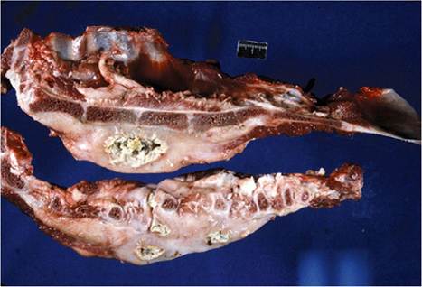
Figure 4.8 Cross - section of the sternum of a goat showing a chronic abscess surrounded by much fibrous connective tissue, and with a tract penetrating the thorax. Source: Courtesy of Dr.M.C. Smith.
The prognosis is poor in these cases. Reports of successful treatment of osteomyelitis in goats with antibiotic therapy are lacking.
Other instances of osteomyelitis are sporadic. Infection of the femur has been reported in an 8-month-old Nubian buck secondary to septic gonitis. Lameness in the affected limb was severe. Pseudomonas aeruginosa was isolated from the stifle joint. The case was successfully managed by amputation of the affected limb (Ramadan et al. 1984). Another case of osteomyelitis in the femoral head of a young goat with a putative diagnosis of polyarthritis due to E. rhusiopathiae was thought to result either from the extension of infection from the hip joint or from an initial septicemia (Wessels 2003).
Multifocal osteomyelitis due to Corynebacterium renale has been diagnosed in an 18-month-old French Alpine kid (Altmaier et al. 1994). The goat was lame on the right forelimb and radiographs revealed a fracture of the distal scapula associated with focal demineralization of bone and multifocal, lytic lesions in the ribs (Figure 4.9). At the time of necropsy, osteomyelitis was confirmed and evidence of septicemia was present, with abscesses noted in intestine, lymph nodes, and liver.
An unusual multifocal Rhodococcus equi osteomyelitis of the skull and thoracic vertebrae has been recorded in a 2-year-old Saanen doe in Australia. The clinical
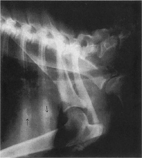
Figure 4.9 Radiographic evidence of osteomyelitis in the ribs of a goat (note arrows). Source: Courtesy of Cummings School of Veterinary Medicine at Tufts University.
presentation was progressive posterior paralysis due to spinal cord involvement (Carrigan et al. 1988). Suppurative vertebral osteomyelitis and diskospondylitis have been reported in a 3-week-old goat with acute hindlimb paresis, but the cause was not identified (Alexander et al. 2005).
Lyme Disease
Lyme disease or Lyme borreliosis is a tick-borne spirochetal disease of humans and animals originally described in people in Lyme, Connecticut, in 1977. The predominant clinical sign is arthritis. The disease is now known to occur in the northeastern, midwestern, and northwestern portions of the United States, as well as in Europe, Russia, China, Japan, and Australia, with a higher prevalence in forested regions. The causative agent is the spirochete Borrelia burgdorferi and it is transmitted mainly by Ixodes spp. ticks, which normally complete their life cycle on deer and mice. Fragmentation of forest habitat and a concomitant increase in white-footed mouse (Peromyscus leu- copus) populations relative to other small mammalian tick hosts is believed to be responsible for an increasing prevalence of the disease, at least in the United States (Ostfeld and LoGiudice 2003). Larval, nymph, and adult stages of Ixodes ticks are capable of transmitting the infection. While the different stages have different host feeding preferences among small and large mammals, all stages feed on and can infect humans. Risk of transmission is highest in spring and fall when nymphs and adults are most active.
Seroepidemiologic studies on Lyme disease have been carried out around the world on various mammals and many of these surveys have included goats. The seroprevalence of antibodies to B. burgdorferi in goats has been reported as 5% in Bolivia (Ciceroni et al. 1997), 8.5% in France (Doby and Chevrier 1990), 17.2-19.4% in Slovakia (Travnieek et al. 2002), 18% in Egypt (Helmy 2000), 36.8% in Italy (Ciceroni et al. 1996), 48% in Bulgaria (Angelov et al. 1993), and between 19.1% and 61.3% in different provinces of China, with goats in mountainous regions having much higher seroprevalence than those in plains regions (Zhang et al. 1998; Long et al. 1999). A serosurvey of humans in the Canary Islands noted that male goat farmers had significantly higher seroprevalence than men of a similar age who were not goat farmers, and that the rate of seropositivity in goat farmers was three times higher than that of the general population. This suggests that goats may serve as a reservoir for human infection or that goat herders at pasture are more frequently exposed to infected ticks (Carranza et al. 1995). Assays for detection of antibody to B. burgdorferi currently in use include ELISA, the indirect immunofluorescent antibody test, and the WB.
Clinical Lyme disease is well described in humans and dogs and also occurs in horses and cattle (Steere 1989). The disease in humans (Hengge et al. 2003) and in horses (Divers et al. 2018) has been reviewed. Cattle with acute Lyme disease show fever, a stiff gait, swollen joints, and decreased milk production. They may also show edematous lesions on the hairless skin of the udder and chronic weight loss (Constable et al. 2017).
While goats and sheep commonly show serologic evidence of infection, definitive confirmation of clinical Lyme disease, manifested as arthritis, remains elusive in small ruminants. Two cases of suspected Lyme borreliosis in lambs were reported from Norway in 1992. The lambs were from flocks in a district heavily infested with Ixodes ricinus ticks and both had high serum immunoglobulin (Ig)G antibodies to B. burgdorferi by ELISA test, but attempts to isolate spirochetes were unsuccessful (Fridriksdottir et al. 1992). Presumptive cases of Lyme disease in goats and sheep were reported from endemic regions of Connecticut (Baldwin 1990). Putative signs in affected goats include depression, fever, pain in joints sufficient to preclude weight bearing, back pain, a bow-legged stance, an unsteady gait in the hind end, and a stiff neck. Ticks may be difficult to find on the animal because the infective nymphal stages are only pinhead size. Alternate-day benzathine penicillin therapy carried out for 21-28 days was reported as successful in these goats. In cattle, daily treatment with either penicillin or oxytetracycline for 21 days is recommended. A vaccine is now commercially available for use in dogs, but is not approved or recommended for use in goats.
To date, the infective organism has not been isolated or identified from any presumed case of Lyme disease in goats. While affected animals had serologic evidence of infection, serologic surveys indicate that many individual animals with antibodies show no sign of clinical disease. Lyme disease may indeed occur in goats; however, definitive proof is still lacking. Presumptive diagnosis of Lyme disease should be made with caution, only in areas where the disease is known to occur in other animal species, and only after other known causes of caprine arthritis, especially CAE and mycoplasmosis, have been definitively ruled out.
Confirmation of the presence of B. burgdorferi in affected tissues is still required for definitive diagnosis. Cultures are difficult to maintain and require several weeks to grow. Concentrations of organisms in affected organs or fluids may be very low, making culture problematic. However, alternatives to culture have become available for identifying the organism. PCR techniques can be used to identify B. burgdorferi in synovial fluid samples from affected joints or milk (Lischer et al. 2000) or from tissues at necropsy.
Clostridial Myositis and Myonecrosis
Blackleg and malignant edema (gas gangrene) are common, costly diseases of cattle and sheep caused by the “tissue-invading” or “gas gangrene” group of clostridial organisms. In contrast, these clostridial diseases are uncommon in goats. One possible explanation is that the causative bacteria are soil-borne organisms that gain entry into livestock primarily through grazing of low-lying, wet pastures. As browsing animals, goats may be less frequently exposed to clostridial spores in soil, though this hypothesis is unproven.
Etiology and Pathogenesis
Clostridium chauvoei, Clostridium septicum, Clostridium sordelli, and Clostridium novyi are Gram-positive, rodshaped, spore-forming, toxin-producing, anaerobic bacteria. They are ubiquitous in soil and persist for long periods under the protection of sporulation. They may also be present in the intestinal tract and liver of normal livestock without producing disease. Classical or true blackleg is caused by C. chauvoei. “False” blackleg, more appropriately referred to as malignant edema, is caused most often by C. septicum, but C. novyi, C. sordelli, C. chauvoei, and even Clostridium perfringens have been isolated from lesions typical of malignant edema.
The pathogenesis of blackleg is incompletely understood. It is thought that grazing animals ingest spores of C. chauvoei that cross the alimentary epithelium and are carried to muscle tissue via the lymphatics and blood. Spores remain dormant until local conditions in muscle favor bacterial multiplication. This might involve muscle trauma or circulatory disturbances that create anaerobic conditions. The proliferation of bacteria is accompanied by release of toxins that produce a necrotizing myositis and a fatal toxemia.
Malignant edema occurs when spores are introduced into tissue by penetrating wounds, which often result from routine management activities such as vaccinations, disbudding, castration, and especially shearing. Fighting and head butting among bucks can also facilitate introduction of spores and produce a type of malignant edema known as “swelled head.” Tissue damage associated with wounds creates anaerobic conditions that allow bacterial proliferation. Toxins are released that produce severe localized inflammation and generalized, fatal toxemia. Local inflammation is often characterized by tissue necrosis with accumulation of edema and gas. Subcutaneous and connective tissue is often more directly involved than muscle, but muscle can be affected.
Epidemiology
Clostridial myositis has been reported infrequently in goats, mostly from southern Africa, Australia, and New Zealand. Angora or feral goats are most often affected, but this may reflect the preponderance of such goats in these locations rather than a breed predisposition. In the United States, it was observed that blackleg and malignant edema did not occur in goats, even where the diseases occurred frequently in other animals on the same premises (Guss 1977).
A review of clostridial diseases in Australia indicated that classical blackleg due to C. chauvoei is virtually unreported and that goats may be less susceptible to the disease than sheep (King 1980a). Mortality due to navel infections with C. septicum is also seen less frequently in kids than in lambs. More common is C. novyi infection in fighting bucks, leading to swelled head. The relationship of C. novyi infection to hepatic “black” disease in goats is discussed in Chapter 11.
In New Zealand, a group of 460 Angora and feral does were given IM injections of cloprostenol to induce abortion. Of these, 14 (3%) died from C. chauvoei infection within six days after injection (Day and Southwell 1979). Gas gangrene after IM injections with various medications is not uncommon in other livestock species.
In South Africa, the shearing of sheep is commonly associated with C. chauvoei infection, while shearing of Angora goats is not (Van Tonder 1975). It is presumed that the nature of the fleece and the relative absence of skin folds and body pleats in goats produce fewer shearing wounds, with less secondary invasion of clostridial spores. When postshearing clostridial infections do occur, they are more often caused by C. septicum. Gangrenous metritis caused by mixed clostridial infections with C. septicum, C. novyi, and/or C. chauvoei is the most common clostridial problem seen in Angora goats in South Africa. The condition is associated with the penning or kraaling of does during the kidding period (Bath et al. 2005). Morbidity is variable, but case mortality rates can approach 100% (Van Tonder 1975). In Namibia, spontaneous deaths of grazing adult cattle, sheep, and goats have been recognized as caused by C. sep- ticum (Wessels 1972).
Clinical Signs
In blackleg, the clinical course is short and affected animals are usually found dead or recumbent and moribund. A sanguinous discharge from the nostrils or anus may be present. If there is a chance to observe animals early in the course of disease, lameness or a stiff gait may be noted when limb musculature is involved. An outbreak of putative blackleg (black quarter) was recorded in India in which 19 adult and kid goats in a herd of 142 showed dullness, fever, and lameness in the hindlimbs, with hot, painful swellings and palpable crepitation (Manokaran 2005).
In malignant edema, which is associated with wounds or injections, there is usually heat, swelling, pain, and erythema at or around the wound site from 12 to 48 hours after introduction of the infection. As the infection progresses, skin becomes discolored and cold and subcutaneous gas may be identifiable as crepitus. Animals rapidly become weak and shocky, exhibit a high fever exceeding 41.1 °C (106 °F), and will die within hours. Malignant edema (gas gangrene) was definitively diagnosed in nine pregnant hair goats showing subcutaneous swellings and crepitation in a herd in Turkey, with C. septicum confirmed in culture and by PCR (Gazioglu et al. 2018).
Swelled head is seen primarily in group-housed bucks that head butt. Large edematous swellings occur, usually beginning around the eyes and extending down the face and neck, sometimes to the chest. The swelling is quite disfiguring. The taut skin cracks and yellows, and edematous fluid seeps from the cracks. Affected bucks become extremely depressed and weak, with heads held low. They fall over and remain recumbent, dying within one to two days of onset of signs (King 1980a).
Clinical Pathology and Necropsy
Impression smears and swabs taken from wound sites or subcutaneous aspirates may aid in the antemortem diagnosis of malignant edema or swelled head. Gram staining, fluorescent antibody testing, and anaerobic culturing can all be performed. In blackleg and many cases of malignant edema, necropsy is the primary approach to diagnosis. For bacteriologic confirmation, it is imperative that fresh necropsies be performed, preferably within one hour of death. Otherwise, clostridial organisms present in the alimentary tract or liver can invade tissues post mortem and produce false-positive diagnoses.
At necropsy, goats with blackleg may exhibit crepitant swellings over heavy muscle groups, as well as gas bub - bles in liver, kidney, and uterus and excessive accumulations of serosanguinous fluids in all body cavities (Pauling 1986). Goats with malignant edema may demonstrate generalized and advanced decomposition of the carcass, even when necropsy is performed shortly after death. There is a characteristic smell of clostridial decomposition and gelatinous infiltration of the pericardium. Pericarditis and myocarditis may be observed (Wessels 1972). In malignant edema and swelled head, there is marked accumulation of yellow or serosanguinous edema fluid in subcutaneous and intermuscular spaces, especially around wound sites.
Diagnosis
Definitive diagnosis of clostridial myositis in goats depends on confirmation of the presence of the causative clostridial organisms from tissues or wound sites of affected animals before or immediately after death. This can be accomplished by anaerobic culture, fluorescent antibody staining, or multiplex PCR (Sasaki et al. 2002).
When goats are found dead in the field, the differential diagnosis should include tympany, lightning strike, poisonings, and anthrax, especially because the latter condition is also associated with postmortem nasal and anal san- guinous discharges. If there is any suspicion of anthrax, the carcass should not be opened.
Treatment
In general, Clostridium spp. are sensitive to penicillins and cephalosporins. Opportunities to treat individual cases of blackleg are limited due to the rapid, fatal outcome; however, prophylactic treatment of herd mates may prevent the development of additional cases. In one report, benzathine penicillin, at a dose of 15 000 IU/kg bw, was used to effectively limit an outbreak in a goat herd (Manokaran 2005). Individual cases of malignant edema, swelled head, or genital gas gangrene are more likely to be detected early enough for therapeutic intervention. In malignant edema and swelled head, the skin over affected regions should be liberally incised and serial fasciotomy performed on underlying muscles to allow aeration and drainage of infected tissues. Intravenous penicillin therapy at a dose of 20 000 IU/kg every six to eight hours should be initiated immediately and maintained until the patient is stabilized. The animal can then be placed on IM procaine penicillin G for continued treatment. As animals are toxemic and often in shock, parenteral fluid therapy, steroids, and non-steroidal anti-inflammatory drugs may also be indicated. The prognosis in advanced cases is always guarded.
Control
Vaccination in the face of an outbreak is indicated when blackleg or malignant edema occurs in a flock or herd. Numerous multivalent clostridial vaccines manufactured throughout the world protect against the common causes of clostridial myositis. When vaccinating in the face of an outbreak, animals at risk should be treated with long- acting penicillin preparations at the same time that they are vaccinated, to reduce losses while immunity develops.
Ideally, animals should be vaccinated routinely before outbreaks occur. However, the incidence of blackleg and malignant edema in goats is so low relative to sheep and cattle that widespread vaccination of goats may be difficult to justify economically, except in those circumstances where the disease is predictably known to occur. As goats should be routinely vaccinated against enterotoxemia caused by C. perfringens, use of multivalent clostridial vaccines may fit readily into the existing vaccination program. Where genital gas gangrene is known to occur, does should be vaccinated annually with appropriate multivalent clostridial vaccine three weeks before kidding. To control swelled head, bucks should be routinely vaccinated and housed separately to minimize fighting.
Foot Scald, Foot Rot, Foot Abscesses, and Digital Dermatitis
Infectious foot problems are not as widely reported in goats as in sheep, but the pattern of disease is similar. Foot scald, also known as interdigital dermatitis, is defined as an infection with Fusobacterium necrophorum confined to the interdigital epidermis. Benign foot rot occurs when the feet of animals with foot scald become coinfected with weakly virulent strains of Dichelobacter (Bacteroides) nodosus, leading to some underrunning of the soft horn of the hoof. Virulent foot rot, also known as infectious foot rot, is defined as a co-infection of the interdigital epidermis with virulent strains of D. nodosus and F. necrophorum, with extension of inflammation and infection into the horny and laminar structures of the foot. Foot abscess is an infection of the deep structures of the foot caused by bacteria other than D. nodosus, usually F. necrophorum or Trueperella (formerly Arcanobacterium, formerly Actinomyces, formerly Corynebacterium) pyogenes.
Another distinct disease of the feet of sheep believed to be caused by treponemes and known as contagious ovine digital dermatitis (CODD) was first described in the United Kingdom in the 1990s. Treponema infections of the feet also have been recognized in housed dairy goats in the United Kingdom, and this condition must be differentiated from foot scald and foot rot. As a formal name for the disease in goats has not yet been proposed, it will be referred to as CODD-like disease in the following discussion.
Etiology
F. necrophorum, a Gram-negative, anaerobic bacterium, is a common environmental organism that can colonize the interdigital epidermis when epithelial integrity is compromised and by itself can produce foot scald. F. necrophorum is also a co-pathogen with D. nodosus in benign and virulent foot rot. Additionally, F. necrophorum can be introduced into the deep structures of the foot by trauma and puncture wounds. F. necrophorum and T. pyogenes are the most common organisms associated with foot abscesses.
Dichelobacter nodosus is also a Gram-negative, anaerobic bacillus. In smears of exudate, it can be recognized by its characteristic morphology as a large, slightly curved rod with bulbous ends. It is an obligate anaerobe that is adapted to the interdigital epidermis of ruminants and survives only to a maximum of four days in the environment when passed in discharges from infected feet (Plant et al. 2017). Therefore, while the presence of F. necrophorum is required for virulent foot rot to develop, D. nodosus is considered the inciting pathogen because it must be introduced into a naive herd or flock for foot rot to develop, whereas F. necro- phorum is generally already present in the environment. Different strains (serogroups) of D. nodosus vary in their virulence based on their degree of keratolytic and elastol- ytic activity. The presence of proteases and elastases contributes to the invasiveness of horny hoof tissues. Caprine isolates of D. nodosus with high elastase activity were demonstrated to be virulent for sheep (Claxton and O'Grady 1986). The potential for cross-transmission of D. nodosus between goats and sheep must be taken into account when devising control programs.
Eight major serogroups of D. nodosus have been identified on the basis of agglutination responses to surface fimbria found on the organism. This is important regarding the efficacy of vaccination when vaccines comprised of fimbrial antigens are used, because the degree of crossprotection varies between different serogroups and is generally not strong.
The etiology of CODD is not fully elucidated. As in bovine digital dermatitis, various species of treponeme spirochetes are involved. In sheep these are Treponema medium-like, Treponema phagedenis-like and Treponema pedis. However, D. nodosus and F. necrophorum are frequently isolated from cases of CODD and likely play a yet undetermined role in its pathogenesis, or indicate that foot rot is occurring concurrently with cases of CODD (Duncan et al. 2018). In the reported cases of lameness and atypical foot lesions of dairy goats in the United Kingdom associated with treponemes, T. medium/Treponema vincentii- like, T. phagedenis-like, Treponema denticola/Treponema putidum-like or T. pedis was detected, sometimes in association with D. nodosus (Groenevelt et al. 2015; Sullivan et al. 2015).
Epidemiology
Foot scald, foot rot, and foot abscesses are most common in temperate regions of the world and occur most frequently in the spring and early summer in association with warm temperatures and heavy rainfall. The combination of wet pasture and warm temperatures (above 10 °C [50 °F]) facilitates transmission of the disease by softening and moistening the skin of the foot, thus predisposing to dermatitis and traumatic injury, and also by allowing the bacteria to persist away from the host for longer periods on pasture. In contrast, foot rot is rarely observed in hot, arid regions, even when large numbers of sheep or goats are maintained.
Other environmental and management factors predispose goats to infectious foot problems. These include wet, muddy yards or poorly drained pastures, overgrown hooves, overcrowding, the introduction of infected goats or sheep to a susceptible herd or flock, return of animals from shows or breeding stations, and turnout of goats onto contaminated pastures (Baxendell 1980). Goats are prone to excessive growth of the hoof when maintained in confinement. When the abaxial hoof wall overgrows the sole, it may cause inward compression of the digits, leading to excessive irritation of the interdigital skin (Claxton and O'Grady 1986). Dairy goat breeds are considered to be more susceptible to foot rot than are fiber and meat goat breeds, and some family lines of dairy goats are particularly prone to having “bad feet,” requiring more frequent trimming (Skerman 1987). Heritability estimates have been reported in different breeds of goats regarding susceptibility to foot rot, suggesting that improved resistance is possible through selection (Banik and Bhatnagar 1983). A study in India indicated that purebred goats of the indigenous Malabari and Attappady Black breeds had significantly lower rates of foot rot infection than goats of those same breeds crossed with Alpine, Saanen, or Boer breeds and grazed together under the same conditions (Thomas et al. 2011).
Transmission occurs by contact when infected and susceptible animals are commingled together at pasture under suitable conditions of moisture and temperature. The organism is present in discharges from animals with clinical lesions and can invade the damaged epidermis of the foot of susceptible goats. In sheep, the carrier state is known to be important in the spread of infection into susceptible flocks when carrier animals are newly introduced. This is also presumed to occur in goats. It has been reported that at least some strains of D. nodosus transmit readily between sheep and goats, so the risk of cross-species transmission must be considered, and control efforts must be applied to both goats and sheep when the animals are kept together (Ghimire et al. 1996).
There may be differences in susceptibility between sheep and goats. In one study, few goats showed marked underrunning lesions of the hoof, while sheep developed more severe lesions consistent with virulent foot rot when infected with the same strains. It was suggested that this might be because the stratum corneum of the interdigital skin is considerably thicker in goats than in sheep, and therefore goats may be more resistant to maceration of the interdigital skin and subsequent invasion of D. nodosus (Ghimire et al. 1999).
Foot rot is less prevalent in goats than in sheep, and until 1985 confirmation that the condition in goats was caused by D. nodosus was notably lacking (Merrall 1985; Claxton and O'Grady 1986; Egerton 1989). Foot scald and, to a lesser extent, foot rot are increasingly recognized as economic constraints on both fiber and milk goat production in the wetter regions of Australia and New Zealand (Anonymous 1987a). While morbidity and mortality information for goats is scant, foot scald was cited by New Zealand goat farmers as the second most important disease of goats after enteric diseases (Merrall 1985).
Foot scald and foot rot can be important causes of economic loss in Angora goats in South Africa during periods of unusually wet weather (Van Tonder 1975). A seasonal prevalence of foot abscesses in Angora and Boer goats in South Africa is related to seasonal increases in populations of ticks with long mouth parts, notably Hyalomma spp., Amblyomma spp., and Rhipicephalus glabroscutatum. Adult ticks produce deep wounds in the interdigital space while feeding, and these wounds become secondarily infected, mostly by T. pyogenes (McIvor and Horak 1987; Bath et al. 2005). Foot rot occurs sporadically in dairy goats in the United States and Europe when they are maintained under conditions of poor management, as described above (Guss 1977; Pinsent 1989). Foot rot has been confirmed in farmed goats in the semi-arid region of northeastern Brazil during the season of heavy rainfall, with the goats pastured during the day and confined to pens at night (Aguiar et al. 2011). Migratory goats in northwestern India have also been identified with foot rot while grazing in common mountainous pastures during monsoon season (Wani et al. 2015)
Currently, CODD-like disease in goats associated with treponemes has been reported only from the United Kingdom and has involved housed dairy goats. The related disease in sheep, chronic ovine digital dermatitis, was first described in the United Kingdom and may be occurring in sheep elsewhere in Europe, as indicated by a recent report from Sweden (Bernhard et al. 2019).
Pathogenesis
The pathogeneses of foot scald, foot rot, and foot abscesses have not been expressly studied in goats. The development of these diseases is presumed to be similar to that described in sheep (Constable et al. 2017). Wet conditions and softened hooves allow for infection of the interdigital skin and skin-horn junction with F. necrophorum. This can produce inflammation and hyperkeratosis and the relatively benign condition known as foot scald. However, the inflammation produced by F. necrophorum can facilitate the introduction of D. nodosus if that organism is present in the flock. In virulent foot rot, D. nodosus attaches to the foot with the aid of adherence pili (fimbria) and colonizes the interdigital epithelium. When these D. nodosus strains are virulent and keratolytic, the keratolytic activity allows invasion and underrunning of the horny hoof tissue. The interaction of D. nodosus and F. necrophorum causes marked inflammation and affected tissues are severely compromised, leading to necrosis. As a result, adherence of the hoof corium to the basal epithelium may be destroyed, resulting in detachment of the horny hoof from the underlying soft tissue. The transmission and pathogenesis of CODD are not yet fully understood and it is not clear if the pathogenesis of CODD-like disease in goats will be the same, as there are some differences in the clinical presentation so far between sheep and goats (Duncan et al. 2018).
Clinical Findings
In food scald, there may be a mild lameness. Careful inspection of the feet reveals erythema or swelling of the interdigital epidermis. There is usually little or no odor, and minimal underrunning of the horn at the coronet.
In foot rot, lameness can be quite pronounced when there is necrotic underrunning of the horn. Severely affected goats may walk on their carpi and, when all four feet are involved, may refuse to walk at all. In general, lesions of caprine foot rot are less severe than those commonly seen in sheep (Claxton and O'Grady 1986). Swelling of the interdigital epidermis or possible separation of the hoof at the skin-horn junction may be noted. A small quantity of pus also may be visible at the coronet and detached horn can be readily pared or pulled off. The characteristic necrotic smell of foot rot may be detected.
Chronically affected animals may show a marked loss of body condition and decreased production. In addition, myiasis and tetanus are two possible sequelae of foot rot. Chronic cases may show hoof deformities after healing.
Foot abscesses may affect the heel or the toe. In contrast to foot rot and foot scald, only one foot may be involved, and only one claw of that foot. There may be a history of trauma or puncture. The foot is hot and swollen and very painful to the touch. The animal may carry the leg rather than bear weight. Swelling or draining pus may be present above the coronet.
Relatively few cases of the CODD-like disease associated with treponemes in dairy goats have been described and the reported findings are variable. Lesions in the interdigital space as seen in foot rot are relatively uncommon. In some cases the condition presents similarly to that of CODD, with initial skin lesions along the coronary band rather than at the interdigital space. This may progress to separation of the hoof capsule at the coronary band, with the underlying tissue appearing granulomatous and hemorrhagic. In other cases, lesions appear to occur primarily on the sole, with loss of solar horn and again the presence of granulomatous and hemorrhagic tissue. Extensive ulcers of the sole are reported as characteristic in affected goats (Crosby-Durrani et al. 2016). Cases might involve only a single hoof or a single claw on a single hoof, and signs of lameness can be pronounced, with refusal to bear weight on affected limbs (Groenevelt et al. 2015; Sullivan et al. 2015).
Clinical Pathology and Necropsy
Gram-stained smears of exudate from underrun horn of affected feet can be used to identify the characteristic D. nodosus organisms in foot rot cases, but such identification may require a very experienced technician. Samples can be submitted for anaerobic culture on hoof agar. Serotyping of isolates can be important to determine the potential effectiveness of commercial vaccines, and this is done through culture and PCR in the laboratory. In early cases, evaluation of the virulence of isolates by measurement of proteolytic activity can be valuable in anticipating the severity of an impending outbreak. In the case of CODD-like disease in goats, the presence of treponemes can be identified in swabs or biopsies of hoof lesions by culture or PCR (Groenevelt et al. 2015; Sullivan et al. 2015).
Radiography may be helpful in diagnosing goats with foot abscess and may assist in prognosis by identifying the presence of secondary osteomyelitis in the distal phalanx. At the time of necropsy, lesions are limited to the affected feet and reflect what is observed ante mortem. Sagittal section of the distal limb may indicate the extent of foot abscesses and the occurrence of secondary osteomyelitis in the distal phalanx.
Diagnosis
Presumptive diagnosis of foot rot and foot scald is based on the onset of lameness, swelling at the interdigital space, and underrunning of the horn of the foot. Definitive diagnosis is based on culture of D. nodosus or identification by PCR from lesions. Diagnosis of foot abscess depends on identifying a pus pocket in the deeper structures of the foot by paring of the sole or heel. Until the clinical presentation of CODD-like disease in goats is more fully characterized, the disease should be considered in housed dairy goats when foot lesions include evidence of ulceration and granuloma formation and swabs or biopsies of lesions confirm the presence of treponemes.
The differential diagnosis for the sudden onset of lameness in groups of goats should include FMD and bluetongue. Lesions at the coronet may be seen in both diseases, but oral and muzzle lesions also occur in some affected individuals with bluetongue and possibly FMD. Clinical bluetongue is uncommon in goats. Laminitis after grain engorgement can occur in groups of goats, but other signs such as diarrhea and bloat accompany or precede the lameness. Selenium toxicosis produces cracks in the hoof that become secondarily infected and may mimic foot rot, except that the course is more chronic. The differential diagnosis for foot abscesses, when one leg is affected, include fractures, subluxations, and soft tissue injuries of the distal limb.
Treatment
Because of the highly contagious nature of foot scald and foot rot, recognition of even a single case of the disease calls for examination and potential treatment of all feet of all goats in the herd. As in sheep, several elements of therapy need to be considered, namely paring of feet, use of antibacterial footbaths, topical and/or parenteral antibiotic therapy, and vaccination, which can be used as an aid for control of outbreaks as well as for prevention. These various approaches can be used alone or in combination, depending on the number of goats involved, their value and use, and the severity of the disease. A comparison of the cost-effectiveness of various treatments used in sheep has been reported (Salman et al. 1988).
Historically, aggressive paring of affected feet was considered to be an important component of therapy in advanced cases of foot rot, because removal of dead tissue was thought to expose deep-seated D. nodosus organisms to oxygen and the antibacterial effects of subsequent footbaths or topical antibiotics. However, current thinking is that in foot rot, paring of feet should be limited to what is necessary for assisting in diagnosis and for removing loose horn (Winter 2008). In one study the use of foot trimming as a treatment for foot rot in sheep was assessed to be a risk factor rather than a limiting factor for the condition (Green et al. 2007). In another study, the use of foot trimming as a treatment intervention for foot rot in sheep actually delayed healing, compared to the use of parenteral, long-acting oxytetracycline (Kaler et al. 2010).
In the case of foot abscesses, paring feet is important to find pus pockets and drain them to relieve pressure in the sore foot. As foot rot is painful for affected animals, consideration should be given to treatment with non-steroidal anti-inflammatory drugs such as aspirin, flunixin meglumine, or meloxicam.
Disinfectant footbaths are a useful way of treating large numbers of goats. However, goats are high jumpers and have a distinct aversion to walking in water. Footbath design, then, must be considered carefully. A circular bath with a central feeding trough is commercially available in New Zealand. This design encourages goats to wade into the bath to be rewarded with grain (Yerex 1986). Footbath solutions must be non-irritating if the owner expects the goats to enter the bath a second time. Zinc sulfate solutions of a 10-20% weight-to-volume ratio are both mild and effective and are currently the solution of choice, though they may cause abomasal necrosis if the goats drink it, as discussed further in Chapter 19. The efficacy of the solution may be enhanced by addition of surfactants such as sodium lauryl sulfate at a concentration of 2% volume-to- volume ratio. In severe cases, one-hour foot soaks repeated weekly for three treatments may be required.
Formalin and copper sulfate footbaths are less desirable. A 5% formalin solution is an effective treatment, but has several drawbacks including the production of irritating fumes, painful stinging of aggressively pared feet, hardening of the hoof, and the potential for permanently damaging the skin if concentrations are not strictly regulated. If formalin is used, the solution should be changed daily to ensure that evaporation has not resulted in excessive concentration. Goats stung by formalin footbaths are unlikely to enter them again voluntarily. Copper sulfate solution at a 10% weight-to-volume ratio is also effective, but stains the hair or fleece blue-green and is potentially toxic if consumed. Splashing of solutions on the goats' bodies can be reduced by lining the footbath with wood chips, peat moss, or wool tags. After dipping, goats should be turned out on dry ground or slotted floors to permit drying of footbath solutions on hooves.
A number of topical disinfectants and antibacterials can be used after paring, in lieu of footbaths. Zinc sulfate, copper sulfate, copper naphthenate, the quaternary ammonium compound cetrimide (20% alcohol tincture), oxytetracycline, penicillin, and other compounds are available in different countries as paints, powders, or sprays for direct application to the hoof. Putting a bandage on the hoof after application may enhance healing. The feeding of zinc sulfate at a rate of 0.5 g/head/day for 21 days during the treatment period has also been recommended when the diet is known to be deficient in zinc.
Parenteral antibiotic therapy has been used effectively in sheep. The in vitro effectiveness of beta-lactam antibiotics against caprine isolates of Dichelobacter spp. and Fusobacterium spp. has been reported (Piriz Duran et al. 1990). Azlocillin, mezlocillin, and piperacillin were most effective against the largest number of isolates. Penicillin G and ampicillin were almost as effective, and cefuroxime, cefoperazone, cefotaxime, cefoxitin, and imi- penem were generally less effective. Keep in mind that antibiotic treatment of milking goats will result in a period of milk withdrawal with its own associated costs, and that in the United States few antibiotics are specifically approved for use in goats.
For consideration of extralabel drug use for foot rot in goats, current recommendations for antibiotic treatment in sheep are a single IM dose of penicillin/streptomycin at 70 000 U/kg bw penicillin and 70 mg/kg dihydrostreptomycin; a single IM dose of erythromycin at 10 mg/kg bw; a single SC dose of lincomycin/spectinomycin at 5 mg/kg bw lincomycin and 10 mg/kg bw spectinomycin; a single SC dose of gamithromycin at 6 mg/kg bw; a single IM dose of long-acting amoxicillin at 15 mg/kg bw; or a single IM dose of long-acting oxytetracycline at 20 mg/kg bw (Constable et al. 2017). In goats, however, IM oxytetracycline can be extremely irritating to muscle tissue and the preferred route of administration is SC. Tilmicosin has been used for the treatment of CODD in sheep at a single SC dose of 10 mg/kg bw (Winter 2011). While effective in treatment of individual cases, tilmicosin was not effective as a control intervention for eradicating CODD from flocks (Angell et al. 2016). In goats, tilmicosin injections greater than 10 mg/kg IM or SC can cause toxicity and it is recommended that the drug not be used in goats (Papich 2016). Long-acting amoxicillin given once IM at a dose of 15 mg/ kg bw has also been reported as an effective treatment for CODD in sheep (Duncan et al. 2012).
Treated animals should not be turned back into wet or muddy yards, nor should they be put out to pasture that has not been free of infected animals for at least seven days. When feet are pared for diagnostic or therapeutic purposes, parings from trimmed feet should be considered as fomites and removed from the premises or burned. Paring knives should be disinfected in 10% formalin between goats to avoid additional spread of infection, and gloves should be worn for paring and regularly disinfected. It is recommended that goats with foot rot be given tetanus antitoxin or reimmunized, as the anaerobic environment of the affected foot may predispose to tetanus.
Where vaccines against D. nodosus are available they can be used in the management of outbreaks, but should be considered as a useful adjunct to the interventions described above and not a substitute for them, since the efficacy of commercially available vaccines may be limited. Issues around vaccination are discussed further in the next section on control.
Control
Premises with no history of foot rot or foot scald may remain free by maintaining completely closed herds. This means not purchasing any new stock and not taking animals to shows or breeding stations. Transport vehicles and common housing facilities can be contaminated with D. nodosus if not thoroughly cleaned and disinfected. If new animals are purchased, they should be from sources free of foot rot. Feet of purchased animals must be carefully inspected for lesions and if any doubt exists, the animals should be run through a foot bath or treated with parenteral antibiotics, quarantined for four weeks, and reexamined before joining the new herd.
When foot rot already exists on the premises, control is directed toward maintenance of proper hoof care, prophylactic use of footbaths, antibiotics, vaccination, and breeding programs. Individual animals with severe or recurring cases should be culled from the herd. Vaccination for tetanus should be up to date.
Feet should be monitored for excessive overgrowth. Provision of rock piles for climbing and playing may help goats to naturally wear off excessive hoof growth. Regular trimming may be necessary to keep the hoof wall from overgrowing the sole and to keep toes from growing too long. However, foot trimmers must be knowledgeable about the procedure. Feet that are aggressively trimmed, especially to the point that bleeding occurs, may actually contribute to the development of other foot problems, such as granulomas at the toe (Winter 2004).
Use of walkthrough footbaths once a week during high- risk periods of wet, warm weather helps to control the spread of infection. Zinc sulfate baths are preferred for repetitive use.
A multivalent, commercial, oil-adjuvanted foot rot vaccine based on multiple fimbria of different serogroups of D. nodosus is available in the United Kingdom, France, New Zealand, and elsewhere for use in sheep, though not in the United States. The vaccine is initially given twice at an interval of four to six weeks. The duration of immunity may be limited, so vaccination should be timed to precede those periods when transmission of the disease is most likely to occur (Egerton 2007). The vaccine should be given SC, but still may produce a sizable reaction at the injection site, particularly after the second injection. Owners should be so advised, particularly if show animals are involved.
The multivalent commercial D. nodosus vaccine contains 10 different strains of the organism to represent the most common serogroups associated with foot rot, as there is little cross-protection conferred between different sero- groups. In practice the vaccine produces variable results, and it is believed that inclusion of so many strains and their associated fimbrial antigens may actually diminish the immune response against the particular strains present in a given herd, which usually is considerably fewer than 10. To maximize the effectiveness of D. nodosus vaccines, they must contain a strain or strains immunogeni- cally related to those occurring in the affected flock. This was illustrated in Nepal, where an autogenous vaccine containing only two virulent serogroups isolated from local sheep and goats was more effective in reducing foot rot in both species than an imported, multivalent commercial vaccine in a controlled field study (Egerton et al. 2002).
Additional vaccination trials using outbreak-specific vaccine formulations were conducted in Australia, with excellent results (Dhungyel et al. 2013). The value of outbreak-specific vaccines is sufficiently well established that a public-private partnership has been established in Australia for veterinary practitioners to submit samples from outbreaks to laboratories for D. nodosus serogroup/ strain identification, followed by production of a custom vaccine at a private vaccine production facility based on the strains identified. A maximum of two strains are included in any custom vaccine. If the outbreak involves more than two strains, separate, additional mono- or bivalent vaccines are prepared for the remaining strains for administration at an interval of three months (Anonymous 2017). The evolution of D. nodosus vaccine development for foot rot and the application of new vaccine technologies involving recombinant fimbrial vaccines and other innovations have been reviewed (Dhungyel et al. 2014). A F. necrophorum bacte- rin is available in the United States, approved for use in cattle but not small ruminants. Its safety and efficacy for use in goats for foot scald or foot rot are not established and its use is not recommended.
The selection of goats with apparent resistance to foot rot for breeding programs is considered to offer long-range benefits to the goat industry for foot rot control. Culling goats with poor foot conformation is a good place to begin.
Control and eradication of foot rot are possible, but it can be a long, challenging, and costly process, and veterinarians need to communicate well with clients in this regard. At the outset, it is essential that producers understand that foot rot is a herd problem and not an individual animal problem. There is no magic bullet, and no single intervention will successfully eliminate the condition once established on a farm. Additional details on eradication and control of foot diseases in sheep and goats are available elsewhere (Winter 2011).
As CODD often occurs concurrently with foot rot in sheep, it is recommended that control efforts be aimed at both diseases together and should include prompt individual treatment, foot rot vaccine, improving underfoot hygiene, culling, and biosecurity measures (Duncan et al. 2018).