Causative Agents
Like other dimorphic fungi, Histoplasma organisms are characterised by their temperature-dependent transition from a saprophytic mould phase to a parasitic yeast form in host tissues.
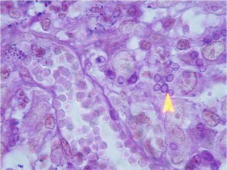
The ability to convert from the mould to the yeast form is a prerequisite for virulence. In the tissues of mammalian hosts, Histoplasma yeasts are oval and small in size (2 x 4 μm) with a thin wall and a narrow-based budding process. They are extracellular or may be found inside host macrophages (Fig. 5.1). In culture or in environmental conditions, Histoplasma organisms form branched septate filaments (1-2 μm in diameter). From these filaments, two types of conidia can be produced: round- or pear-shaped micro-aleurioconidia (2 x 4 μm) and tuberculate and refringent wall macro-aleurioconidia (6 x 15 μm) (de Hoog et al. 2009).Histoplasma organisms can be found in soils in temperate and subtropical areas, but some regions are recognised as areas of hyperendemicity. This is the case for midwestern and southern USA and regions along the rivers (Missouri, Ohio or Mississippi) (Kauffman 2009). Traditionally, the single species H. capsulatum comprises three distinct subspecies with variable geographical distribution, host preferences and associated clinical signs (de Hoog et al. 2009). Histoplasma capsulatum var. capsulatum may be found in many different regions all over the world. The subspecies capsulatum is responsible for pulmonary and systemic infections with small-sized yeastlike cells in the macrophages of many mammals, including humans. Histoplasma capsulatum var. duboisii is present in Western and Central Africa. It develops as large (6 x 15 μm) yeasts with a thick wall in tissues of primates. The subspecies duboisii is responsible for lymphadenopathy with a possible dissemination to the skin and bones. Histoplasma capsulatum var.
farciminosumFig. 5.1 Photomicrograph of subcutaneous tissue in a cat. Large numbers of Histoplasma capsulatum organisms filling the cytoplasm of macrophages are visible (arrow). Periodic acid stain (courtesy from Georges Plassiart, Metz, France)
can be found in many countries from Africa, Asia and South America. This subspecies infects the skin and the subcutaneous lymphatic system of horses, donkeys and mules. It has also been recovered from humans, dogs, cats and badgers. Using multilocus sequence typing (MLST), Kasuga et al. (2003) divided the species H. capsulatum into the following eight geographically separate groups: North American-1, North American-2, Latin American (=A), Latin American (=B), Australian, Netherlands (of Indonesian origin), Eurasian and African. The subspecies farciminosum was composed of three phylogenetic groups, supporting the hypothesis that this taxon is a collection of strains from different clades rather than a true phylogenetic species. Many strains from Europe and Asia were supposed to represent a single clone because they shared the same alleles at all four investigated loci. As no resolution of the branching order of the clades could be obtained, Kasuga et al. (2003) suggested that H. capsulatum radiated rapidly over a short period, most probably 3-13 million years ago.
Isolates of H. capsulatum were classified in two chemotypes according to the composition of the cell wall (Reiss et al. 1977). Chemotype I includes isolates without polysaccharide α-(1,3)-glucan in their cell wall. All isolates from this chemotype belong to the North American-2 clade defined by Kasuga et al. (2003). Isolates from other H. capsulatum clades are corresponding to chemotype II. Their cell wall contains α-(1,3)-glucan, which might be required for virulence and immune evasion. However isolates from chemotype I are recovered from lesions despite the absence of α-(1,3)-glucan (Rappleye et al. 2007).
5.2