Causes of Chronic Weight Loss
In this section, likely causes of weight loss in goats are given by etiologic category. The main intent is to provide a list for differential diagnosis, including only that information that may help to determine its relevance to the case at hand.
Note that this text is international in scope and the conditions proposed for inclusion in the differential diagnosis must be adjusted by readers to their own location. Additional information about the diagnosis, treatment, and prevention of each disease is provided elsewhere in this volume along with references. In general, nutritional deficiencies, parasitisms, and dental disease are the conditions most amenable to treatment.Nutrition -Related Causes
Nutrition-related causes of chronic weight loss fit in either of two categories: primary cases resulting from the unavailability of appropriate feed or specific nutrients, or secondary cases caused by the inability of animals to obtain or use feed when available.
Primary Nutritional Problems
In extensive systems, feed availability can be curtailed by sustained, severe weather conditions such as drought or heavy snow cover, and also by overgrazing, overstocking, or failure to provide supplemental feed when nutrition requirements are not being met. Weight loss, emaciation, or even outright starvation may occur under these circumstances. In confinement systems, inadequate feeder space can lead to weight loss. This should become apparent if animals are observed during feeding. In any management system, specific nutritional deficiencies may be caused by inappropriate feedstuffs, unexpected feed shortages, unbalanced rations, inexperience with livestock feeding, and occasionally neglect.
Regarding specific deficiencies, inadequate energy and protein levels are most commonly involved in cases of primary nutritional weight loss. Feed analysis may be necessary to establish the availability of protein and energy in the overall ration.
The results of such analyses must be compared with established nutrient requirements for goats in the relevant type and stage of production and climatic region.Cobalt deficiency may be a specific cause of progressive emaciation with no localizing signs in goats. In the United States, portions of New England, the upper midwest, and the southeast have cobalt-deficient soils, as do parts of New Zealand, Australia, the United Kingdom, Germany, the Netherlands, and Kenya. Goats grazing cobalt-deficient pastures show loss of appetite, rough haircoat, weakness, muscle wasting, and anemia, and may die after several weeks to months. In pastured animals, cobalt deficiency must be differentiated from gastrointestinal helminthiasis, which produces similar signs. Diagnosis of cobalt deficiency can be established by response to parenteral vitamin B12 administration or oral cobalt chloride. Appetite returns within a few days of treatment.
Copper deficiency can cause inadequate weight gain in growing goats. Diagnosis may be aided by the presence of other clinical signs including anemia, pigmentation defects in the haircoat, conjunctivitis, prolonged diarrhea, and incoordination in newborn (swayback) and young goats (enzootic ataxia). Copper deficiency can also increase susceptibility to gastrointestinal parasitism and adult goats with copper deficiency and parasites may be emaciated, anemic, and hypoproteinemic.
Where forage quality is extremely poor, with little or no fresh green forage available, hypovitaminosis A may contribute to progressive emaciation. Other signs of vitamin A deficiency may include night blindness, corneal cloudiness, ocular discharge, and neurologic signs including convulsive seizures.
Secondary Nutritional Problems
A variety of possibilities must be considered that could limit the animal's ability to obtain or use feed. These include the following.
Behavioral Problems
Even when feeder space is adequate, goats with a low social standing in the herd hierarchy may have their attempts to eat thwarted by more dominant goats who drive them away from feeders.
This can be determined by observation at feeding time. Regrouping animals or feeding affected individuals separately may resolve the problem.During the breeding season, bucks, though otherwise healthy, often become quite thin, presumably because of less time spent eating and more time and energy spent in amorous pursuits. It is imperative that bucks enter the breeding season in good health and good body condition, because a certain amount of weight loss is inevitable.
Oral Problems
Goats with marked brachygnathism may be unable to pre- hend and masticate efficiently and may not do well under extensive management. In confinement feeding systems, affected animals are usually able to consume adequate feed.
The excessive wear or loss of teeth, a common cause of thin ewe syndrome, can also occur in goats, though it is less frequent. This is because goats are more likely than sheep to browse in range and pasture, and therefore less likely to wear away teeth by grazing low to the ground on abrasive soils. Nevertheless, thin does, particularly older does, should be checked for tooth loss. Tooth wear may be aggravated by inadequate dietary calcium or calcium/phospho- rus imbalance resulting in softer teeth, or by fluoride toxicity. When cutting and grinding surfaces of teeth are excessively worn, prehension and mastication become inefficient and feed intake and utilization decrease. Water intake can also diminish because worn teeth are more sensitive to cold temperatures. Animals with dental attrition become progressively weaker and emaciated and can succumb to secondary disease processes, particularly pneumonia and pregnancy toxemia.
Periodontal disease can be an additional cause of tooth loss. Coarse feeds or awns of certain plants may pack between gums and teeth, initiating a progressive cycle of infection, tooth loosening, tooth socket deterioration, and additional impaction of foreign material until teeth are dramatically loosened or expelled. Osteomyelitis of the jawbones may also result.
Signs of molar loss include pouching of the cheeks from feed impaction in empty molar sockets, dribbling of fluid from the mouth, excessive staining of the lips and teeth, and a necrotic or stale feed odor to the breath. When a molar is missing, the normally apposing molar grows excessively long because of a lack of occlusal wear, causing trauma to soft tissues in the mouth.
Extensively managed animals with severe dental attrition or periodontal disease should be culled. The productive life of animals with moderate dental disease can be prolonged by placing them on lusher pasture, feeding processed feeds in confinement, and floating sharp teeth with a flat file. Supplementing calcium and balancing it with phosphorus are indicated as preventive and therapeutic measures in herds in which the diet was previously inadequate.
Soft Tissue Infections
Contagious ecthyma is common in goats worldwide and can produce painful, scabby lesions on the lips that persist from three to six weeks or longer if secondary bacterial infections occur. These lesions can impair feed intake and lead to weight loss. Less common or geographically restricted causes of stomatitis that can cause dysphagia, inappetence, and weight loss are discussed in Chapter 10. Peste des petits ruminants causes severe stomatitis, affecting the gums, tongue, lips, and palate. These lesions may be slow to heal in non-fatal cases, leading to prolonged reduction in feed intake and weight loss, and even animals that recover from the disease tend to remain thin. Foot and mouth disease can also affect goats, but foot lesions tend to be more common and severe than oral lesions.
Blindness
Animals fed in confinement can readily adapt to sudden blindness if surroundings are unchanged. Grazing animals on sparse rangelands, however, probably depend heavily on sight for adequate feed intake. Angora goats with excessive hair covering of the face, an inherited trait, graze inefficiently because of impaired vision.
They may become thin and often succumb to secondary disease problems.Blindness in goats may be caused by gid, hypovitaminosis A, permanent corneal damage after outbreaks of infectious keratoconjunctivitis, or as a sequel to polioencephalomalacia.
Locomotor Problems
Foot and leg problems can interfere with efficient grazing or produce sufficient pain to keep animals recumbent instead of standing at the feeders. Weight loss associated with a reluctance to ambulate and eat may precede outward signs of lameness.
Numerous conditions can lead to impaired locomotion. Foot rot, foot scald, and foot abscesses occur in goats. Chronic bacterial polyarthritis can occur in very young kids after navel infection. Mycoplasma spp. are an important cause of arthritis in goats worldwide and the CAE virus has been recognized as a major cause of arthritis and lameness since 1980. Where it occurs, foot and mouth disease can affect goats and produce lameness with impaired locomotion. Non-infectious conditions that cause impaired mobility and reduced intake include fractures, other traumatic limb injuries, peripheral nerve damage, or rickets secondary to mineral imbalances.
Viral and Prion Causes of Chronic Weight Loss
Caprine Arthritis Encephalitis Virus
It is the arthritic form of CAEV infection that is generally associated with weight loss. This form most often appears clinically between 1 and 2 years of age, with great variability in the progression and severity of signs. The first sign of CAE may be unexplained weight loss, followed by stiffness and reluctance to move. Periods of recumbency increase, food intake diminishes, and weight loss becomes more pronounced. Some goats become severely crippled within a few months, while others may show only intermittent lameness or stiffness for years, and remain that way indefinitely.
Far less commonly, a pneumonic form of CAE occurs in mature goats that produces long-term interstitial pneumonia with intermittent secondary bacterial pneumonia, exercise intolerance, and weight loss.
Weight loss may occur independently of arthritic or pneumonic signs in goats that are infected with the virus as demonstrated by serology. Because CAEV infection is a chronic infection, wasting may be an effect of endogenous cytokine release, and may not depend only on decreased ambulation and feed intake. Because many serologically positive goats never show any signs of disease, however, other possible causes for weight loss must also be considered in thin, otherwise normal goats with positive CAE serologic tests.
Other Viral and Prion Diseases
Contagious ecthyma is discussed above under oral problems. Foot and mouth disease usually produces mild clinical signs in goats, but lameness and oral lesions may impair feed intake sufficiently to cause weight loss.
Scrapie, a prion infection of small ruminants, is characterized by an extended incubation period of months to years, so that most sheep and goats with scrapie are sub- clinically infected. If they ultimately do show signs, the clinical presentation is quite variable and may involve a variety of neurologic signs and/or pruritis, as discussed further in Chapter 5 and summarized in Table 5.4. Progressive weight loss can be an early sign or an only sign of clinical scrapie in goats, so scrapie must be considered in the differential diagnosis of progressive weight loss, particularly in areas or herds where scrapie is known to exist.
Peste des petits ruminants can cause pronounced weight loss and debilitation in goats during the prolonged convalescent periods that may follow recovery from the acute diarrheal and respiratory phase of the disease. As mentioned above, severe stomatitis in the acute phase may also contribute to decreased feed intake.
Pulmonary adenomatosis, or jaagsiekte, is a neoplastic transformation of the lung caused by a retrovirus distinct from CAEV. It is primarily a sheep disease, but has been reported in goats. The disease produces a syndrome of progressive dyspnea and weight loss with pronounced nasal discharge over a period of several months. Moist lung sounds are audible and considerable fluid drains from the lungs if the animal is held vertically with the head hanging down. Diagnosis is by histopathologic examination of the lungs.
Bacterial Causes of Chronic Weight Loss
Paratuberculosis (Johne's Disease)
It is very likely that paratuberculosis is a frequent but undiagnosed cause of progressive weight loss in adult goats. Failure to diagnose this condition usually results from three factors. First, chronic diarrhea, the cardinal sign of paratuberculosis in cattle, is rarely observed in goats, so veterinarians familiar with the cattle disease may not consider paratuberculosis when confronted with a thin goat. Second, rapid, reliable laboratory tests historically have not been readily available as aids to clinical diagnosis, though the situation has improved in recent years. Last, gross postmortem lesions of intestinal thickening, common in cattle, are uncommonly seen in affected goats. In most cases, histopathologic examination of intestinal tissue and associated lymph nodes with acid-fast staining and/or mycobacterial culture of feces or intestine must be performed to establish paratuberculosis as the cause of chronic weight loss. Such examinations are warranted in unexplained caprine wasting disease because of the relative importance of paratuberculosis as a cause of weight loss that is commonly overlooked in goats.
Caseous Lymphadenitis
This important chronic infectious disease of goats caused by Corynebacterium pseudotuberculosis (Corynebacterium ovis) is best known by the superficial lymphadenopathy it produces. However, abscesses of internal lymph nodes and viscera also occur, though less commonly. While the impact of superficial lymph node abscesses on general health in goats is not remarkable, a stronger association exists between progressive emaciation and internal abscesses.
Antemortem diagnosis of internal abscesses can be difficult. Goats with internal abscesses often do not have concurrent superficial abscesses. Physical exam rarely establishes proof of an internal lymph node abscess, although large abscesses in the chest may alter normal heart and lung sounds and large abdominal abscesses are sometimes palpable. Radiography or ultrasonography may be helpful if large or focally calcified abscesses are present. Neutrophilia and hypergammaglobulinemia may support the diagnosis, but are not pathognomonic. At necropsy, thoracic and abdominal organs should be examined and abscesses cultured because other pyogenic organisms may be involved. Tuberculosis may present similarly.
Tuberculosis
Tuberculosis is uncommon in goats, but sporadic cases continue to be reported. Mycobacterium bovis is the most common cause, but Mycobacterium avium and Mycobacterium tuberculosis also occur, and the zoonotic potential of the disease should be respected. Tuberculous goats often are commingled with cattle. The presentation of weight loss is variable. It may precede other clinical signs, accompany them, or not occur at all. Clinical tuberculosis most often manifests as chronic pneumonia with a moist, persistent, productive cough. Intestinal tuberculosis also occurs with diarrhea as a presenting sign. Antemortem diagnosis is by intradermal skin test. At postmortem examination, obvious caseated, sometimes calcified, parenchymal or lymph node granulomas are present. Tuberculosis coincident with paratuberculosis in goats has been reported from Spain. In such cases, definitive diagnosis depends on mycobacterial culture of granulomatous lesions.
Melioidosis
This zoonotic infection caused by a soil saprophyte, Burkholderia (Pseudomonas) pseudomallei, is reported from tropical regions of the world, primarily Southeast Asia and Australia. It may be endemic in some regions and outbreaks are associated with flooding of pastures. Acute and chronic forms are seen in goats. Clinical manifestations are varied and include high fever, nasal discharge, pneumonia, lymphadenopathy, mastitis, orchitis, polyarthritis, and frequently neurologic signs including ataxia, head tilt, nystagmus, or circling. Acute deaths may occur within 24-48 hours after fever, weakness, and recumbency. Animals that survive may have any of the signs noted in addition to pronounced weight loss. Asymptomatic infections can also occur in the herd. The organism may be transmitted in goat milk and the disease is zoonotic.
Antemortem diagnosis may be accomplished by bacterial culture of milk, nasal discharges, or aspirates of swollen lymph nodes; skin testing with meliodin; or serologic methods, including complement fixation and indirect hemagglutination assays. Necropsy usually reveals multiple abscesses in numerous organs, especially spleen, liver, and lungs. These abscesses should be cultured (see Chapter 3).
Chronic Bacterial Infection and Cachexia
Any prolonged infection may contribute to a process of progressive debilitation and muscle wasting, referred to as cachexia, which in and of itself can contribute to an adverse patient outcome. Chronic inflammation is considered to be a major contributor to the evolution of cachexia. The mechanisms of cachexia are multifactorial and not fully elucidated. Alterations in carbohydrate, protein, and lipid metabolism all contribute to the loss of tissue and body mass. These metabolic changes are driven by an inflammatory response mediated in part by the upregulation of production of pro-inflammatory cytokines, including but not limited to interleukin 1, interleukin 6, gamma interferon, and tissue necrosis factor alpha (TNF-α), also referred to as cachexin. TNF-α has a direct catabolic effect on skeletal muscle and adipose tissue through the ubiquitin proteas- ome pathway. A second proteolytic pathway, the autophagylysosome system, also contributes to muscle breakdown. Advances in knowledge on the mechanisms of cachexia derive mainly from studies aimed at better understanding the condition in human cancer patients, but may also apply to cachexia associated with chronic infection. The mechanisms of cachexia and potential opportunities for therapeutic management in human patients have been reviewed (Samant et al. 2019). There are no studies on the pathogenesis of cachexia involving goats, but the process is certainly at play in chronic caprine infections such as CAE, paratuberculosis, tuberculosis, and melioidosis, as discussed above. One experimental study of CAEV infection in goats identified elevated levels of TNF-α in infected goats compared to uninfected controls (Mdurvwa et al. 1994).
Other chronic bacterial infections likely to be associated with weight loss in goats are as follows.
Pasteurella and Mannheimia pneumonias of goats are common and often become chronic due to failure of early detection coupled with inadequate therapy. Chronic pasteurellosis is often complicated by secondary lung abscessation due to Trueperella (Arcanobacterium) pyogenes. Careful auscultation and radiology or ultrasonography may confirm the presence of chronic pneumonia and abscessation.
Chronic mastitis cases are generally culled from commercial goat dairies in a timely fashion. Hobbyists, however, may show a marked reluctance to cull mastitic does. Typically, affected does with chronic mastitis appear thin and depressed, and have a pendulous udder containing extensive fibrotic areas and multiple large firm abscesses, often caused by T. (Arcanobacterium)pyogenes (Figure 15.2).
Peritonitis can occur secondary to a number of conditions. These include rumenitis resulting from grain overload, migration of liver flukes through abdominal organs, administration of irritating drugs intraperitoneally, the rupture of internal abscesses, and, as sequelae to dystocia, abdominal surgery or septicemia. Abscesses, adhesions, and bowel stenosis can occur as a result. Diagnosis is based on history of predisposing conditions, evidence of fever and abdominal pain, digestive dysfunction, an inflammatory blood picture, and supportive abdominocentesis findings. Thin animals with evidence of peritonitis should be culled.
Salmonellosis and enterotoxemia present most often as causes of acute, frequently fatal, enteritis or toxemia. Chronic forms of both diseases, however, occasionally occur in adult goats, and their clinical presentations are quite similar. Both conditions are characterized by periodic intermittent diarrhea accompanied by fever, loss of appetite, and depression. Sporadic recurrences may be observed over several months, accompanied by progressive weight loss. Notably, the chronic form of enterotoxemia can occur even in animals with a strong vaccination history against Clostridium perfringens type D. Definitive diagnosis depends on isolation of Salmonella spp. during episodes of active diarrhea or, in the case of enterotoxemia, identification of epsilon toxin in diarrheic feces or gut content.
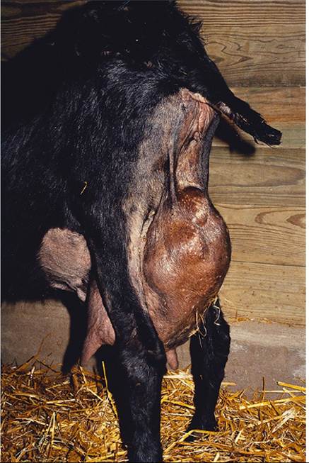
Figure 15.2 A thin doe with chronic mastitis due to Trueperella (Arcanobacterium) pyogenes. Note the characteristic, large, nodular, multiple abscesses in the udder. Source: Courtesy of Dr. David M. Sherman.
Parasitic Causes of Chronic Weight Loss
Both ectoparasites and endoparasites can contribute significantly to progressive weight loss in goats, either primarily or in association with malnutrition or concurrent disease.
Gastrointestinal Parasitism
Coccidiosis
Coccidiosis is easily diagnosed as a cause of diarrhea, dysentery, dehydration, and often death in young kids. However, less obvious is the fact that animals that survive clinical coccidiosis often suffer sufficient damage to the intestinal mucosa that normal growth and development are permanently impaired, presumably due to occurrence of intestinal malabsorption. Specific diagnosis of this condition is difficult. A history of coccidiosis on the farm and exclusion of other likely causes of poor growth or inadequate weight gain may lead to a presumptive diagnosis of chronic intestinal damage secondary to coccidiosis.
Nematodiasis
Nematodiasis is the most important parasitism contributing to ill-thrift. Young animals between 2 and 24 months of age are most often affected, but age-related resistance to parasitism is not as strong in goats as it is in cattle, and clinical parasitism is not uncommon in older animals, particularly when concurrent disease or poor nutrition is present. Clinical evidence of gastrointestinal parasitism includes pale mucous membranes, fluid accumulation between the jaws (bottle jaw), weakness, decreased growth rate or milk production, rough haircoat, and progressive weight loss or emaciation. Diarrhea is a common though inconsistent finding.
Cestodiasis
Tapeworms occur in goats worldwide. Their contributory role in the development of weight loss is controversial, but in general they are not considered to be clinically important unless heavy infestations are found in animals younger than 6 months of age, in which case they may contribute to poor growth. An inordinate amount of importance is placed on tapeworm infestation by goat owners because they can readily observe tapeworm segments (proglottids) in goat feces.
The goat can serve as an intermediate host for tape - worms of the genera Taenia and Echinococcus, resulting in cysticercosis and hydatidosis, respectively. These conditions usually remain subclinical, producing no signs other than poor weight gain in young animals. Sometimes they produce peritonitis with accompanying fever, depression, and weakness. Diagnosis is usually accomplished at slaughter or necropsy when numerous, large, larval cysts are found in abdominal viscera, mesentery, and lungs.
Trematodiasis
Four species of liver flukes affect goats: Fasciola hepat- ica, Fasciola gigantica, Fascioloides magna, and Dicrocoelium dendriticum. While acute and chronic forms of liver fluke disease occur, it is the chronic form caused by Fasciola spp. that is associated with weight loss. This results from localization and persistence of adult flukes in the bile ducts, where they consume blood and produce bile duct irritation with resultant liver dys - function. The clinical syndrome produced may be indis - tinguishable from gastrointestinal nematodiasis, although icterus is sometimes seen. Ova. may be found in feces using sedimentation techniques, but diagnosis is easiest at necropsy, where adult flukes and their excretions are found in the bile ducts. Note that F. magna infestation can cause weight loss and death without the appearance of eggs in the feces.
Pancreatic flukes of the genus Eurytrema can also produce emaciation in goats. The condition is discussed further in Chapter 11.
Schistosomosis can produce ill-thrift in goats in Asia, Africa, the Middle East, South and Central America, and the Mediterranean region. The disease occurs where livestock are exposed to aquatic snails, the intermediate parasite hosts. In the nasal form, weight loss is accompanied by snoring, sneezing, and dyspnea, and in the intestinal form by diarrhea and anemia. This form may be clinically indistinguishable from gastrointestinal nematodiasis. Diagnosis is based on identifying parasite eggs in feces, urine, liver biopsy specimens, or nasal discharges.
Lungworm Infestations
Three species of lungworms affect goats in the United States. Dictyocaulus filaria has a direct life cycle and is considered the most pathogenic. Protostrongylus rufescens and Muellerius capillaris both have snail intermediate hosts and are considered less pathogenic. All are likely to occur in cool, wet, autumnal weather when younger stock on lowland or irrigated pastures ingest infective larvae or snails. While adult Dictyocaulus and Protostrongylus cause obvious coughing in affected goats by irritating airways, adult forms of M. capillaris do not because they do not reach the airways. They reside in the alveoli, producing small, gray-green nodules visible at necropsy on the surface of the diaphragmatic lobes. Weight loss may accompany respiratory signs in Dictyocaulus infection, but may be the only clinical sign of severe Muellerius infection.
External Parasites
Ectoparasites can contribute to ill-thrift when infestations are severe and concurrent diseases are present. Biting lice such as Damalina caprae may cause irritation to affected hosts, leading to reduced feeding efficiency. The blue louse of goats, Linognathus stenopsis, feeds on host blood using piercing mouth parts and causes anemia and weight loss. Lice are more common in winter months and are promoted by confinement and overcrowding. Parasites are visible when the hair is parted and the skin carefully observed.
Among the mange mites affecting goats, Sarcoptes, and to a lesser extent Chorioptes, can produce intense pruritus that torments the host and results in decreased feeding activity and weight loss. Generalized crusting and flaking of the skin with alopecia are apparent and skin scrapings should confirm the diagnosis. The sheep ked, Melophagus ovinus, also affects goats and can produce intense pruritus, particularly in winter. A period of ill-thrift has been observed in goats with demodectic mange and also in association with heavy ear mite infestation.
In tropical regions, heavy infestations of goats with fleas of the genus Ctenocephalides can cause anorexia, anemia, weight loss, and even death. Kid goats are most severely affected.
Hemoparasites
Trypanosomosis affects goats in the tsetse fly regions of A if ica. Acute, subacute, and chronic forms occur. Scvijre emaciation is associated with chronic disease. idditional clinical signs may include anemia, depression, anorexia, lack of rumen motility, and peripheral lymphadenopathy Diagnosis is by identifying the hemoparasite on stained blood smears.
Rarely, eperythrozoonosis may be a cause of anemia and ill-thrift in goats. Infection is usually subclinical. Caprine infection has been reported in Hungary, Turkey, Egypt, South Africa, Pakistan, China, Malaysia, the Philippines, Australia, Brazil, and Cuba. The disease is discussed further in Chapter 7.
Miscellaneous Causes OfChronicWeight Loss
Plant Toxicoses
A wide variety of poisonous plants may affect goats on pasture or range. Most plant intoxications produce sudden death or an acute disease syndrome that does not involve weight loss. One important exception is toxicity due to locoweed (Astragalus spp.), a chronic intoxication characterized by mild nervous signs, muscular incoordination, fetal abnormality or abortion, and chronic, progressive weight loss. Locoism is seen most commonly in the western United States. Goats are susceptible, though the disease is seen more often in sheep.
Lupinosis, or phomopsin poisoning, can be a cause of weight loss, anorexia, depression, and jaundice. It is associated with the consumption of lupine stubble contaminated with the fungus Diaporthe toxica, which produces pho- mopsin mycotoxin. It has been reported in Europe, Australia, New Zealand, and South Africa. The condition is seen most commonly in cattle and sheep, but has been reported as well in goats in Western Australia.
In Germany, Austria, and Switzerland (Braun et al. 2000), a syndrome of vitamin D toxicity causing progressive weight loss, lameness, decreased milk production, and calcification of soft tissues has been observed in goats consuming yellow oat grass (Trisetum flavescens), which contains 1,25-dihdroxycholecalciferol. This golden oat grass intoxication may also cause cardiovascular abnormalities and is discussed further in Chapter 8.
Gastrointestinal Foreign Bodies
In South Africa, phytobezoars consisting of seed hairs of karoo bushes can cause abomasal impaction in grazing goats. Progressive weight loss accompanied by progressive abdominal distension suggests this diagnosis. Anorexia and progressive weight loss of six weeks' duration has been reported in three Angora goats, subsequently confirmed by necropsy of two and rumenotomy of the third to have multiple hair balls (trichobezoars) in their rumens (Baillie and Anzuino 2006).
Consumption of plastic trash bags is common where rubbish disposal is uncontrolled and where goats forage in urban areas, as is common in parts of Africa, the Middle East, and South Asia (Figure 15.3). Baling twine can be ingested when confined goats are fed baled hay. These foreign bodies can lead to partial gastrointestinal obstructions, chronic indigestion, abdominal distension, and weight loss. In a study from Jordan, 4.5% of 722 adult goat cases presented at a veterinary teaching hospital over a 3.75-year period were diagnosed with soft foreign bodies (plastic) in their rumens and reticuli (Hailat et al. 1998).
Neoplasia
In general, tumors are an uncommon finding in goats and therefore are a relatively infrequent cause of progressive emaciation. However, neoplasia still should be considered in a differential diagnosis of chronic weight loss, especially in older goats kept as pets, because they are often kept to very advanced ages, with a concurrent increase in the likelihood of neoplasia. Sporadic cases of neoplasia, including intestinal adenocarcinomas, have been diagnosed at necropsy in adult goats after a prolonged course of ill-thrift. More consistently, enzootic intranasal tumors of goats have been reported, primarily in Europe. Affected goats show persistent, profuse, often unilateral, serous nasal discharge; progressive dyspnea; and chronic weight loss. It has been demonstrated by transmission studies that these nasal adenocarcinomas are caused by a retrovirus (De las Heras et al. 1995). The disease is discussed further in Chapter 9.
Amyloidosis
Goats are used extensively in research and industry to produce antibodies for commerce or research. Repeated immune stimulation in these animals with adjuvanted antigens can stimulate the production and deposition of renal amyloid. Renal amyloidosis manifests clinically as progressive emaciation with weakness, anorexia, depression, and possible edema and ascites caused by hypoproteinemia subsequent to proteinuria. Diarrhea may also be present, caused by hypoproteinemia or concurrent intestinal amyloidosis. Diagnosis is aided by urinalysis and kidney biopsy. There is no treatment for the condition. Although amyloidosis is an unlikely cause of chronic weight loss in the general goat population, it may occur frequently in this specialized group of antibody-producing goats.
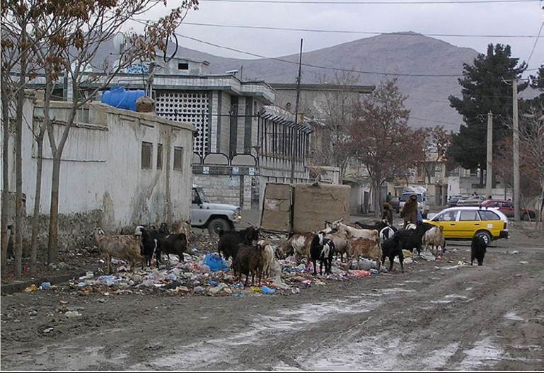
Figure 15.3 Urban goats in Kabul, Afghanistan, foraging in an open trash pile containing plastic bags. Consumption of plastic bags can lead to blockage of the reticulo - omasal orifice, resulting in chronic indigestion and weight loss. Source: Courtesy of Dr. David M. Sherman.
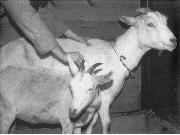
Figure 15.4 Two 7- month - old Saanen doeling sisters. The stunted goat in the foreground was diagnosed at presentation with a cleft palate. Source: Courtesy of Dr. David M. Sherman.
Other Sporadic Causes
Though an uncommon occurrence, congenital ventricular septal defect results in poor growth in young affected goats. Stunted growth may also result from chronic aspiration pneumonia associated with cleft palate or other causes (Figure 15.4). In Nubian goats, the inherited lysosomal storage disease mucopolysaccharidosis IIID can cause muscle wasting in homozygous affected individuals, resulting in weakness and ill-thrift. The condition is discussed further in Chapter 5.