Clinical Examination for Diagnosis ofWeight Loss
Clinical examination of the goat is covered in detail in Chapter 1. Aspects relevant to the problem of ill-thrift are briefly reviewed here.
A number of the causes of weight loss are infectious diseases with chronic, subclinical, or carrier forms, notably caprine arthritis encephalitis (CAE), caseous lymphadenitis, and paratuberculosis.
Therefore, it is important to establish how long the herd or flock has been in existence, whether or not the owner knows the original sources of animals in the herd, the disease status of those herds of origin, and whether any prepurchase testing for these or other diseases was performed on animals brought into the herd.Determine if and when there have been any previous cases of weight loss. If similar cases occurred in previous years, it suggests a possible endemic condition. Any patterns of occurrence within a certain age group or with regard to season, housing, pasturing, feeding, or other management conditions should be evaluated. Questions regarding current cases should include information about the possibility of recent stressful situations such as parturition, purchase, and introduction into the herd, transport and attendance at sales or shows, or episodes of acute disease, since such stresses can trigger latent paratuberculosis or expose the animal to new infections. Evaluation of parasite control programs is important. Determine that control efforts are directed at all important types of internal and external parasites. Establish that the anthelmintics
Goat Medicine, Third Edition. Mary C. Smith and David M. Sherman. © 2023 John Wiley & Sons, Inc. Published 2023 by John Wiley & Sons, Inc.
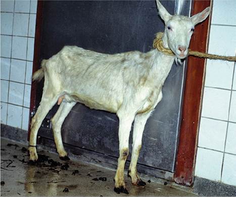
Figure 15.1 Wasting goats such as this one, with no other localizing signs, present a diagnostic challenge for the veterinary practitioner.
This particular doe was ultimately diagnosed with paratuberculosis. Source: Courtesy of Dr. Daan Dercksen.employed are appropriate and effective and determine how available pastures are used for grazing, including information about rotation and stocking rates.
The history related to feeding may be critical in many cases. Solicit information on the basic ration fed to the group or groups showing weight loss and any recent changes in feedstuffs, feeding methods, feeder space, feeder design, or number of animals fed per group. Be sure that feeding programs are appropriate for the level and type of production being called for in each group. Nutrient requirements for different levels and types of production are given in Chapter 19.
Physical Examination
All animals in contact with the thin animal should be examined at least from a distance to detect any clinical signs, such as coughing or diarrhea, which might represent conditions that could manifest in a chronic form as weight loss. An assessment of body condition in herd mates by digital palpation of the back, ribs, and sternum is essential to establish the body condition perceived as normal by the owner and to determine the prevalence of thin goats in the group. This is particularly important in Angora goats, in which the fleece may hide body condition. For veterinarians more familiar with dairy breeds, the normal Angora goat feels thin because these animals are more slightly built by nature. Details on body condition scoring in goats are presented in Table 19.6.
On direct examination, weakness, dullness, lack of appetite, and a rough haircoat are all non-specific signs associated with ill-thrift that may accompany weight loss. Careful physical examination may reveal additional abnormalities undetected by the owner that can help in diagnosis. Some specific points to consider are as follows.
Examine the skin for lice, keds, or fleas by carefully parting the hair on the face, body, and limbs, and look in the ears and under the tail for ticks.
A thorough oral examination may reveal brachygna- thism, tooth loss, gum or alveolar abscesses, painful sores, or other abnormalities that might interfere with normal prehension, mastication, or swallowing. Evaluate the gums, conjunctiva, and other mucous membranes for anemia or icterus suggestive of chronic helminthiasis or liver fluke infestation. Check for nasal discharges that could indicate lung disease or nasal adenocarcinomas.
Routine palpation of all superficial lymph nodes is essential, because several causes of weight loss may cause lymph node enlargement. These include caseous lymphadenitis, melioidosis, tuberculosis, trypanosomosis, CAE and lymphosarcoma. In thin goats, however, be aware that lymph nodes may be more readily palpable, giving a false impression of enlargement. In this case, asymmetrically sized nodes are of greater significance. Careful auscultation of the lungs may reveal abnormalities consistent with chronic pneumonia. Displacement of the heart by a mediastinal mass may alter normal heart sounds. Because congenital heart disease may occasionally occur and lead to ill-thrift, cardiac murmurs should be noted when present.
Establish the presence of normal rumination and gut motility. Assess the animal for ascites or rumen impaction by ballotment or observation of the abdominal contour.
Foot and joint problems should not be overlooked as a cause of inefficient grazing that can result in weight loss. Check the interdigital space for foot scald and trim the feet to assess for foot rot. Palpate all joints carefully. Forced exercise may help identify signs of limb problems, chronic pneumonia, or anemia.
Environmental Examination
The level of general sanitation is important in confinement operations. Manure buildup can contribute to the spread of coccidiosis, paratuberculosis, and chronic mastitis. Overcrowding can facilitate transmission of lice and pneumonia and predispose to herd outbreaks of enteric diseases, some of which can assume chronic forms.
Poorly drained paddocks and barns can contribute to foot problems and coccidiosis.All components of the ration should be inspected for spoilage, molds, and palatability. Look for fouled or frozen water supplies, which can reduce water and feed intake. Chronic weight loss can be related to the way animals are fed. If young animals can climb or defecate in feeders, then fecal contamination promotes the spread of coccidiosis. Keyhole-type feeders common in goat dairies may facilitate the rupture of lymph node abscesses on the head and neck during feeding, causing contamination of feed and spread of caseous lymphadenitis. Inadequate feeder space in either keyhole-type or trough-type feeding systems may deprive individual animals, low in the social pecking order, of adequate feed intake.
In range systems the stocking rate; the availability and quality of forage, water, and supplemental feeds; and the prevailing weather conditions must be considered when thin animals are observed. Angora goats have specialized needs for supplemental protein and energy at different times in their fiber production and reproductive cycles and these requirements can be heightened by harsh, wet, and cold weather.
Laboratory Diagnosis and Necropsy
Despite thorough clinical examination and knowledge of the likely causes, the definitive diagnosis of chronic weight loss may remain elusive. Hematology and clinical chemistry may show evidence of anemia and or hypoalbumine- mia, but tests results are often unrewarding, merely confirming the presence of undifferentiated chronic disease. Serologic testing, however, can be helpful in the diagnosis of some infectious causes of weight loss, especially CAE and paratuberculosis. Fecal examination can assist in the diagnosis of various endoparasitisms.
When multiple animals are affected in commercial herds or flocks, sacrifice of an individual animal for postmortem examination is justifiable on economic grounds. The animal should be checked thoroughly for evidence of gastrointestinal, pulmonary, and hepatic parasites, and for internal abscesses. The degree of rumen fill and the general body condition should be noted. Emaciated goats with obvious loss of muscle mass and perirenal fat, paradoxically, may still have noticeable fat deposits in the mesentery. This should not be interpreted as evidence of good body condition, because goats preferentially store whatever fat they have intra-abdominally rather than subcutaneously.
For field necropsy, tissues submitted for histopathologic examination in addition to any gross lesions should include kidney, carpal synovial tissue, and ileocecal lymph node and valve for diagnosis of amyloidosis, caprine arthritis encephalitis virus (CAEV) infection, and paratuberculosis, respectively, because these conditions may not be evident from gross findings.