Clinical Signs
6.5.1 PCM Infection
Paracoccidioidin-positive infected individuals who do not have symptoms or laboratory evidence of the disease have developed the primary complex, but do not show disease progression (Wanke and Londero 1994).
6.5.2 PCM Disease in Humans
The disease occurs as a consequence of the progression of the primary complex, the reactivation of a quiescent focus (endogenous reinfection), or exogenous reinfection. Three main forms are observed: (1) acute-subacute, also called juvenile type or juvenile, which affects mainly children and young adults under 30 years of age; (2) chronic, also called adult type, usually observed in patients over 30 years of age; and (3) sequelae, usually chronic obstructive pulmonary disease, dysphonia, and laryngeal scarring (Franco et al. 1987).
Acute-subacute PCM (juvenile type) affects both sexes and represents about 10% of the general distribution of the disease. Among these patients, about 5% are children between 10 and 14 years old. Although the disease is rare in very young children, its occurrence in that age range is serious and potentially fatal. The juvenile form can be subdivided into mild or severe, according to the degree of dissemination. Clinical manifestations are caused by rapid and progressive involvement of the mononuclear phagocyte system with diffuse lymphadenopathy, hepatosplenomegaly, and bone marrow dysfunction in severe cases. Cutaneous manifestations and bone lesions may also be observed in acute PCM. Fever and weight loss also accompany this clinical picture that quickly leads to general commitment of the patient (Del Negro et al. 1994; Mendes 1994; Shikanai-Yasuda et al. 2006).
Chronic adult PCM is responsible for most cases of PCM in humans (about 90%). It is observed mainly in males between 30 and 60 years old, mainly rural workers with frequent contact with or disturbance of soil, e.g., farmers, construction workers, tractor drivers, and armadillo hunters (Cadavid and Restrepo 1993; Wanke and Londero 1994; Shikanai-Yasuda et al.
2006). Among women the disease is less frequent (F/M ratio 1:10), since the female hormone 17-β estradiol provides protection by preventing the transformation of inhaled conidia in yeast in the lung parenchyma (Restrepo et al. 1984; Salazar et al. 1988). Factors such as smoking, alcohol consumption, and malnutrition are also associated with chronic adult PCM. Probability of patients with PCM clinically becoming ill was 14 times higher among smokers and 3.6 times among individuals who ingested >50 g/day of alcohol (Martinez and Moya 1992; Santos et al. 2003; Bellissimo-Rodrigues et al. 2011). Chronic PCM can be subdivided into unifocal and multifocal with mild, moderate, and severe degrees of disease (Franco et al. 1987; Shikanai-Yasuda et al. 2006).6.5.3 PCM Associated with Immunosuppression
Association of PCM with human immunodeficiency virus (HIV) infection is reduced when compared, for example, with histoplasmosis. In general patients with PCM/AIDS have prolonged fever, substantial weight loss, lymphadenopathy, hepatosplenomegaly, lung damage, and skin and neurological injuries. A prevalence of PCM/AIDS in 1.4% of patients has been reported in the southeastern region of Brazil; most of them have CD4 counts lower than 200 cells/mL, they are young, and few of them work in agriculture (Goldani and Sugar 1995; Bellissimo-Rodrigues etal. 2011).
6.5.4 PCM Sequelae
PCM induces a granulomatous inflammatory response in the host, which often leads to fibrosis. Advanced stages of the disease present substantial increase in cytokines, such as TNF-α and TGF-β, which induce the accumulation of collagen. Fibrosis has been observed in the lungs of ~50% of patients, of whom a small percentage develop chronic obstructive pulmonary disease (COPD). Studies show that 15-50% of patients show reduced adrenal gland function and about 3% develop Addison's disease and require hormone replacement therapy (Do Valle et al. 1993; Faigal et al. 1996; Onate et al. 2002).
The CNS is also affected by PCM sequelae in about 6-27% of patients, leading to motoric deficits, seizures (epilepsy), and/or hydrocephalus (Nobrega and Spina-Franga 1994; Shikanai-Yasudaet al. 2006).Another important aspect in PCM sequelae is the presence of fibrotic lesions in mucosa and skin. Accordingly, humans with this disease have presented chronic changes of the voice (dysphonia due to the injury of the vocal cords), laryngeal obstruction that leads to tracheostomy, and microstomia due to perioral lesions (Paniago et al. 2003; Weber et al. 2006). Abdominal lymphatic obstruction, reported in acute PCM, has led to malabsorption syndromes, protein loss, and obstructive jaundice (Cazzo et al. 2015).
6.5.5 PCM Disease in Animals
In armadillos, animals that are frequently infected, occasional granuloma formation was observed in the liver and lungs, which may indicate that the animals develop active PCM disease (Bagagli and Bosco 2008). The generalized PCM reported in one two-toed sloth leads to progressive lethargy, anorexia, dehydration, and fatality. Necropsy revealed granulomatous lesions in the lungs, liver, spleen, and kidneys containing numerous fungal cells as shown in silver-stained histopathological sections (Trejo-Chavez et al. 2011).
As already mentioned, P. brasiliensis infection in dogs seems to be common; however, PCM disease with clinical symptoms has been reported only in three dogs, in which the main clinical sign was generalized lymphadenomegaly. In the first report, an adult female, non-neutered, Doberman, presented swelling of submandibular lymph nodes and poor general condition. The diagnosis was confirmed by histopathology, immunohistochemistry, and molecular detection of GP43 gene in lymph node biopsy. The animal was treated with ketoconazole producing total regression of lymphadenopathy. However, clinical recurrence was observed after 18 months, and the dog was euthanized without being subjected to autopsy (Ricci et al.
2004). The second case, also in an adult female non-neutered Doberman, showed lymphnodes enlargement (Fig. 6.4), and the diagnosis was confirmed by culturing P. brasiliensis from popliteal lymph node biopsy. The animal was successfully treated with itraconazole for 2 years (Farias et al. 2011). The third report was described in a 5-year-old female Labrador with lymphnodes enlargement and skin lesion in the left side of the upper lip. The diagnosis was confirmed by cytological, serological, and molecular identification of P. brasiliensis isolated from fine needle aspirate from lymphnode. The dog was treated with itraconazole and is under observation for 18 months with remission of the symptoms (Headley et al. 2017).A single case of feline PCM was reported in a cat from Chile, which developed neurological and renal symptoms. The host was an 8-month-old Persian cat with anorexia, weakness, fever, and neurological signs (depression, nystagmus, and tremors). The diagnosis was confirmed by cytopathological detection of the fungus in cerebrospinal fluid and urine, with no fungal culture (Gonzalez et al. 2010). This report should be considered with caution, since (1) the animal is from Chile, a country that PCM has never been recorded for human, and (2) the images of micromorphology of the fungus in the publication are not completely compatible with P. brasiliensis. Likewise, the same precaution should be considered to the reported case of Mycobacterium bovis and dimorphic fungi (P. brasiliensis and B. dermatitidis) co-infection in beef cattle in Kenya, Africa, by the observation of yeasts cells in Ziehl-Neelsen smears (Kuria and Gathogo 2013).
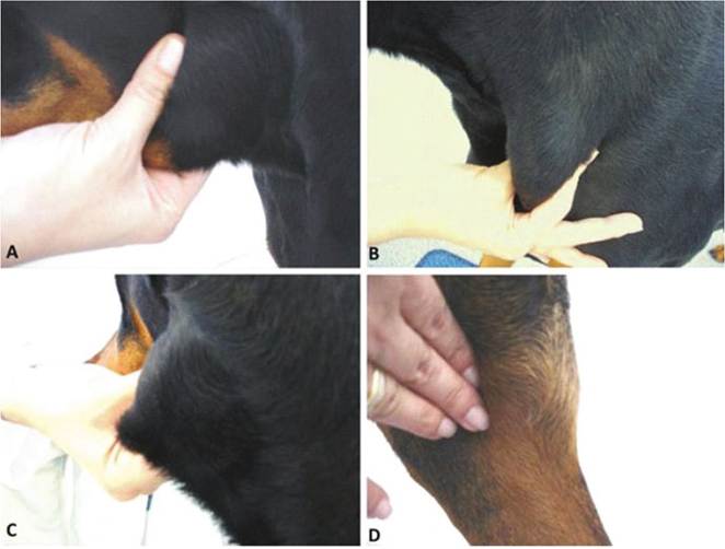
Fig. 6.4 Generalized Iymphadenomegaly in a 6-year-old female Doberman. (a) Submandibular lymph node; (b) prescapular lymph node; (c) inguinal lymph node; (d) popliteal lymph node. “Copyright Mycopathologia (172(2):147-152, 2011), reprinted with permission”
P. lutzii has never been isolated directly from armadillos or any other animal species. To date, there is one study showing serological evidences about P. lutzii infection in domestic and wild animals (Mendes et al. 2017). The authors evaluated 481 animals (horses, dogs, and wild mammals) from South region of Brazil, and 105 reacted positively for P. lutzii. Among these seropositive animals, 54 showed crossreaction with P. brasiliensis antigens and only 51 animals (11 horses, 30 dogs, and 10 wild mammals) were considered infected by P. lutzii, confirming the existence of this species in South region of Brazil (Mendes et al. 2017).
6.6