Clinical Signs and Lesions
The disease in humans and dolphins is usually chronic and takes time to develop into large lesions (Bossart et al. 2015; Reif et al. 2006; Rodriguez-Toro 1993; Tapia et al. 1978; Talhari and Talhari 2012).
Typical lesions are monomorphic or multimorphic and painless but may develop mild pruritus. The most affected anatomical sites in humans are ears, shoulders, limbs, back, and abdominal areas. Human lacaziosis clinical features have been subjected to extensive reviews since the first case was reported (Cardoso de Brito and Quaresma 2007; Francesconi et al. 2014; Talhari and Talhari 2012). The first effort to describe the polymorphic clinical characteristics of the disease in humans came from Silva and Brito (1994). These authors described five clinical forms including the typical parakeloidal granuloma and the gummatous, infiltrate, ulcerated, and verrucous form. The same year Machado (1972) described two basic forms. The first one is a hyperergic state that includes the macular, gummatous, and nodular forms described by Silva and Brito (1994). The second form is a hypoergic state including the parakeloidal and verrucous forms (Fig. 9.3). In the polymorphic form, the host immune system may react to the antigens presented during infections in a similar way as in the paucibacillary and multi- bacillary forms of M. leprae infection (Eichelmann et al. 2013). In a revision of 40 cases in Acre, Brazil, Opromolla et al. (2000) also described the above forms and indicated that the most frequent anatomical area was the ear (Fig. 9.3a). Differential diagnosis in humans includes chromoblastomycosis, leishmaniasis, leprosy, neoplasia, and paracoccidioidomycosis (Eichelmann et al. 2013; Lacaz et al. 1986).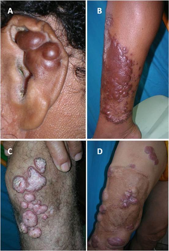
Fig. 9.3 Cases of human Iacaziosis from Acre, Brazil.
(a and d) Some of the clinical manifestation of the infection described by Silva and Brito (1994). (a) The most common anatomical site followed by different type of granulomatous lesions on the limbs. (d) An example of recurrence after surgical removal of old lesions (Courtesy of Dr. P. Rosa)The infection has been diagnosed in several dolphin species including Tursiops truncatus, T. aduncus, Sotalia guianensis, and Lagenorhynchus obliquidens (Bossart et al. 2015; De Vries and Laarman 1973; Kiszka et al. 2009; Minakawa et al. 2016; Van Bressem et al. 2009). The terms “lacaziosis-like” and “lobomycosislike” are currently used to identify the clinical features of putative cases of the disease based only on gross anatomical observations, but lacking histopathological confirmation (Daura-Jorge and Simoes-Lopes 2011; Kiszka et al. 2009; Reif et al. 2006; Sacristan et al. 2016; Tajima et al. 2015). However, due to the new proposed names for the disease, the use of paracoccidioidomycosis ceti-like is recommended. The clinical features of the disease in dolphins are characterized by the formation of white, gray to reddish nodular or verrucous lesions (cauliflower-like), sometimes with prominent elevation over non-infected skin areas (Fig. 9.4) (Bossart et al. 2015; Minakawa et al. 2016; Rotstein et al. 2009; Reif et al. 2006; Ueda et al. 2013). These areas could ulcerate and form papillary nodules becoming large plaques. The lesions bleed easily after small traumas (Fig. 9.4a). The most frequently affected anatomical areas are anterior dorsum, dorsal and pectoral fins, flukes, rostrum, dorsal cranial surface, and the mid body (Bossart et al. 2015; Reif et al. 2006). Photographic
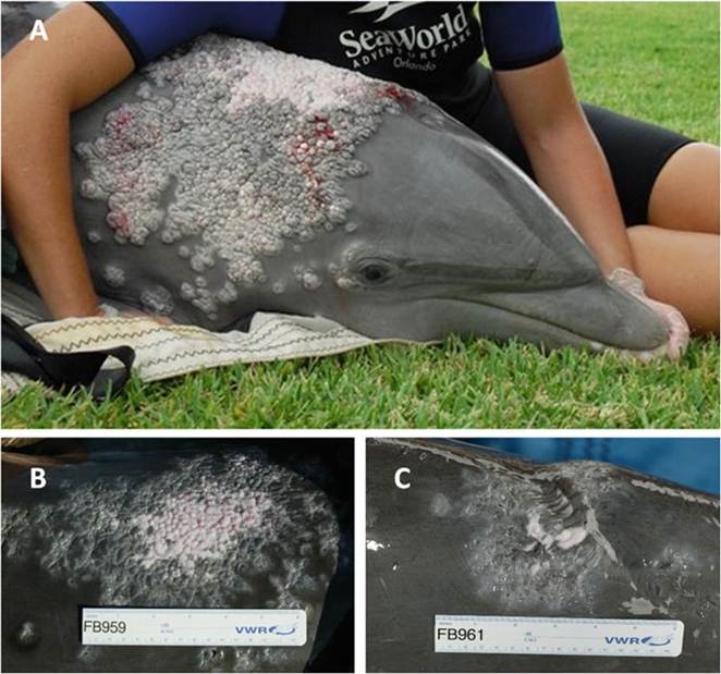
Fig. 9.4 (a) A common bottlenose dolphin (Tursiops truncates) with extensive plaques over the frontal anterior section of the dorsal fin caused by Paracoccidioides brasiliensis var. ceti. Note numerous elevated verrucous gray and white plaques some of them bleeding. New nodular small satellite lesions adjacent to the large plaque are observed (Courtesy of Dr. J. St. Leger). (b and c) Two different dolphins with large nodular white plaques typical of paracoccidioidomycosis ceti (Courtesy of Drs. G.D. Bossart, P.A. Fair and J.S. Reif)
observations for long periods revealed that small lesions in dolphins slowly progressed to form extensive granulomatous plaques. Most veterinarians agreed that environmental factors might play a key role in some epizootic reports of the disease in the coastal areas of Florida and North Carolina, USA (de Moura et al. 2014; Lane et al. 2014; Reif et al. 2006, 2009; Tajima et al. 2015).
9.5