Pathological Findings
The diagnosis of the lacaziosis in humans is confirmed after biopsy collection from the infected areas. Hematoxylin-eosin (H&E)-stained tissue samples show the presence of atrophy of the epidermis with extensive fibrosis interspersed with microabscesses containing numerous lymphocytes, plasma cells, foamy histiocytes, and multinucleated giant cells, some enclosing the pathogens (Fig.
9.5) (Baruzzi et al. 1981; Carneiro et al. 2009; Francesconi et al. 2014; Lacaz et al. 1986; Talhari and Talhari 2012). Acanthosis with hyperkeratosis is commonly observed. The Splendore/Hoeppli phenomenon enclosing one or more L. loboi yeast-like cells has been occasionally reported (Opromolla et al. 2000). In H&E L. loboi yeastlike cells appear as unstained hyaline structures resembling ghost-like bodies (Fig. 9.5a). In Gomori methenamine silver (GMS) stain, L. loboi cells appear uniform in shape (4-12 μm in diameter) developing one or more chains of three or more yeast-like cells connected by slender tubes (Fig. 9.5b). In some instances, few chains of yeast-like cells are present forming individual or budding spherical structures. The yeast-like cells shape varied between spherical to oval (lemon shape).Dolphins are protected mammalian species; thus the collection of material for histopathology is strictly regulated. Biopsies are collected after the diseased dolphin is restrained and precautions are taken to avoid traumas that may have future repercussions. In H&E, the inflammatory process and the pathogen seem very similar to that in cases of human lacaziosis (Esperon et al. 2012; Minakawa et al. 2016; Rotstein et al. 2009; Ueda et al. 2013). Minor differences in the pathogen cell size in humans and dolphins have been mentioned (Haubold et al. 2000; Minakawa et al. 2016). Acanthosis, hyperkeratosis, hyperpigmentation, and extensive progressive fibrosis are the main pathological features in dolphins (Bossart et al.
2015; Tajima et al. 2015; Ueda et al. 2013). Microabscesses containing numerous unstained thick-walled hyaline yeast-like cells are often present (Fig. 9.5c, d). Histiocytes, giant cells, few lymphocytes, and plasma cells can also be observed. In GMS, the presence of numerous dark, 2-10 μm wide, oval yeast-like cells connected by short isthmuses is diagnostic of dolphin cases (Fig. 9.5e, f).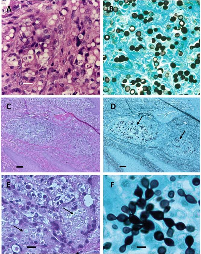
Fig. 9.5 (a) Unstained Lacazia Ioboi yeast-like cells (H&E) (arrows) from a case of human Iacaziosis Acre, Brazil (bar = 18 μm). (b) A Gomori methenamine silver stain sample of panel A (bar = 18 μm). Small yeast-like cells chain of more than two cells connected by slender isthmuses are visible (Courtesy of Dr. Patricia Rosa). (c and e) H&E stained histological sections of a dolphin with paracoccidioidomycosis ceti. Presence of microabscesses containing inflammatory infiltrate, extensive fibrosis, acanthosis (Panel C; bar = 65 μm), and numerous unstained Paracoccidioides brasiliensis var. ceti yeast-like cells (Panel E; bar = 20 μm) (arrows). (d) A low magnification of panel C stained with Gomori methenamine silver. Presence of numerous black chains of yeast-like cells (arrows) (bar = 65 μm). (f) A close-up of Panel E. Presence of the yeast-like cells uniform in size and formation of branching chain connected by small bridges (bar = 10 μm)
9.6