Critical Care and Fluid Therapy Monitoring Techniques
K. Gary Magdesian
Monitoring tools provide for advanced critical care in the large animal intensive care unit. Several of these tools are pertinent to fluid therapy and provide guidance, end points, and safety limits.
Many of these tools allow for direct monitoring of critical patient status during administration of therapy, whereas others provide indirect information through hemodynamic data. Both of these factors are important to overall case management of the critically ill equine patient.Central Venous Pressure
Central venous pressure (CVP) is the pressure within the vena cava; the term most commonly refers to that within the cranial vena cava in most species. It is determined by blood volume, venous tone, and cardiac contractility. Increases in blood volume or venous tone increase CVP, as does suboptimal cardiac function. The use of CVP as a measure of adequacy of fluid resuscitation has been much debated, but less controversial is its utility as a limit on fluid administration. With serial measurements CVP can provide a limit to the administered volume of fluid. Normal CVP is approximately 2 to 12 cm H2O (1.5 to 8.8 mm Hg) in foals and 5 to 15 cm H2O (3.7 to 11 mm Hg) in adults.1-4 Subnormal to negative CVP values indicate hypovolemia; however, normal values do not necessarily imply euvolemia. This is because CVP is affected by compensatory responses such as venoconstriction and by cardiac contractility and intrapleural pressures, among others.
CVP is easily measured in neonatal foals using 20- to 30-cm central venous catheters, including long-term single- to triple-lumen polyurethane intravenous catheters placed in the jugular vein. Placement can be easily confirmed through thoracic radiography, with the radiopaque catheters being visualized in the cranial vena cava just cranial to the cardiac silhouette.
Measurements can be obtained in adult horses with the use of 55-cm commercial CVP catheters placed in the lower jugular vein. CVP can also be measured by passing a smaller gauge polyurethane catheter or polyethylene tubing through a 14-gauge, 5/4-inch standard intravenous catheter placed in the jugular vein. The actual measurement can be recorded with a disposable water manometer (for spot or intermittent readings of the CVP value) or using a pressure transducer for continuous waveforms. Interpretation of CVP waveforms requires knowledge of component waves and descents, including the “a,” “c,” and “v” waves, and the “x” and “y” descents. The “a” wave represents atrial contraction. The mean of the “a” wave is the appropriate point at which to measure CVP (during end-expiration in a spontaneously breathing patient). The “c” wave is associated with tricuspid valve closure and a bulging of the valve into the right atrium, and the “v” wave is caused by atrial filling from venous return. The “x” descent occurs after the “a” wave and represents the fall in pressure associated with atrial relaxation. The “y” descent occurs after the “v” wave, representing a drop in CVP caused by ventricular relaxation and reopening of the atrioventricular valves.High CVP readings occur with fluid overload, as well as with pericardial tamponade, pleural effusion, or pneumothorax. False increases may result from catheter occlusion and air within the lines. For accurate and consistent serial results, a zero reference point should be selected and used for each measurement. The top of the sternal manubrium is a good reference point for the level of the pressure transducer or water manometer. The pressure transducer or water manometer can be taped to a fluid pole, providing a fixed zero point for repeated measurements in standing horses. Precision and reliability are limited by variable zero reference points, the effects of afterload and ventricular compliance, and alterations in intrathoracic pressure.5
It should be noted that adequacy of intravascular volume cannot be guided by any one CVP level.
There is not a linear relationship between intravascular volume and filling pressures, and CVP should be regarded as only an indirect estimate of volume. Serial measurements and trends over time are more useful than single numbers. CVP can be used on a clinical level for monitoring fluid therapy as follows: If the patient responds to fluid boluses without increases or with only minor increases (2 to 3 cm H2O) in CVP, it is appropriate to continue infusions until signs of hypoperfusion are reversed or improved, as long as other signs of fluid overload are not present.5 Additional guidelines used in human patients are as follows: If the CVP increases to 3 to 7 cm H2O (2 to 5 mm Hg), the infusion should be paused and perfusion reevaluated after a 10-minute wait. If the change in CVP was an increase of 7 cm H2O or greater (≥5 mm Hg) after the fluid bolus, the infusion is stopped.5 In this regard CVP is a better “upper limit” to fluid therapy rather than an actual goal to strive for, other than normalization of CVP in horses with subnormal values.Arterial Blood Pressure
Arterial blood pressure measurements provide some insight into perfusion status, particularly when used in conjunction with clinical signs and lab work, such as blood lactate concentration. It can be measured either directly by using arterial catheters or indirectly with a blood pressure cuff on the tailhead. Arterial lines can be placed in the great metatarsal artery of recumbent foals and the transverse facial artery in standing adult horses for direct measurements. Indirect measurements are best performed with oscillometric blood pressure monitors that provide systolic, diastolic, and mean arterial pressure (MAP). Cuffs are provided by the manufacturer and vary in width and length with the size of the patient.
Arterial blood pressure in healthy neonatal foals is highly variable and depends on breed, gestational age, and size of foal.
Reported mean arterial blood pressure (direct) in neonatal foals has been reported to be 84.4 ± 3.7 mm Hg at 1 day of age and up to 101.3 ± 4.4 mm Hg at 14 days of age.2 Indirect blood pressure readings in Thoroughbred foals has been reported as 144 ± 15, 74 ± 9, and 95 ± 13 mm Hg for systolic, diastolic, and mean pressures, respectively.6 Direct blood pressure has been reported for adult horses: 126 to 168 (systolic)/85 to 116 (diastolic) with a range for the mean arterial pressure of 110 to 133 mmHg.7-10 Indirect blood pressure in adult horses is reported as 111.8 ± 13.3 mm Hg for systolic pressure and 67.7 ± 13.8 mm Hg for diastolic pressure.11Blood pressure values should not be regarded as the sole criteria for intervention. Rather, they should be used in conjunction with perfusion parameters, blood lactate concentration, and urine output in deciding whether there is need for further fluid administration or inotrope and vasopressor support. The exact blood pressure value that should trigger intervention in horses and foals is unknown and likely varies with the individual patient; however, end-organ perfusion requires a mean arterial pressure of 60 mm Hg, thereby making that a reasonable general minimal target.
Blood or Plasma Lactate Concentration
Blood or plasma lactate concentration is very useful in monitoring fluid support, as well as perfusion and metabolic status. Increased blood lactate concentration may be a result of hypoperfusion and reduced oxygen delivery (hypovolemia, hypotension, anemia, hypoxemia, heart failure). Other considerations for hyperlactatemia should include SIRS, sepsis, catecholamine surges, liver or renal failure, thiamine deficiency, alkalosis, hyperglycemia, exercise, seizure activity, and the action of drugs such as salicylates and theophylline.12-15 Sepsis and SIRS may increase circulating lactate concentrations independent of perfusion status; inflammatory mediators and cytokines activate pyruvate dehydrogenase kinase, an inactivator of pyruvate dehydrogenase, resulting in reduced activity of the citric acid cycle.
Normal lactate concentrations in horses are less than 2 mmol/L, with most horses having concentrations less than 1 mmol/L.16-18 Neonatal foals have decremental values after birth, with reported concentrations of 4.9 ± 1.02, 2.25 ± 0.6, and 0.89 mmol/L at birth, 12 hours of age, and 24 hours of age, respectively.18-20 In a study performed by the author, foals had values of 2.4 ± 1.0, 1.2 ± 0.3, and 1.1 ± 0.3 mmol/L at 0 to 2, 24, and 48 hours of age, respectively, compared with 0.6 ± 0.2 mmol/L in adult horses.21Cardiac Output
Advanced monitoring in equine critical care includes cardiac output measurement. A number of methods of cardiac output monitoring have been described in horses, including the Fick principle, indicator dilution methods (such as lithium dilution), Doppler and volumetric echocardiography, pulse contour analysis, and partial carbon dioxide rebreathing. The most practical of these include lithium dilution and echocardiographic techniques.22-26
Lithium dilution precludes the need for placement of cardiac catheters. A small bolus of lithium chloride is injected into a peripheral vein or the cranial vena cava (via a central line). Arterial blood is sampled using an arterial catheter at a constant rate and passes through a lithium electrode to generate a lithium concentration-time curve. Cardiac output is calculated from the area under the curve for lithium over time. Advantages of lithium dilution include only moderate invasiveness, good accuracy, and requirements for only small volumes of injectate. Disadvantages include the need for continuous arterial blood and limitations on repetitive measurements owing to lithium accumulation.25-27
Noninvasive cardiac output measurements can be made using the Bullet method through volumetric echocardiography.24 McConachie and colleagues28 reported that the Bullet method provided an accurate estimate of cardiac output in anesthetized foals.
The Bullet method, as well as the four-chamber arealength, Simpson, and right ventricular outflow Doppler methods, had reasonable agreement with lithium dilution in adult horses.28 For the Bullet method, cardiac output is calculated using heart rate and stroke volume (CO = HR ? SV), where stroke volume is estimated as follows: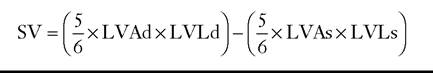
where LVAd is the left ventricular area in diastole (short-axis view), LVLd is the left ventricular length in diastole (long-axis view), LVAs is the left ventricular area in systole (short-axis view), and LVLs is the left ventricular length in systole (long-axis view).
Cardiac output in healthy adult horses (400 to 500 kg) is 32 to 40 L/min.22 Normal cardiac index, which is cardiac output expressed per unit of body weight, is 72 to 88 mL/kg/min. In the neonatal foal, cardiac output has been reported to be 7.1 ± 0.4 L/min (cardiac index = 155.3 ± 8.1 mL/kg/min) in 2-hour-old foals.2 At 24 hours of age, cardiac output was determined to be 9.0 ± 0.5 L/min (cardiac index = 197.3 ± 12.0 mL/kg/min). This increased further to 15.7 ± 1.5 L/min (cardiac index = 222.1 ± 21.6 mL/kg/min) at 14 days of age.2 A general guideline for normal cardiac index is 100 to 300 mL/ kg/min in neonatal foals.24
Blood Glucose
Blood glucose concentration has been the focus of much study in human critical care in recent years. A number of initial studies demonstrated improved survival and reduced complications with tight glucose control using intensive insulin therapy in critically ill humans.29,30 Maintenance of euglycemia (80 to 110 mg/dL) resulted in reduced mortality in surgical intensive care unit patients as compared with hyperglycemia (180 to 200 mg/dL). Hyperglycemia may be associated with detrimental effects in pediatric intensive care patients as well.31 However, after these initial studies, additional studies, including metaanalyses, did not document similar benefits and revealed some adverse events with tight glycemic control, especially develop- 3233
ment of severe hypoglycemia.32,33 Current guidelines therefore are more relaxed and recommend using a glucose concentration of 180 mg/dL or less as the target.34 How these findings relate to critically ill horses is unknown, but prevention of hyperglycemia is likely warranted, and its effects in the equine intensive care unit require further study. In one study evaluating blood glucose in horses with acute abdominal disease, hyperglycemia in the first 48 hours of hospitalization was associated with a worse prognosis for survival to hospital discharge.35 A study in hospitalized neonatal foals showed that foals with blood glucose concentrations less than 50 mg/dL or greater than 180 mg/dL at admission were less likely to survive.36
Monitoring Urine Analytes
Urine indices are useful for monitoring responses to fluid therapy. Successful production of urine output in response to fluid administration is a primary goal of fluid therapy, and measurement of urine production allows determination of “fluid balance” (input-output). Another important means of assessing the response to fluid loading is measurement of urine specific gravity and/or osmolarity. In the absence of renal failure, progressively dilute urine is a positive response to fluid administration. Horses on feed and water should be making 0.4 to 2.0 mL/kg/h urine, whereas those off feed and water will produce far less (0.13 to 0.5, up to 1 mL/kg/h depending on duration).37,38 Horses on fluids should therefore be producing a minimum of 0.5 but optimally 1 to 2 mL/kg/h of urine.
Urinalysis should be performed, and fractional excretion of electrolytes, particularly of sodium, provides information regarding tubular function and can aid in differentiation of prerenal and renal azotemia. Normal fractional excretion is less than 1% in adult horses and 0.31% ± 0.18% in neonatal foals.39-42