Diagnosis of Gastrointestinal Disease by Presenting Sign
The purpose of this section is to assist in the differential diagnosis of gastrointestinal diseases on the basis of common presenting signs. The possibilities offered are not geographically restricted, so some of the diseases listed for a given presenting sign may not apply to the sick goat at hand.
The major infectious, parasitic, and metabolic diseases of the digestive system are mentioned when appropriate for a given presenting sign, but are discussed in detail later in the chapter. Other diseases that occur infrequently or sporadically, that are very localized geographically, or that are not well described in goats are discussed fully in this section and are not addressed again later in the chapter. For conditions discussed elsewhere in the text outside of this chapter, the reader is referred to the appropriate chapter number. In 2011, after the last edition of this text appeared, the disease rinderpest was globally eradicated (Njeumi et al. 2012). The authors have chosen to keep references to it here so that readers will continue to recognize the signs of that dread disease and include it in their differential diagnosis, just in case there were to be an unexpected reemergence of the disease in the future.
Inappetence
A history of inappetence often tempts the clinician into presuming a primary digestive disorder. However, many multisystemic diseases, particularly if they produce fever, reduce appetite. The likelihood that inappetence reflects a digestive dysfunction increases if it is accompanied by changes in rumen motility, vomiting, the development of abnormal feces, tenesmus, or distortions of the abdominal contour.
Frothing at the Mouth
Frothing at the mouth may be seen independently of excessive salivation, slobbering, and drooling, as discussed below. In all species, frothing is commonly associated with uncontrolled chewing or lip smacking during convulsive activity.
In goats, frothing is probably seen most commonly as a mild adverse reaction to administration of the anthelmintic levamisole. Frothing or excessive salivation, depression, and hyperesthesia may occur in some goats shortly after administration of the drug, even at the suboptimal dose of 8 mg/kg orally or parenterally. Frothing, along with dyspnea, frequent urination and defecation, and clonic convulsions may occur when levamisole is overdosed, particularly by parenteral administration.Idiopathic frothing at the commissures of the lips associated with cud chewing has been observed by the author (DMS) in several individual French Alpine and Nubian does in different herds. The animals were normal in all other respects and were not receiving medications of any kind. Owners reported that the condition occurred intermittently in individuals over periods of months to years.
Frothing at the mouth in conjunction with dyspnea, diarrhea, and ataxia was reported in goats from the Sudan poisoned by the plant Cadaba rotundifolia (El Dirdiri et al. 1987). In Kenya, frothing at the mouth in conjunction with bloat, incoordination, and death was caused by ingestion of Cestrum aurantiacum, a shrub used for hedges and windbreaks (Mugera and Nderito 1968). At necropsy, there was severe hemorrhagic gastroenteritis. Frothing at the mouth has been seen in conjunction with signs of neurologic disease in goats with organophosphate toxicity and in experimental rabies as discussed in Chapter 5.
Excessive Salivation, Slobbering, or Drooling
Stomatitis, or inflammation of the mucosa of the oral cavity, is most often responsible for excess salivation, slobbering, or drooling in the goat. A foul odor to the breath may accompany salivation if necrosis of the oral mucosa has occurred. If pain is severe, dysphagia may also occur.
Infectious causes of stomatitis include contagious ecthyma, goat pox, unclassified viral dermatitis, foot and mouth disease, bluetongue, vesicular stomatitis, rinderpest, peste des petits ruminants (PPR), caprine herpesvirus, necrotic or ulcerative stomatitis caused by Fusobacterium necrophorum infection, and alveolar periostitis.
Contagious ecthyma (soremouth, orf) is the most common cause of stomatitis in goats worldwide. The virus produces papular eruptions, mainly at the commissures of the lips and sometimes on the hard palate that may become secondarily infected with bacteria. The lesions heal spontaneously within six weeks, but excess salivation, reluctance to eat, and mild dysphagia leading to weight loss can precede resolution.
Goat pox may produce ulcerative lesions on the lips and in the mouth in addition to the characteristic skin nodules, pyrexia, and conjunctivitis. The disease is absent from the Americas and Australia and has been largely eliminated from Europe. A highly fatal, unclassified viral dermatitis has been observed in India since 1946 (Haddow and Idnani 1948). The condition is distinct from goat pox clinically and pathologically, although a pox virus has been isolated from clinical cases (Patnaik 1986). Widespread cutaneous eruptions may include the lips, gums, and tongue. Rubbery, non-exudative nodules and papules develop on the oral surfaces and lead to necrotic ulcers in 7-10 days.
Foot and mouth disease may have drooling or lip smacking as presenting signs resulting from vesicles in the oral cavity. In goats and sheep, as compared with cattle, however, foot lesions are more common than oral lesions. The disease is endemic in much of Asia, Africa, and South America.
Bluetongue rarely causes clinical disease in goats, although serologic evidence of exposure occurs extensively. When clinical signs do occur, they include stomatitis associated with edema and congestion of the oral mucosa and ulceration of the lips and dental pad. These signs may be accompanied by high fever, nasal discharge, diarrhea, and lameness caused by coronitis.
Vesicular stomatitis, caused by a rhabdovirus, occurs only in the Americas. Goats are less susceptible than cattle, horses, and swine. In goats, the only sign may be salivation with vesicles at the commissure of the lips, and the condition should be differentiated from contagious ecthyma by virus isolation or polymerase chain reaction (PCR).
Rinderpest, now eradicated, produces a necrotic stomatitis involving the gums, cheeks, and tongue. However, salivation is not a dominant sign; it can be overshadowed by profuse, mucopurulent, oculonasal discharge, and, later, diarrhea. Historically, goats were less severely affected than cattle.
PPR is similar to rinderpest in clinical presentation, causing necrotic stomatitis, diarrhea, and bronchopneumonia. It affects goats severely. It occurs in much of Africa, the Middle East, and south and east Asia.
Caprine herpesvirus has been reported as a cause of severe illness and fatality in kids in Switzerland. Marked oral erosions were observed in conjunction with nasal discharge and conjunctivitis, and at necropsy erosions of the cecum, colon, and urinary bladder (Mettler et al. 1979).
Necrotic stomatitis, caused by F. necrophorum and other secondarily invasive bacteria, has been reported in 4-6-week-old Boer goat kids in South Africa (van Tonder et al. 1976). Clinical signs included fever, salivation, frothing, lip smacking and chewing movements, mucopurulent nasal discharge, anorexia, weight loss, and a mortality rate of 21%. Lesions were confined to the mouth, tongue, and throat and consisted of well-circumscribed necrotic ulcers up to 4 cm in length. Treatment with parenteral chloramphenicol twice daily for five days was effective. Sodium sulfadimidine, penicillin, streptomycin, and tetracycline are all effective treatments for this condition in calves. A similar outbreak in India involved adult goats and kids. Pregnant does aborted and there was evidence of F. necro- phorum septicemia (Nayak and Bhowmik 1988).
Dental disease, particularly alveolar periostitis, can produce pain in the mouth and inflammation of the gums, leading to dysphagia and salivation. Oral examination should include careful inspection of the molars. Radiographs of the dental arcades may be helpful, because thorough visual examination of the teeth is awkward.
Non-infectious causes of stomatitis include chemical irritants, traumatic injuries, plant and chemical poisonings, and possibly neoplasia.
If caustic dehorning pastes are used to remove horn buds on young kids, their licking the paste from each other's polls leads to chemical stomatitis. Caustic soda used to clear drains on farms may also cause stomatitis if the drains are backed up and goats drink the standing water. Battery acid from carelessly discarded automotive storage batteries can taste salty and has been consumed by curious goats, leading to chemical stomatitis (King 1980c).Giant hogweed (Heracleum mantegazzianum) has caused ulcerative stomatitis with anorexia and profuse salivation in a goat in Britain (Andrews et al. 1985). Other specific references to plant or chemically induced stomatitis in goats are rare. Agents known to cause stomatitis in other species may presumably affect the goat. These include mercurial and arsenical compounds and plants containing ranunculin, such as buttercup and crocuses.
Traumatic stomatitis with salivation can result from sharp dental points on the buccal aspect of the upper molars and lingual aspect of the lower molars. Abrasion and ulceration of the mucosa can lead to secondary bacterial infections and abscesses, particularly in the cheek. Certain plant awns, such as barley, foxtail, and thistle, can also traumatize the oral mucosa.
Rough or careless administration of medications using dose syringes and balling guns is a common cause of traumatic stomatitis of the hard palate and pharynx. Resulting injuries can produce salivation, dysphagia, regurgitation of feed, and possibly aspiration pneumonia.
Neoplasia of the oral cavity is rare in goats, but three cases of lymphosarcoma or adenocarcinoma have been reported to involve oral structures, leading to loosening and displacement of molar teeth (Baker and Sherman 1982; Lane and Anderson 1983; Craig et al. 1986). A rare, primary parotid salivary gland tumor of mixed epithelial and mesenchymal elements has been reported in a 2-month-old Jamnapuri crossbred female goat (Omar and Fatimah 1981).
Salivation may occur independent of stomatitis, usually caused by neurologic disease, systemic poisonings, or obstructions of the digestive tract distal to the oral cavity. Neurologic diseases associated with salivation or drooling in the goat are listeriosis, caprine arthritis encephalitis (CAE) virus infection, migrations of the nematode parasite Parelaphostrongylus tenuis, and trauma to the facial nerve. Rabies and polioencephalomalacia may also produce excessive salivation, but usually in conjunction with other neurologic signs that are more general or progressive than those seen with the focal conditions just mentioned. Botulism produces a general weakness of the muscles, including the tongue and muscles of mastication. Therefore, dysphagia and drooling can be seen along with recumbency. Neurologic diseases are discussed in detail in Chapter 5. Nutritional muscular dystrophy, or white muscle disease, can produce necrosis of the muscles of the tongue and pharynx, leading to dysphagia and drooling. The condition is discussed in detail in Chapter 4.
Profuse salivation may be one of the first recognized signs of organophosphate and carbamate poisoning. Additional signs include muscle stiffness and fasciculation, frequent urination and diarrhea, pupillary constriction, colic, dyspnea, nervousness, and sudden death. Some of these same signs, including salivation, may be seen in urea toxicosis, as discussed in Chapter 19. Acute chlorinated hydrocarbon poisoning also produces an abundant flow of ropy saliva in conjunction with signs suggesting neurologic dysfunction, such as belligerence, muscle twitching, clonic and tonic convulsions, and death. Xylazine produces excessive salivation at doses used for tranquilization.
Some plant poisonings produce salivation without stomatitis in goats. Plants of the family Ericaceae produce salivation and vomiting, as discussed later in the section on regurgitation. Cotyledon orbiculata is common in South Africa. The plant is cardiotoxic, but excessive salivation is an important presenting sign after ingestion (Tustin et al. 1984).
Consumption of Prosopis Juliflora (mesquite) and Prosopis glandulosa (honey mesquite) may be toxic to goats. When ingested in sufficient quantities, the unknown plant toxin can produce signs of salivation, dysphagia, protrusion of the tongue, and tremors of the mandible (Washburn et al. 2002; Misri et al. 2003). Vacuolation of neurons in the trigeminal motor nucleus and neuronal necrosis and loss in the trigeminal ganglia are associated with these clinical signs (Washburn et al. 2002). Expression of toxicity may depend on duration of exposure. Goats fed P. glandulosa at up to 90% of their total ration for two weeks did not show toxicity (Cook et al. 2008).
“Slobbers” is caused by the fungus Rhizoctonia legumini- cola, or “black patch,” that grows on legume forages, particularly red clover, during wet weather and high humidity. It can persist on stored forages. The fungus produces an alkaloid, slaframine, which produces copious salivation within one hour of ingestion and may last for 24 hours after a single exposure. The condition is seen primarily in the United States and Japan and is reported in goats (Isawa et al. 1971). Slobbering may be the only clinical sign, but lacrimation, frequent urination, diarrhea, dyspnea, abortion, and even death may be seen with prolonged exposure. There is no specific treatment, except for rapid diagnosis of the problem and removal of goats from the offending feed.
Cyanide poisoning, associated with ingestion of numerous plant species, can have salivation as the initial sign. The disease is discussed in Chapter 9.
Physical obstruction of the esophagus or pharynx can prevent the normal swallowing of saliva and lead to excess salivation. Bloat accompanies salivation when obstruction of the pharynx or esophagus is complete. Foreign bodies likely to produce obstruction are fruits, tubers, and roots.
No specific treatment for excessive salivation is recommended. Atropine may reduce saliva flow, but the additional effects of reduced gastric motility argue against its use. The major goal is to identify and treat the underlying cause of salivation. It should be remembered, however, that saliva is produced in very large amounts and contains many electrolytes, including bicarbonate. Prolonged loss of saliva may lead to dehydration, electrolyte imbalance, and acidosis. These sequelae should be managed with fluid and electrolyte therapy.
Dysphagia
Dysphagia may be manifested by prolonged chewing, retaining feed in the mouth, dropping feed from the mouth (quidding), and, as just discussed, drooling or slobbering. Many of the causes of stomatitis also can produce dysphagia, depending on the extent and severity of the oral lesions present.
Focal neurologic diseases such as listeriosis, brain abscess, parasitic larval migration, and CAE can produce dysphagia when damage to the cranial nerve roots VII, IX, X, or XII occurs. Nutritional muscular dystrophy affecting the tongue and pharyngeal muscles can mimic these neurologic deficits. Accumulation of feed in the buccal space can be associated with the loss of one or more molars secondary to aging or alveolar periostitis. This can also occur with facial nerve paralysis, resulting in flaccidity of the cheek muscles. Holding feed for prolonged periods in the mouth without chewing may be observed in rabies, tetanus, botulism, and polioencephalomalacia.
Developing teeth are subject to structural modification by chronic exposure to fluoride. Fluorosis can produce pitting, a rough chalky enamel, discoloration, and accelerated wearing of teeth (Milhaud et al. 1980). The excessive tooth wear and associated pain can produce signs of dysphagia. The third and fourth pairs of incisors are most often affected. Exostoses of the mandible and maxilla may also occur. Fluorosis is discussed in detail in Chapter 4. Excessive tooth wear is also associated with grazing of sandy soils by goats.
Additional dental problems that may lead to dysphagia include overeruption of teeth; broken teeth; vestigial teeth; and displaced, rotated, or migrated teeth (Rudge 1970). Fibrous osteodystrophy, discussed in Chapter 4, can lead to severe inward rotation of the dental arcades.
In recent years, there have been unsubstantiated reports from Kenya that goats grazing on the seed pods of P. juli- flora (mesquite) are losing their teeth as a result of the seeds sticking between the teeth and gums, leading to gingivitis, alveolitis, disfigured jaws, impaired ability to eat, and inanition (Mwangi and Swallow 2005). While tooth loss is unsubstantiated, Prosopis spp. can produce salivation and dysphagia without a direct effect on teeth, as discussed above in the section on salivation.
Presence of milk or chewed feed at the nares and coughing in kids may result from cleft palate. Retropharyngeal abscess caused by Corynebacterium pseudotuberculosis in older goats may block normal swallowing by compression of the soft palate, leading to coughing and nasal expulsion of feed. Balling gun or dose syringe injuries may initiate these abscesses. Attempts at surgical drainage carry a guarded prognosis. There is a reported case of a goat whose tongue was encircled by the neck of a broken bottle, leading to trauma to the tongue and dysphagia. To correct the condition, the glass ring was snapped with a bone forceps and with supportive treatment the goat recovered (Alhendi et al. 1999).
Treatment of animals with dysphagia should include broad-spectrum parenteral antibiotics until normal swallowing resumes, because the risk of aspiration pneumonia in association with dysphagia is high. Correction of dehydration and nutritional supplementation through a stomach tube must also be considered during prolonged disease.
Regurgitation, Retching, or Projectile Vomiting
Partial or complete obstructions of the pharynx or esophagus can lead to regurgitation of feed in goats (Fleming et al. 1989). Acute copper toxicosis in a herd of goats given an overdose of oral copper sulfate resulted in repeated attempts to vomit, as well as abdominal pain, muscle fas- ciculations, labored breathing, tachycardia, and frothing at the mouth before death (Shlosberg 1978).
Megaesophagus is a rare condition in ruminants, but several idiopathic cases have been reported in goats. Regurgitation after eating was a presenting sign in most but not all caprine cases. Other signs include anorexia, weight loss, moderate bloat, and swelling of the neck ventrally. Diagnosis of the condition is confirmed by fiberoptic examination, contrast radiography, or necropsy (Ramadan 1993; Mozaffari and Vosough 2007; Silva Junior et al. 2011; Nascimento et al. 2016).
The toxic plants most often associated with vomiting in goats are members of the heath family (Ericaceae) and include rhododendrons, azaleas, laurels, lily of the valley tree (Clethra arborea), and Japanese pieris. They contain the toxic principle grayanotoxin (also referred to as andromedotoxin), which acts primarily on the autonomic nervous system, stimulating the vomiting center via the vagus nerve and producing hypotension (Smith 1978; Knight 1987; Gibb 1987). Clinical signs can occur when as little as 0.1% of the animal's bodyweight is ingested as fresh leaves.
Within six hours of ingestion of Ericaceae, goats may show signs of depression, weakness, anorexia, salivation, abdominal pain, vomiting, and possibly bloat and diarrhea. Tenesmus and blood in the feces were reported in a goat consuming Pieris formosanum (Hollands and Hughes 1986). While the condition may be self-limiting within days if only small amounts of the plants are eaten, ingestion of large quantities may cause death (Visser et al. 1988). Exposure of 18 goats to clippings of Rhododendron macrophyllum resulted in convulsions and death in two, weight loss in all, and agalactia in seven (Casteel and Wagstaff 1989). Sublethal ingestion of Japanese pieris has been associated with fetal mummification in a goat (Smith 1979).
If known exposure has occurred, rumenotomy before the onset of signs is recommended. After onset of signs, treatment should include intravenous fluid therapy to counter hypotension, relief of bloat by stomach tube if necessary, oral magnesium hydroxide and activated charcoal, and parenteral calcium gluconate, as well as parenteral antibiotic to minimize the risk of aspiration pneumonia associated with vomiting. The antispasmodic hyoscine-n- butylbromide has also been used to control vomiting. More recently, administration of an intravenous lipid emulsion (60 mL to an adult goat) has proven beneficial in treating animals poisoned with Japanese pieris (Bischoff et al. 2014).
Prevention involves educating goat owners regarding the potential danger of these plants to livestock. Members of the heath family are widely used as ornamental plantings, and goats should be kept away from plantings and not fed prunings, because these will be readily eaten.
Other plants may cause vomiting in goats. A spontaneous case of vomiting in conjunction with constant bellowing, rumen atony, and depression was reported in a goat ingesting Raphanus sativus, a member of the Cruciferae, or mustard, family (Drahn 1951). Vomiting, depression, and a staggering gait were observed in a goat in England after ingestion of Solanum nigrum, or black nightshade (Gunning 1949). Gladiolus corms eaten by goats produced bleating or moaning, vomiting, staggering, and depression within hours. Exposure was fatal in some cases. The prominent finding at necropsy was watery-brown rumen content (Anonymous 1988b).
Rumen Atony
Normal rumen mixing contractions may be reduced in frequency when goats are fed rations low in coarse roughage and high in finely ground concentrate. Contractions may disappear when goats are excited or fearful, when parasympathomimetic drugs such as atropine are administered, or when central nervous system anesthetics and depressants such as barbiturates have been given.
Pathologic processes occurring outside the digestive system can inhibit rumen motility. These include pain from any source, severe dehydration, electrolyte or acid-base imbalances, hypocalcemia, high fever, and toxemias. Peritonitis as a cause of rumen atony is uncommon in goats compared with cattle, mainly because of the infrequent occurrence of traumatic reticuloperitonitis in goats. There are few references to this condition in the goat (Maddy 1954; Sharma and Ranka 1978; Tanwar and Saxena 1984). It is presumed that the more selective, discriminating food prehension behavior of goats limits the ingestion of foreign bodies capable of penetrating the reticulum. When traumatic reticulitis does occur in goats, signs are similar to those in cattle. There is a report of experimentally induced traumatic reticuloperitonitis in 17 goats fed wires, nails, and/or needles (Roztocil et al. 1968). The clinical presentation reflected what is seen in naturally occurring bovine cases, namely fever, anorexia, depression, cessation of rumen motility, elicitation of pain with application of pressure in the region of the xiphoid cartilage, and neutrophilia with a left shift. Some individuals also developed traumatic pericarditis, pneumonia, and/or hepatitis, depending on the ultimate locations of the metal foreign bodies.
Pathologic processes within the digestive system leading to rumen atony include simple indigestion, bloat, rumen acidosis caused by acute carbohydrate engorgement, and rumen alkalosis associated with urea poisoning. In general, plant and chemical poisonings frequently include rumen atony as one of the presenting signs.
Abdominal Distension
There is considerable variation in the normal abdominal silhouette. Pygmy goats are achondroplastic dwarfs and, as such, their abdominal girth is proportionately greater for their height and weight compared with other breeds. As young pygmy goats develop full rumen capacity, they sometimes appear to inexperienced owners to be abnormally distended in the abdomen. An inherited condition identified as “dropped stomach” has been reported in Toggenburg does. It involved permanent stretching of the ventral abdominal musculature in late pregnancy. After parturition, the ventral abdomen remains pendulous, but affected animals are otherwise normal (Wirth 1980). The condition has also been observed in other breeds (Figure 10.1).
Ruminal tympany, or bloat, is not uncommon in goats, and both the frothy (nutritional) and free gas forms can occur. In all types, abdominal distension is initially most
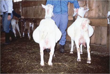
Figure 10.1 Dropped stomach in a mother and daughter pair of Saanen goats. Note prominent abdominal distension. Neither goat is pregnant. Source: Courtesy of Dr. David M. Sherman.
prominent in the left paralumbar fossa, and may also involve the entire left side of the abdomen and the right ventral abdomen if severe.
Two cases of left displaced abomasum have been reported in the goat. The first was associated with intermittent bloat (West et al. 1983). The second showed abdominal distension and a presumptive diagnosis of rumen impaction was made. However, at surgery, the gas-filled abomasum was discovered between the rumen and the left body wall and obstruction of the pylorus with a single phytobezoar was found (Parizi et al. 2008). Acute carbohydrate engorgement can also produce a similar pattern of abdominal distension due to both gas accumulation and fluid pooling in the rumen. The presence of fluid can be established by ballot- ment of the ventral rumen and by reflux through a stomach tube.
Rumen impaction produces a distended rumen, palpable as a firm mass low on the left side of the abdomen behind the ribs. The causes and management of rumen impaction are discussed later in this chapter. Abdominal distension predominantly in the lower right quadrant is most commonly associated with late pregnancy, as the rumen displaces the expanding uterus to the right. Herniation of the gravid uterus through the right abdominal floor causing prominent right ventral abdominal distension occasionally occurs (Horenstein and Elias 1987).
Abnormal distension of the right ventral abdomen also can be associated with abomasal disorders. Abomasal impaction with metal shavings has been reported in an adult goat from India (Purohit et al. 1986). Numerous abo- masal impactions with phytobezoars have been reported from South Africa. The concretions are composed of the pappus hairs of the seeds of Karoo bushes. Goats are more often affected than sheep and Boer goats more often than Angora goats (Bath 1978). The clinical syndrome is one of progressive abdominal distension and weight loss. Occasionally animals are found dead due to rupture of the stomach. The phytobezoars, which are multiple and round, may be felt in the abomasum by deep palpation behind the xiphoid cartilage. Early recognition and quick slaughter for salvage are recommended, because no satisfactory treatment has been identified (Bath and Bergh 1979).
The feeding of high-fiber roughages of low digestibility, mentioned as a cause of rumen impaction, can also produce abomasal impaction, particularly in pregnant does. In these cases, the lower right abdomen is distended, or a doughy abomasal content can be palpated. Animals are thin and feces are soft, fibrous, and malodorous. Treatment includes dietary improvement and the use of laxatives such as mineral oil. The value of abomasotomy has not been reported. Pregnancy toxemia is a possible sequela, and the prognosis is guarded.
Abrupt changes in feeding, from milk to milk replacer or milk pellets, are associated with abomasal bloat. Calf milk replacers that are high in lactose (and consequently low in fat and protein) have been associated with abomasal bloat in small ruminants. The pathogenesis of abomasal bloat may involve the rapid fermentation of sugars by gasproducing anaerobic bacteria present in the abomasum, such as Sarcina spp., which were identified during an epidemic of abomasal bloat in kids 6-10 weeks of age that occurred in a large goat dairy in California (DeBey et al. 1996). Abomasal bloat is also seen predictably in young kids in artificial rearing systems, particularly when milk is fed directly from a trough or bucket without a nipple (Thompson 1987). This is presumed to be caused by too rapid ingestion followed by excessive fermentation of milk. Because the abomasum in the suckling goat is the largest stomach, gas distension may occur on the left and right sides of the abdomen. The condition can be fatal. Alternative methods of feeding using nipple feeders or feeding ad libitum should be considered (Morand-Fehr et al. 1982; Thompson 1987). Successful treatment of a series of abomasal bloat cases in kids 1-2 weeks of age has been reported using a single intramuscular injection of hyoscine at 0.3 mg/kg bodyweight (bw), plus metoclopramide at 0.5 mg/kg bw and a vitamin E/selenium compound at 0.1 mg/kg bw (Kojouri 2004).
Overfeeding of milk can also lead to ruminal bloat when excessive volumes of milk reflux into the developing rumen (Chennells 1981). Overfeeding may also predispose to acute enterotoxemia with signs of hemorrhagic enteritis, nervous dysfunction, or death.
Bilateral ventral distension of the abdomen due to chronic indigestion secondary to abomasal impaction, as described above, may be seen in goats. It also has been observed in a goat with acute duodenal obstruction caused by a phytobezoar (Sherman 1981). Accumulation of ascites fluid secondary to conditions causing hypoproteinemia or cardiac insufficiency also may produce such bilateral distension. There is a report of marked fluid distension of the abdomen of an 11-year-old pygmy goat secondary to extensive abdominal fat necrosis attributed to tall fescue toxicity in North Carolina (Smith et al. 2004). In young goats, gastrointestinal nematodiasis and tapeworm infestations can lead to a marked “pot-bellied” appearance, as can low- quality roughage diets with a slow transit time through the rumen.
In female goats of breeding age, pseudopregnancy or hydrometra is a common cause of bilateral ventral abdominal distension. In male goats, the most common cause is urine accumulation in the abdomen after ruptured bladder caused by obstructive urolithiasis. This condition is discussed in detail in Chapter 12.
Intestinal and ovarian adenocarcinomas have been associated with accumulations of as much as 30 L of intraabdominal fluid in aged, female goats with progressive abdominal distension and discomfort (Haibel 1990; Memon et al. 1995). There is also a single case report of abdominal mesothelioma involving the peritoneum in an 8-year-old female Toggenburg goat in Austria that presented with marked ascites and a distended, pear-shaped abdomen (Krametter et al. 2004). Peritoneal mesothelioma was also diagnosed in a goat in South Africa (Bastianello 1983).
Generalized distension of the abdomen may occur secondary to conditions causing generalized ileus, such as severe peritonitis and sporadic intestinal accidents. General abdominal distension in young kids may occur before the onset of diarrhea in the early stages of infectious bacterial enteritis, when the causative organisms are potent gas producers. Congenital atresia ani and atresia coli or recti occur in newborn kids. Atresia can usually be diagnosed by a history of progressive abdominal distension, straining to defecate with no fecal output, depression, and waning appetite in kids from 1-4 days old. However, kids as old as 14 days have been presented for veterinary attention with a history of no defecation. Atresia ani has been successfully corrected surgically in goats by creation of anal patency (Ali et al. 1976). Atresia coli or recti may require a colostomy to salvage the affected animal (Philip 1973). In the goat, atresia ani also can occur associated with rectovaginal fistula, which allows for fecal voiding and may not be recognized until doelings approach breeding age or vaginitis occurs (Johnson et al. 1980). Animals with congenital anomalies of the anus, rectum, and colon should not be used for breeding if salvaged by surgical repair. A high incidence of atresia ani has been reported in the Shami breed of goat in Jordan (Al-Ani et al. 1998).
Focal distortions of the abdominal silhouette can be seen. Causes include umbilical hernia and spontaneous or traumatic ruptures of the abdominal wall, sometimes associated with fighting between horned goats (Figure 10.2). Ventral abdominal herniation in the doe has been associated with stretching of the ventral musculature during pregnancy and the subsequent increased weight of the enlarging udder (Misk et al. 1986). In Saudi Arabia, 193 goats with hernial swellings were presented to a veterinary teaching hospital over a five-year period. Umbilical, ventral abdominal, inguinal, scrotal, and perineal hernias were all diagnosed, some with the assistance of contrast radiography (Abdin-Bey and Ramadan 2001).
Abdominal Pain or Colic
Abdominal pain in goats may be manifested by depression, restlessness, bleating, teeth grinding, reluctance to move,
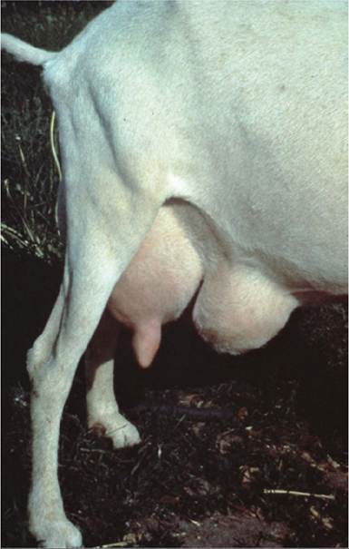
Figure 10.2 Herniation of the abdominal wall secondary to fighting with a horned goat. Source: Reproduced by permission of Dr. C.S.F. Williams.
increased shallow respiration, increased heart rate, tenesmus, or an abnormal posture with an arched back and tucked-up abdomen. Overt colic signs such as kicking at the belly or rolling are less common.
Mechanical Causes
In young kids, abdominal pain may be caused by feeding very cold milk or too much milk. Cecal torsion, intussusception (Mitchell 1983), and torsion of the root of the mesentery are sporadically occurring intestinal accidents more common in young goats than mature goats and are associated with pain. Torsion of the root of the mesentery is most common in unweaned kids fed by bottle or in ad libitum systems (Thompson 1985). In pregnant does, uterine torsion can produce abdominal pain. There is a report of rumen impaction in a goat in which extension and stretching of the fore- and hindlimbs and muscular rigidity, typical of a colicky horse, were presenting signs (Otesile and Akpokodje 1991).
Infectious Causes
Signs of severe abdominal pain may occur in conjunction with diarrhea, screaming, and convulsions before death from acute Clostridium pefringens type D enterotoxemia. Episodes of abdominal discomfort or pain may be observed during or immediately after eating in young goats with acute coccidiosis, before the onset of diarrhea. In young kids, enterotoxemia and coccidiosis must be differentiated from intestinal accidents such as torsion of the root of the mesentery at necropsy.
Peritonitis
Peritonitis can produce abdominal pain in addition to ileus, abdominal distension, and fever. Causes of infectious peritonitis in goats include rumen trocharization, uterine tears associated with dystocia, and extension of metritis from the uterus to the abdominal cavity. It can also occur as a sequela to rumenitis after acute carbohydrate engorgement. Organisms commonly associated with infectious peritonitis are Escherichia coli, streptococci, staphylococci, F. necrophorum, and Clostridium spp. Systemic mycoplasmosis in goats can produce a serositis-arthritis disease complex that includes peritonitis (DaMassa et al. 1983). Filarial worms of the genus Setaria may be found in the peritoneal cavity of goats, but are considered non- pathogenic (Subramanian and Srivastava 1973).
Non-infectious causes of peritonitis in goats include chemical irritation caused by intraperitoneal injections of sulfa drugs or calcium solutions, talc from surgical gloves (Hall 1983), and bile.
Toxicoses
Several ingested plants and chemical toxins can produce abdominal pain. The Ericaceae can produce signs of colic, as can acute oral copper poisoning, acute carbohydrate engorgement, and organophosphate insecticide toxicosis.
Urolithiasis
Perhaps the most common cause of abdominal pain in goats is obstructive urolithiasis seen in male goats. The appearance of pain is characterized by frequent and strained attempts to urinate. This may be misinterpreted by owners as straining to defecate caused by constipation.
Absence of Feces or Constipation
A history of no fecal output in association with tenesmus, abdominal pain, or abdominal distension strongly suggests an intestinal blockage caused by volvulus, intussusception, incarceration, or luminal obstruction by foreign body. Digital palpation of the rectum may reveal feces that arrived at the rectum before the obstruction occurred. The fecal matter should be removed and the animal rechecked later. If the rectum contains mucus only or mucus mixed with blood, this is strong evidence of intestinal obstruction. In neonates, atresia ani and atresia coli must be considered when there is no feces.
Constipation may be observed as a side effect of normal pregnancy or as a sign of pregnancy toxemia during late gestation (Pinsent and Cottom 1987). It may also be seen when access to water is limited or when diets are fibrous and of poor quality.
Extraluminal compression of the intestine by intraabdominal abscesses due to C. pseudotuberculosis may impede fecal output in goats. While clinical coccidiosis is noted for producing diarrhea, subclinical coccidiosis, which is common in young goats, produces constipation in conjunction with anorexia and reduced growth rate (Aumont et al. 198-4).
Diarrhea
There are numerous known infectious and parasitic causes of diarrhea and their frequency of occurrence varies to a large extent with the age of goats affected, as presented in Table 10.3. Diarrhea in young goats results from a complex interaction of etiologic, immunologic, and husbandry factors. The topic is discussed separately at the end of this chapter under neonatal diarrhea complex.
Non-infectious causes of diarrhea include overfeeding in kids, simple indigestion, acute carbohydrate engorgement (lactic acidosis), copper deficiency, and intoxications. Though not as well documented, it is reasonable to assume that toxic agents known to produce diarrhea in other species, such as arsenic and organophosphates, would also do so in goats.
A number of plant intoxications have been documented as causing diarrhea in goats. These include Parthenium hysterophorus, Nerium spp. (oleanders), Euphorbium spp. (spurge), members of the Ericaceae family, Aristolochia bracteata, Ipomoea sericophylla, Citrullus colocynthis, Lagneria siceraria, Jatropha spp., Sesbania vesicaria, Tephrosia apollinea, Malus sylvestris (crab apple), Abrus precatorius, Brachiaria mutica (para grass), Cassia senna (coffee senna), and Cassia italica (Prasad et al. 1981; El Sayed et al. 1983; Galal et al. 1985; Shaw 1986; Dobereiner et al. 1987; Barri et al. 1990).
Goats are considered resistant to the toxic effects of oak ingestion and are often used to clear oak from pastures to allow grazing by cattle and sheep. Nevertheless, hemorrhagic gastroenteritis with bloody diarrhea caused by oak ingestion (Quercus floribunda) has been reported in goats in India and resistance of goats to oak toxicity cannot be considered absolute (Katiyar 1981).
Selenium toxicosis related to the ingestion of selenium- concentrating plants can also produce diarrhea. Experimental daily dosing of goats with sodium selenite at
Table 10.3 Reported infectious causes of diarrhea in young kids.
| 1 day to 4 weeks of age | 4 weeks to 12 weeks of age | Over 12 weeks of age |
| Viruses | Viruses | Viruses |
| Rotavirus | Rotavirusa | Peste des petits ruminants |
| Coronavirus | Peste des petits ruminants | Rinderpest |
| Adenovirus | Rinderpest | Bacteria |
| Herpesvirus | Border disease virusb | Salmonella spp. |
| Peste des petits ruminantsc | Bacteria | Yersinia spp. |
| Rinderpestc | Salmonella spp. | Clostridium perfringens |
| Bacteria | Yersinia spp. | Protozoa |
| Escherichia coli | Clostridium perfringens | Eimeria spp. |
| Salmonella spp. | Protozoa | Nematodes |
| Clostridium perfringensc | Eimeria spp. | Trichostrongylus spp. |
| Yersinia sppc. | Cryptosporidiuma | Ostertagia spp. |
| Protozoa | Nematodes | Cooperia spp. |
| Cryptosporidium | Trichostrongylus spp. | Nematodirus spp. |
| Eimeria sppc. | Ostertagia spp. | Strongyloides papillosus |
| Giardia | Cooperia spp. | Trematodes |
| Nematodes Strongyloides papillosus | Nematodirus spp. Strongyloides papillosus Trematodes Paramphistomes | Paramphistomes |
a Reported, but more common in kids younger than 4 weeks of age.
b Single report of occurrence of persistent diarrhea in 2-4-month-old kids in herds in two provinces of eastern China (Li et al. 2013). c Reported, but more common in kids older than 4 weeks of age.
6 mg/kg produced constipation followed by diarrhea with blood and mucus, as well as polydipsia, polyuria, lacrimation, coughing, and nasal discharge (Pathak and Datta 1984). Diarrhea has also been associated with cobalt deficiency.
Oral ochratoxin administration produced diarrhea and death in a doe fed 3 mg/kg for five days (Ribelin et al. 1978). This is less than one-fourth the lethal dose rate in cattle.
Weight Loss
The major causes of weight loss associated with the alimentary tract are inadequate nutrition, gastrointestinal nemato- diasis, and paratuberculosis. In addition, numerous diarrheal diseases such as enterotoxemia, salmonellosis, rinderpest, and PPR have chronic forms that lead to weight loss. However, there are numerous causes not associated with the alimentary tract. Progressive weight loss is a clinical presentation so common in goats that the differential diagnosis of this condition is discussed in detail in Chapter 15.