Diagnosis ofSkin Disease
The ideal approach to the diagnosis of skin disease is a logical progression from history, to an overall clinical examination of the goat, to a detailed examination of the skin, and finally to confirmatory testing or diagnosis by response to therapy.
The experienced clinician often performs these steps subconsciously and in an abbreviated fashion. For instance, if contagious ecthyma has been diagnosed on the farm in past years and now three otherwise sleek and healthy kids have proliferative scabs restricted to the lips and muzzle, the prior probability that these kids also have contagious ecthyma is so high that no additional testing is justified. On the other hand, when the skin disease is unusual, chronic, or refractory to initial therapy, the entire sequence should be followed for best results.History
Historical information to gather includes details on feeding and management, health history of the affected animals, date when signs were first noticed, and any apparent spread to others in the herd. It is also important to determine if there has been any contact, however brief, with goats or other ruminants from other farms, and what treatments have already been applied, with what results (Jackson 1986).
ClinicalSignsofSkin Disease
A reasonably short differential diagnosis list can usually be generated if close attention is paid to primary lesions (those directly reflecting the underlying disease). Primary lesions include papules, vesicles, pustules, and nodules. Secondary lesions such as scales, crusts, and alopecia are often the result of self-trauma or superimposed bacterial infections. Secondary lesions are less helpful for making a diagnosis (Corke and Matthews 2018), but may suggest the need for Symplomaltc therapy. Subcutaneous lesions are discussed in Chapter 3.
Papules
A papule (pimple) is a circumscribed solid mass less than 1 cm in diameter that is usually elevated and erythematous.
Follicular papules suggest bacterial, fungal, or parasitic infection, whereas papules without a hair follicle at the center are typical of allergy and ectoparasites. A large flattopped lesion, usually arising from confluent papules, is termed a plaque.Vesicles and Pustules
A vesicle is a papule-shaped fluctuant elevation containing serum. Vesicles are transient and suggest autoimmune, irritant, or viral etiologies. A pustule is a pus-filled vesicle and indicates infection if follicular in orientation, but may be autoimmune (pemphigus) if non-follicular. Demodicosis is a common pustular disease in goats. Pox lesions (contagious ecthyma, capripox) follow a typical progression from papule, to vesicle, to pustule, to a crust or proliferative lesion.
Hyperkeratosis
Hyperkeratosis is an increased thickness of the stratum corneum. The term is often used in place of the more precise term orthokeratotic (anuclear) hyperkeratosis. Parakeratotic hyperkeratosis (often called parakeratosis) differs in that nuclei remain in the keratinized layer of the skin. Both of these conditions are common and nondiagnostic histologic findings in chronic skin diseases of many sorts. Diffuse parakeratosis suggests ectoparasitisms, seborrhea, zinc-responsive disease, dermatophytosis, and dermatophilosis (Scott 1988). During physical examination, hyperkeratosis is used to refer to accumulations of adherent keratinized material.
Scales and Crusts
Scales (squames, flakes) are loose fragments of stratum corneum. Admixture with sebaceous and apocrine secretion makes the scales yellowish, greasy, and adherent.
Crusts are solid adherent combinations of materials such as serum, blood, pus, keratin, microorganisms, and medications. They indicate that exudation has occurred and thus have multiple causes. Close examination (crust biopsy), however, may reveal diagnostic clues such as dermatophyte hyphae, Dermatophilus, or many acantholytic keratinocytes (pemphigus complex). Crusts are said to be pallisading when layers of keratin and exudate alternate, as is common in dermatophilosis and dermatophytosis.
Bacterial colonies are to be expected in all crusts, whatever the cause, and have no diagnostic significance.Alopecia
Spontaneous hair loss occasionally occurs in Angoras and more often in crossbreds, mainly at the end of win - ter. Shearing at an inappropriate time is thought to increase the risk of hair loss. Nutritional deficiencies or imbalances (such as high calcium with low zinc) have also been incriminated (van der Westhuysen et al. 1988). There are anecdotal reports that hair loss and scaling along the dorsal spine of adult goats resolve when an organic zinc supplement is added to the concentrate portion of the ration. Periorbital alopecia (with mild scaling) is often prominent in vitamin E- or selenium-responsive dermatosis and alopecic exfoliative dermatitis. There are also anecdotal reports of almost complete hair loss (early shedding) in dairy does that have been subjected to photoperiod manipulation for out-of-season breeding. Partial alopecia (hypotrichosis) is a non-specific secondary lesion. Alopecia can be induced by self- excoriation when pruritus is present or can be the result of grooming by pen mates (Figure 2.3).
Mention needs to be made of focal hair loss on the tail tip, as goat owners in the United States call this “fish tail” and diagnose a copper deficiency based on Internet sources
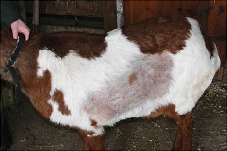
Figure 2.3 Alopecia on the side of a wether caused by grooming by its pen mate, a white - tail deer. Source: Courtesy of Dr. M.C. Smith.
that sell copper oxide wire particles. There is no published evidence that copper deficiency is involved when hair is lost from the tail, and the crusty skin on these tails could be the result of sunburn or trauma. “Fish tails” occur in areas where soil copper is high and there is much anecdotal evidence for copper toxicosis in goats treated with copper oxide wire particles they did not need. Baby oil or ointments containing zinc oxide can be used to address the skin changes on the tail.
Pruritus
Pruritus, or the semblance of itching, frequently leads to excoriations and other secondary lesions. If pruritus is severe, special consideration is given to the possibility of sarcoptic or chorioptic mange. Other conditions that may be pruritic include lice, fleas, hypersensitivity to other insects such as Culicoides, zinc deficiency, pemphigus, and photosensitization. Occasionally, bacterial or fungal dermatitis is mildly pruritic. If vertically oriented linear excoriations develop, migration of Parelaphostrongylus tenuis or Elaphostrongylus rangiferi through the spinal cord or dorsal nerve roots should be considered. Acute pruritus in a goat that dies very soon after clinical signs are noted is suggestive of pseudorabies (Baker et al. 1982). Extreme pruritus but with a longer clinical course has been reported in a single goat with confirmed rabies (Tarlatzis 1954). Pruritus was reported as a clinical sign in 11 of 20 goats in Great Britain with scrapie (Wooldridge and Wood 1991; Wood et al. 1992) and in more than 80% of 500 goats developing scrapie in Italy after infection via a contaminated contagious agalactia vaccine (Capucchio et al. 2001).
Erythema
Erythema, or reddening of the skin, occurs in many acute disease conditions and is thus not diagnostic. It is an early sign in photosensitization. When a chronic disease condition also has crusting and alopecia, response to therapy may be difficult to judge. Subsidence of a previously prominent erythema suggests improvement, even before hair regrowth is noted.
Pigmentary Changes
Few skin diseases in goats are associated with pigmentary changes. Decoloration of hair might occur with copper deficiency, because a copper-containing enzyme is necessary for melanin production. Affected Toggenburg goats and repigmentation with copper supplementation have been reported. Light-skinned Saanen goats normally develop large irregular areas of black pigmentation when exposed to sunlight.
The color fades with confinement away from the sun.Absence of Skin
Cutaneous asthenia, a congenital skin defect seen in sheep where the skin is abnormally fragile and easily torn, has apparently not been reported in goats. There is an anecdotal report of epitheliogenesis imperfecta, where a portion of the epidermis is absent, in a Pygmy goat (Konnersman 2005b).
Body Localization as an Aid to Diagnosis
The entire body surface should be examined. The distribution of skin lesions over the body helps to arrive at a diagnosis. Listed here are some diseases found initially or most severely, but not invariably, in the specified locations. Lesions present only on the ventrum may result from contact dermatitis or parasite invasion. Lesions present only on non-pigmented skin may be caused by photosensitization or sunburn.
Lips, Face, and Neck
• Contagious ecthyma
• Capripox
• Peste des petits ruminants
• Bluetongue
• Staphylococcal folliculitis
• Dermatophilosis
• Dermatophytosis
• Sarcoptic mange
• Zinc deficiency
• Pemphigus foliaceus
• Protothecosis
Ears
• Dermatophilosis
• Dermatophytosis
• Sarcoptic mange
• Ear mites
• Ticks
• Photodermatitis
• Squamous cell carcinoma
• Frostbite
• Pemphigus foliaceus
Feet
• Contagious ecthyma
• Foot and mouth disease
• Staphylococcal folliculitis
• Dichelobacter infection (foot rot)
• Dermatophilosis
• Sarcoptic mange
• Chorioptic mange
• Pelodera dermatitis
• Besnoitia dermatitis
• Zinc deficiency
• Contact dermatitis
• Pemphigus foliaceus
Udder
• Contagious ecthyma
• Staphylococcal folliculitis
• Zinc deficiency
• Hyperpigmentation from exposure to sun
• Neoplasia
Perineum
• Contagious ecthyma
• Caprine herpesvirus
• Staphylococcal dermatitis
• Ticks
• Neoplasia
• Ectopic mammary gland
Clinical Laboratory Examination
Simple observation allows identification of most of the clinical signs of skin disease, and thus many conditions can be diagnosed with reasonable certainty with just the findings of a physical examination.
However, the repertoire of injured skin is limited, and the same sign (such as a pustule or crust) may occur in a variety of conditions of different etiologies and necessitate further testing.Skin Scrapings
When searching for surface-dwelling ectoparasites such as lice and nits or chorioptic mange mites, a flea comb can be used to harvest scales and crusts or hair from extensive portions of the body. The collected sample is then placed in a petri dish or ziplock plastic bag for transport to good light or even a dissecting scope. After initial visual examination, the sample next undergoes a fecal flotation procedure. Mites and eggs come to the surface with centrifugation and can thus be concentrated and separated from the debris that would otherwise obscure their presence.
Repeated, deep scrapings using a scalpel blade dipped in mineral oil are usually necessary to identify sarcoptic mange mites or their eggs. A few drops of 20% potassium hydroxide solution are added to the sample, a coverslip is applied, and clearing of debris allowed to proceed for 15-30 minutes before microscopic examination. Larger samples may be processed by boiling for 10 minutes in 10% potassium hydroxide solution, centrifuging, and performing a sugar flotation on the sediment.
Direct microscopic examination of hair and keratin is useful for demonstrating the presence of dermatophytes.
Ectothrix infections of hair shafts often can be seen if the specimen is placed in mineral oil. Clearing in potassium hydroxide solution, as for mite identification, is another option.
Bacterial Examination
Skin lesions in goats are almost invariably heavily contaminated by bacteria, including Staphylococcus aureus. A culture is most meaningful, then, if material is aspirated from an intact pustule, nodule, or abscess. A punch biopsy obtained after careful disinfection of the skin surface is suitable for culture if intact pus-containing structures are absent. Routine inoculation onto a blood agar plate (aerobic) and into thioglycolate broth (anaerobic) is recommended. More immediate guidance can be derived by making a direct smear of an aspirate or deep aspect of a biopsy specimen and staining with new methylene blue, Gram, or Diff Quik (Siemens Healthineers, Erlangen, Germany) stain. Such a preparation should reveal bacteria within neutrophils and macrophages if they are pathogenic, rather than contaminant bacteria that will be extracellular and clumped in colonies. Gram-positive branching filaments are typical of dermatophilosis.
Fungal Culture
When ringworm is suspected, hairs should be plucked from the periphery of an active lesion after swabbing the area gently with 70% alcohol solution to discourage the growth of bacteria and saprophytic fungal contaminants. Sabouraud's dextrose agar (Sab Duet, Bacti Labs, Mountain View, CA, USA) is routinely used. Most strains of Trichophyton verrucosum require thiamine for growth; this can be supplied by adding 1-2 mL of injectable B-complex vitamins to the culture plate, but products containing alcohol should be avoided. The cultures are incubated at 86 °F (30 °C), with a pan of water in the incubator to maintain adequate humidity. Cultures should be checked every day for 30 days. Standard texts should be consulted for identification of fungal isolates (Scott 1988).
Biopsy for Histology
A biopsy should be performed if a skin disease appears to be unusual or severe, and especially if there has been no response after three weeks of initial therapy.
Several areas are selected as having typical or primary lesions and marked by drawing a circle with a felt-tipped pen. The skin is prepared by clipping hair and injecting lidocaine subcutaneously at each chosen location. The skin must never be scrubbed. In very small kids, dilution of the lidocaine to 1% or 0.5% may be advisable. A 6 mm biopsy punch is used to cut out a full-thickness skin sample, which should be blotted flat, dermis side down, onto a small piece of a tongue depressor. The skin specimen quickly adheres to the wood; it can then be dropped upside down into a vial of 10% buffered formalin, where the wood will keep it suspended in the preservative. The skin defect is closed with absorbable suture. Consideration should be given to the tetanus vaccination status of the animal and tetanus antitoxin or toxoid given if indicated.
The testing laboratory should be consulted if electron microscopic examination is required (as for pox). Glutaraldehyde is usually the preferred fixative.
Immunofluorescence Testing
An autoimmune skin disorder (pemphigus foliaceus) may be suspected from clinical signs or after routine histologic examination of a skin biopsy specimen. If confirmation with direct immunofluorescence testing is desired, a new skin sample (with intact vesicles and pustules) should be procured and fixed with Michel's fixative, which is best obtained from the laboratory that will perform the testing. Glucocorticoids should not be administered for at least three weeks before testing to avoid false-negative results (Scott 1988). Diffuse intercellular deposition of immunoglobulin is found in caprine pemphigus.