Enlarged Lymph Nodes
Diffuse or single lymph node enlargement occurs with infectious (bacterial, viral, fungal) conditions, neoplasia, and, rarely, immune-mediated causes in large animals (Boxes 6.16 and 6.17).
■ BOX 6.16
Causes of Enlarged Lymph Nodes in Horses Common Causes
Strangles
Lymphosarcoma
Upper respiratory infection
Corynebacterium pseudotuberculosis lymphadenitis Uncommon Causes
Ulcerative lymphangitis
Epizootic lymphangitis
Sporadic lymphangitis
Glanders
Melioidosis
Granulomatous lymphadenitis
Plasma cell myeloma
Tuberculosis
Hemolytic uremic-like syndrome
■ BOX 6.17
Causes of Enlarged Lymph Nodes in Ruminants
Common Causes
Caseous lymphadenitis (Corynebacterium pseudotuberculosis) Lymphosarcoma (including bovine leukosis virus)
Abscess or cellulitis of area drained
Uncommon Causes
Tuberculosis
Sporadic bovine encephalomyelitis
Malignant catarrhal fever
Lymphadenopathy may cause obstruction to lymphatic drainage, leading to peripheral edema, pleural effusion, or ascites. The peripheral lymph nodes that are most readily accessible for examination are the submandibular (horses), superficial cervical (ruminants), and superficial inguinal (ruminants) lymph nodes. When there is generalized lymphadenopathy, internal lymph nodes may be enlarged, causing clinical signs such as dyspnea, esophageal obstruction, diarrhea, or other signs of organ dysfunction.
Approach to Diagnosis of Enlarged Lymph Nodes
1. Take history. Note especially history of weight loss, inappetence, depression, lethargy, or lymph node enlargement; inquire about previous illness or wounds; for cattle, determine whether there is a history of lymphosarcoma in the family or herd and whether the cow has a positive BLV test result; for sheep and goats, determine whether there is a history of Corynebacterium pseudotuberculosis abscesses in the flock or herd.
2.
Perform a physical examination. Determine vital signs. Examine peripheral lymph nodes or other swellings, perform a rectal examination to palpate accessible internal lymph nodes, and, when appropriate, examine the uterus in cattle; carefully inspect the skin over the area drained by the lymph node for pain, swelling, or exudate; check mucous membranes for pallor or icterus; determine whether there is jugular venous distention or pulsations or whether there is evidence of pleural or pericardial effusion or ascites.3. Obtain blood for the following:
a. CBC to examine for anemia or leukemic changes; note inclusions, morula, or abnormal appearance of cells
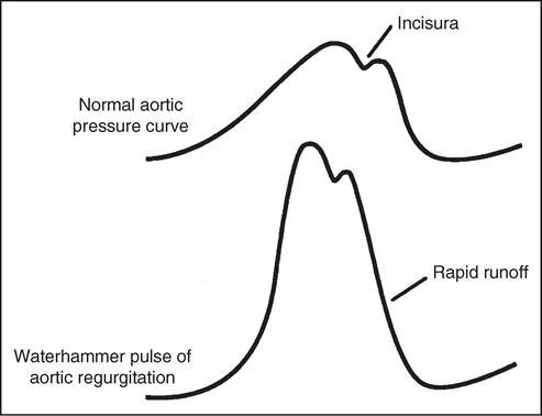
FIG. 6.6 Schematic illustration of the normal arterial pressure pulse. The incisura that occurs during the descending limb is caused by a transient reversal in flow during isovolumic relaxation. Compared with the normal arterial pressure pulse, the waterhammer pulse of aortic regurgitation builds rapidly and has a rapid runoff.
b. Serum chemistry profile to determine if there are signs of other organ dysfunction (e.g., gastrointestinal [hypoproteinemia], liver, or kidney)
4. Test feces for occult blood, if indicated.
5. Perform ultrasonographic examination of the lymph node or swelling.
6. Obtain lymph node or swelling aspirate and biopsy sample for culture and histopathologic examination.
7. Obtain a bone marrow sample for cytologic examination.