Equine Herpesvirus Myeloencephalopathy
Robert J. MacKay
Equine herpesvirus 1 (EHV-1) is a member of the Alphaher- pesvirinae subfamily of Herpesviridae. Along with another alphaherpesvirus, EHV-4, it is a common cause of upper respiratory disease in horses (see Chapter 31); of more importance is that because of a tendency to cause cell-associated viremia and vasculitis, EHV-1 infection is associated with late-term abortion, death of neonatal foals, and spinal cord disease.
Outbreaks of the last of these syndromes, known as equine herpesvirus myeloencephalopathy (EHM), have become more frequent and severe since the 1990s. The U.S. Department of Agriculture has suggested that EHM is an emerging disease on the basis of the following criterion: “A disease [that] changes in severity [or] type of animal that can be infected, or [reflects] other changes in pathogen behavior.”1■ Clinical Signs Acute onset of ataxia and tetraparesis of variable severity most often characterizes the neurologic form of EHV-1.2,3 Signs usually appear between 6 and 10 days after infection. The severity of clinical signs can range from subtle pelvic limb ataxia and weakness to complete recumbency without limb or head movement. Nonneurologic signs, including nasal and ocular discharge, limb edema, colic, ocular lesions, and anorexia, may also be observed.2-7 Horses may be febrile at presentation, but most are normothermic. Fever, often biphasic in profile, is the most consistent premonitory sign of neurologic disease, although not all febrile horses develop myeloencepha- lopathy. Coughing and nasal discharge or abortion sometimes accompany the neurologic signs or may have occurred during the preceding 2 weeks.5,8,9
■ TABLE 35.6
Congenital Diseases of Large Animals With CNS Signs and Known Mode of Inheritance
| Diseases With Predominant Intracranial Signs | Species and Breeds Affected | Clinical Signs/Ancillary Tests | Age at Onset | Mode of Inheritance | References |
| Ceroid lipofuscinosis | Ovine: South Hampshire, Swedish Landrace, Rambouillet Caprine: Nubian Cattle: Devon Horses: breeds not described | Progressive ataxia, blindness, depression, coma | Variable | Autosomal recessive | 1-10 |
| Citrullinemia | Cattle: Holstein | Progressive depression, bellowing, abnormal tongue and facial movements, convulsions, death Increased blood ammonia and citrulline concentrations | Newborns | Autosomal recessive | 11-19 |
| Exophthalmos and strabismus | Cattle: Jersey, Holstein, Brown Swiss, Shorthorn | Protrusion and crossing of the eyes | >6 mo | Unknown | 20, 21 |
| Cerebellar abiotrophy | Cattle: Holstein, Angus, Limousin | Intention tremors, hypermetria, basewide stance; may or may not progress | Variable | Suspected autosomal recessive | 22-26 |
| Bovine familial | Cattle: Angus, | Stillbirth/abortion with | Variable, | Autosomal | 27-33 |
| convulsions and | Charolais, Polled | occasional dorsiflexion of | may | dominant with | |
| ataxia | Hereford, Angus Cross | the spine Intermittent cerebellar signs and convulsions | improve with age | incomplete penetrance | |
| Cerebellar abiotrophy/ hypoplasia | Horses: Arabians, Arabian cross | Head tremor, ataxia, hypermetria | >2 mo | Autosomal recessive | 34-39 |
| Familial ataxia | Cattle: Hereford | Ataxia, abnormal head posture, pupillary dilatation | Newborns | Unknown | 40 |
| Micrognathia/cerebellar hypoplasia | Cattle: Angus | Stillbirth, brachygnathia, cerebellar hypoplasia | Stillborn | Suspected autosomal recessive | 41 |
| Alpha mannosidosis | Cattle: Angus, Murray Grey, Simmental, Galloway, Holstein | Stunted growth, ataxia, head tremor, hypermetria | 1 wk to 15 mo | Autosomal recessive | 42-49 |
| Beta mannosidosis | Cattle: Salers Goats: Anglo-Nubian | Recumbency, deafness, narrow palpebral fissures, intentional head tremor, rotatory head movements, ocular defects, joint abnormalities, domed skull | Newborns | Autosomal recessive | 50-71 |
| Generalized glycogenolysis (beta-galactosidase deficiency, GM1 | Sheep: multiple breeds Cattle: Holsteins | Lethargy, anorexia; head carried low and extended; blind, collapse when head moved | >1 mo | Suspected autosomal recessive | 72-83 |
| gangliosidosis) | |||||
| Bovine generalized glycogenosis (type II | Cattle: Shorthorn, Brahman | Weakness, tremors, ataxia, right-sided heart failure | 2-3 mo | Autosomal recessive | 84-93 |
| glycogenosis, Pompe disease) | |||||
| Globoid cell leukodystrophy | Sheep: Polled Dorset | Depression, head tremor, ataxia, hyperesthesia | >4 mo | Unknown | 94, 95 |
| (Krabbe disease) | |||||
| Neuronal lipodystrophy | Cattle: Angus, Beefmaster Sheep: Breeds not described | Depression, blindness, circling, seizures, coma | >10 mo | Unknown | 96-98 |
| Shaker calf syndrome | Cattle: Horned Hereford | Recumbency, hypermetria, hypertonia, hyperesthesia, muscle tremors | Newborns | Unknown | 99 |
■ TABLE 35.6
Congenital Diseases of Large Animals With CNS Signs and Known Mode of Inheritance—cont'd
| Diseases With Predominant Intracranial Signs | Species and Breeds Affected | Clinical Signs/Ancillary Tests | Age at Onset | Mode of Inheritance | References |
| Maple syrup urine disease | Cattle: Hereford, Polled Shorthorn | Viscous, dark, malodorous urine; fever, depression, opisthotonos, myoclonus, convulsions; spongiform changes in brain | Newborns | Autosomal recessive | 100-116 |
| Hereditary neuraxial Cattle: Polled and Inability to rise; hyperesthesia, edema Horned Herefords, myoclonus, extensor Hereford ? Friesian rigidity, and apnea/dyspnea when stimulated Epilepsy Cattle, horses See text Narcolepsy/cataplexy Horses, cattle, sheep See text Diseases With Predominant Spinal/Motor Nerve/Muscle Signs | Newborns (may be stillborn) | Suspected autosomal recessive | 117-125 | ||
| Inherited myoclonus | Horses: Peruvian Paso | Myoclonus in response to stimuli, bunny-hopping gait; may not be able to stand without assistance | Newborns | Unknown; deficiency of receptors for the inhibitory neurotransmitter GABA | 126 |
| Congenital encephalomyelopathy | Horses: Quarter Horse | Inability to stand; when assisted to stand, hindlimb tremors cause the hindlimbs to bounce off the ground; increased patellar reflexes | Present at birth (i.e., newborn) | Unknown | 127 |
| Systemic neuraxonal dystrophy | Sheep: Suffolk | Progressive hindlimb ataxia leading to recumbency | >1 mo | Suspected autosomal recessive | 128-129 |
| Weaver syndrome (bovine progressive degenerative myeloencephalopathy) | Cattle: Brown Swiss, Angler | Progressive paraparesis, hindlimb ataxia with proprioceptive deficits, muscle tremors, recumbency | >5 mo | Autosomal recessive | 130-146 |
| Progressive spinal myelinopathy | Cattle: Murray Grey | Inability to stand; progressive hindlimb hypertonia and proprioceptive deficits | Newborns | Autosomal recessive | 147-148 |
| Bovine spinal muscular atrophy | Cattle: Brown Swiss, Red Swiss, Braunvieh, Holstein | Progressive muscle atrophy leading to recumbency | >2 wk | Autosomal recessive | 149-157 |
| Spinal dysmyelination | Cattle: Braunvieh, Braunvieh Cross | Inability to stand, generalized tremors, muscle atrophy | Newborns | Autosomal recessive | 158-160 |
| Neuropathy/myopathy/ glomerulopathy | Cattle: Gelbvieh | Progressive hindlimb ataxia and paraparesis, muscle atrophy, reduced anal tone; proteinuria | Few weeks to 1 yr | Unknown | 161-162 |
| Progressive ataxia | Cattle: Charolais, Charolais Cross | Progressive paraparesis and hindlimb ataxia, proprioceptive deficits; muscle/tail tremors on rising | >6 mo | Suspected autosomal recessive | 163-172 |
| Spastic paresis (Elso heel) | Cattle: multiple breeds | Intermittent extensor spasticity and hypertonia of one or both hindlimbs; hyperextension of hock during weight bearing | >3 wk | Unknown | 173-195 |
| Inherited periodic spasticity (crampy syndrome, barn cramps, Krampfigkeit) | Cattle: dairy breeds | Intermittent muscular spasms of hip and upper hindlimb muscles, simultaneous kyphosis | >3 yr | Suspected autosomal dominant with incomplete penetrance | 196-200 |
| Hereditary lethal spasms (Doddler syndrome) Congenital abnormalities of the vertebrae and/or spinal cord | Cattle: Jersey Multiple species | Inability to stand, head and neck spasms when stimulated, convulsions, death See text | Newborns | Suspected autosomal recessive | 201-202 |
CNS, Central nervous system; GABA, gamma-aminobutyric acid.
The neurologic signs reflect injury to the white and gray matter of the spinal cord and include limb and truncal ataxia and paresis, bladder distention, urinary incontinence, weakness of the tail and anus, and reduced perineal sensation.
Affected horses may exhibit either penile paresis or persistent erection (priapism) during the first several days of clinical signs. Pelvic limbs are more severely affected than thoracic limbs, and signs are usually symmetric. In mild and moderately severe EHM, bladder distention is often present, with spastic detrusor tone and without urethral relaxation (dyssynergia). Bladder atony and incontinence also occur, particularly in recumbent horses, and the associated dribbling of urine leads to secondary perineal scalding. Bladder rupture has occurred in such horses. Anal sphincter and caudal rectal tone may also be reduced, with consequent obstipation and gaping of the anus.Signs of brain dysfunction such as seizures, blindness, and cranial nerve dysfunction occur in some severe cases of EHM.10,11 Ocular lesions such as mydriasis, hypopyon, uveitis, and optic neuritis may also be present. The occurrence of multifocal chorioretinopathy, visible several weeks after onset of neurologic signs as foci of depigmentation, appears to be a particularly sensitive indicator of previous EHV-1 viremia.12
The clinical signs typically stabilize within 48 hours, and in most instances recumbency, if it occurs, begins during this period. Some horses continue to deteriorate and eventually die or are euthanized. In survivors, improvement begins within the first 5 to 7 days and may continue for weeks or even months. Many horses make a full clinical recovery; however, an unknown proportion still have residual signs of limb weakness or ataxia. A horse that does not become recumbent during the first 2 days has a much better prognosis for survival.
■ Diagnosis EHM can be diagnosed tentatively on the basis of characteristic neurologic signs, history, and supporting findings on physical examination. A CSF analysis that shows xanthochromic fluid with high protein concentration and normal or slightly high nucleated cell count (albuminocytologic dissociation) is typical and almost pathognomonic for EHM,10,13 although in occasional cases, protein concentration is normal, mononuclear pleocytosis is present, or both occur.
Conventional, nested, or quantitative PCR amplification of EHV-1-specific gene sequences (usually glycoprotein B) in nasal swabs and EDTA-anticoagulated blood is the diagnostic technique of choice for horses with suspected EHM.13 Both nasal swab and blood samples should be submitted at the acute stage of disease; nasal swab samples alone are sufficient to monitor for release from quarantine in some jurisdictions. Nasal swabs are more likely to collect virus than are nasopharyngeal swabs.14 Swabs for this purpose should be approximately 6 inches long with a synthetic shaft. After collection, the swab should be placed in viral transport medium and sent to the diagnostic laboratory. If transport media are not available, the swab can be broken into a sterile tube and transported. Positive samples can be analyzed separately or simultaneously for a single-nucleotide variant in the viral DNA polymerase gene, DNApol D572, which defines the best known and most important neuropathogenic strain of EHV-1. Real-time PCR provides quantification of viral DNA load and allows additional interpretation of the relevance of positive results.13 These molecular diagnostic tests confirm active EHV-1 infection (and viremia when the blood test result is positive) but do not confirm the diagnosis of EHM specifically.
Additional aliquots of nasal swab and anticoagulated blood samples should also be submitted for traditional virus isolation in tissue culture. Virus isolation is important to allow full characterization of outbreak viruses, including susceptibility to candidate antiviral drugs.15
A fourfold or greater increase between acute and convalescent serum virus-neutralizing or complement-fixation (CF) antibody titers in samples taken 7 to 21 days apart is presumptive evidence of EHV-1 infection.13 These tests are of limited value, however, because they do not distinguish between EHV-1 and EHV-4 responses, and results are delayed several weeks.
An ELISA that distinguishes between infections with these two viruses is now available (SVANOVIR EHV1∕EHV4-Ab, Svanova Biotech, Uppsala, Sweden) and is the serologic diagnostic of choice. A practical approach to an outbreak that may involve many horses is to submit serum from both clinical and in- contact∕cohoused horses for serologic study and samples for PCR only from horses with clinical signs of fever or EHM. Such an approach provides additional support for the diagnosis and defines the extent of exposure to the virus during the outbreak.■ Pathophysiology After initial colonization of the epithelia of the upper respiratory tract, EHV-1 spreads to and amplifies in regional lymph nodes before circulating in the blood within leukocytes. The magnitude and duration of the leukocyte-associated viremia are positively related to the risk for EHM.13,16 Virus is presumed to enter endothelial cells of small arterioles of the CNS after binding to its surface class I major histocompatibility complex receptor. Endothelial infection incites destructive innate and specific immune inflammatory responses, which in turn initiate coagulation and widespread arterial microthrombosis and hemorrhage. Viral replication within neurons has not been demonstrated in EHM; rather, neuronal injury is secondary to ischemia that, in turn, is secondary to vasculitis and thrombosis.
■ Epidemiology Herpesvirus infection is enzootic in the horse population. Infection usually occurs through the respiratory epithelium when the animal comes in contact with the virus in aerosolized droplets or via fomites contaminated with fluids from an abortion or in ocular, nasal, or other respiratory tract secretions.3,10 Foals probably become infected with virus shed from their dams during recrudescence of latent infections.13 Most horses in the United States first become infected before 1 year of age and remain infected with latent virus in the trigeminal ganglia or in lymphoid tissues around the head.13,15 Surveys of necropsies of adult horses indicate that more than 50% of normal horses are latently infected by EHV-1 in one or both of these sites.13
EHM is rare, but cases have been reported worldwide.
Outbreaks occur more commonly in Europe and the United States than in Australia and New Zealand.13 Viral polymorphism undoubtedly accounts for differences in neurovirulence. Viruses with one allele of a single-nucleotide polymorphism in the viral DNA polymerase gene, D752,17 were found to be 162 times more likely to cause EHM than were viruses with the alternative allele, N752.18 The mechanism for enhanced neuropathogenicity of D752 is related partly the fact that viremia in infected horses is of higher order than that in horses infected with N752, but it is not due to greater neurotropism. It is common for horses to be latently infected with both variants (D752 and N752), and there is no evidence that horses latently infected with the neuropathogenic strain pose a higher risk of being the source of future EHM outbreaks.13 Polymorphism elsewhere in the genome probably also modulates neuroviru- lence.9 During outbreaks, the virus appears to be spread readily, and both morbidity and mortality rates can be high.Horses of any age, gender, and breed are susceptible. In some studies females in general, or mature pregnant or lactating mares, are most often affected.7,16,19 The principal risk factor for development of EHM is fever, especially the second peak of a biphasic fever.6 The onset of neurologic signs often follows closely the decline from peak temperature. Additional purported risk factors in at least some outbreaks were older age,6,7 cooler seasons (all but summer), introduction of horses to a herd,7,8 and inconsistent outbreak-specific breed susceptibilities.7,8,13 Foals are rarely affected. Mules and donkeys are infected and shed virus but in the United States apparently do not develop EHM.20 In contrast, EHM is relatively common and highly fatal in donkeys and mules in Ethiopia.21 Frequent (up to four times annually6,19) and recent19 vaccination against EHV-1 have been reported as independent risk factors.
Stress-associated recrudescence of latent infections and subsequent shedding without clinical signs has been difficult to confirm experimentally but undoubtedly is important in the initiation of some outbreaks, especially those occurring after long-distance transportation or in closed herds. Experimentally infected horses may nasally shed virus for 3 weeks or longer after infection; however, viral shedding was not found after 9 days after onset of clinical signs in 20 horses with EHM.22Primary attack rates, as judged by occurrence of fever (usually defined as rectal temperature >40.6° C [101.5° F]) among at-risk horses, may be up to 90%; an average of 10% of these infected horses develop EHM, and mortality rates for horses with EHM are between 20% and 80%; a typical rate is 50%.3,13 There is no report of EHM caused by modified live virus vaccines currently approved for use in horses.
■ Necropsy Findings Gross and histologic lesions are not always limited to the CNS. Ocular lesions such as hypopyon, iritis, and chorioretinitis have been reported. Cystitis and scrotal edema may be present. Focal areas of hemorrhage may be found throughout the brain and spinal cord parenchyma and meninges. Vasculitis and thrombosis in the small arteries and veins of the spinal cord and brain result in ischemic lesions in the CNS.23,24 Equine herpesvirus is infrequently cultured from the CNS, but PCR, in situ hybridization, and immunohistochemistry studies can be performed on CNS tissues obtained post mortem to confirm the presence of the virus.13
■ Treatment Acyclovir is guanosine analog with in vitro activity against EHV-1 that has been used successfully for treatment of herpes simplex encephalitis in humans. Adequate blood levels of acyclovir can be achieved in horses by oral dosing with valacyclovir (30 mg/kg q8h for 2 days and then 20 mg/kg q12h) or IV administration of acyclovir (10 mg/kg q12h) but not by oral acyclovir.25 Although valacyclovir is safe and effective when used as preventive treatment, limited data suggest that it is not effective in horses with ataxia (i.e., EHM).26,27 In view of the scant evidence available, however, valacyclovir treatment is still worthwhile when economic circumstances permit. Pharmacokinetic and pharmacodynamic data have been obtained for several other antiherpetic drugs, but their effectiveness for EHV treatment has not been investigated.26
Supportive care is the most important aspect of treatment for EHM. Measures include bladder decompression by sterile catheterization at least twice daily for horses with bladder distention or incontinence, evacuation of the rectum, administration of intravenous or oral fluids, and enteral or parenteral nutritional support. Recumbent horses must be appropriately padded, and decubital lesions must be prevented or treated. Horses that are recumbent but able to support their weight when lifted should be managed with an abdominal sling.
Administration of antiinflammatory drugs soon after the onset of neurologic signs may be beneficial.28 NSAIDs (e.g., flunixin meglumine, 1.1 mg/kg IV q12-24h) should be used especially if the horse is febrile or if a painful condition is clinically suspected. Corticosteroids are often used because of presumed suppression of vasculitis, and although no objective data for their efficacy are available, there is a solid theoretical basis for their use.13,28 Dexamethasone (0.05 to 0.1 mg/kg IV or IM) can be given every 12 to 24 hours for 3 to 5 days; the dosage is then tapered for 1 to 3 days. There are unquantifiable risks related to steroid-induced immunosuppression, including increased susceptibility to bacterial infection of the urinary tract and reactivation of latent virus. Because of uncertainties as to its benefits and risks, steroid treatment should be reserved for horses that are recumbent or in imminent danger of becoming so. DMSO (1 g/kg as a 10% solution in polyionic fluids by slow IV infusion) every 12 to 24 hours is often administered, although its efficacy also has not been documented.
Administration of antibiotics should be considered if the horse is recumbent or has urinary tract or respiratory tract involvement. Chronic cystitis, sometimes associated with bacteria resistant to common antimicrobials, is a common complication of urinary incontinence and bladder catheterization in horses with EHM. Management of such cases is discussed in Chapter 34.
The lesions of EHM are characterized by extensive micro- vascular thrombosis,29 and so aspirin or heparin may be used in an effort to prevent worsening of spinal cord lesions (see next section).
■ Control and Prevention Vaccines currently used to prevent EHV-1 respiratory and abortion syndromes are not claimed to prevent EHM, and there is no reliable evidence that they do. A small study concluded that modified live virus vaccine provided modest, albeit significantly better, protection against experimentally induced EHM than did a killed low-antigen vaccine,30 although the duration and robustness of such protection, if any, are not yet clear. Vaccination with inactivated “high-antigen” vaccines (i.e., those with a label claim for prevention of herpesvirus abortion), enhanced adjuvant vaccines, or modified live vaccine does have the important effect of reducing nasal shedding and thus potentially limiting the spread of infection during an outbreak. This rationale has also been used to justify booster vaccination of at-risk horses during an outbreak. Because of persistent evidence-based concerns about inducing or exacerbating EHM by excessive and recent vaccination, however, this practice remains controversial.6,19
Immunostimulants are sometimes administered to horses at risk for EHM on the basis of the theory that immune system activation will prevent viral reactivation or replication,13 but supportive evidence is lacking. A history of nutritional supplementation with a product containing zinc protected horses exposed during an EHM outbreak from developing the disease according to a retrospective study of horses involved in the 2011 Ogden, Utah, outbreak.19
Valacyclovir and valganciclovir are prodrugs of the antiviral agents acyclovir and ganciclovir that have adequate bioavailability in horses when given orally.25 Although these agents have in vitro activity against EHV-1, valacyclovir and ganciclovir were not effective at preventing EHM when either one was administered 4 to 6 days after viral inoculation (i.e., at the onset of the second temperature rise).31 Ganciclovir, but not valacyclovir, reduced the severity of ataxia. In contrast, vala- cyclovir significantly reduced the severity of neurologic signs when begun either the day before challenge or at the onset of fever and continued for 1 to 2 weeks.27 Therefore if these drugs are used for prevention, they should be begun no later than the onset of the initial fever.
Protocols for management of premises with EHM cases are now well established and tested. Comprehensive and regularly updated guidelines are available from the American Association of Equine Practitioners (AAEP; http:// www.aaep.org/). In addition, the British Horserace Betting Levy Board publishes Codes of Practice on Equine Diseases (https://codes.hblb.org.uk/). In brief, the essentials of these protocols are (1) to isolate affected horses; (2) to monitor temperatures twice daily on all quarantined horses; (3) to submit diagnostic samples; and (4) to implement robust biosecurity procedures.13 There is still uncertainty about the length of quarantine required after the last occurrence of fever in any horse. AAEP guidelines currently call for 28 days. A workable but expensive alternative is to require 14 days after the last fever in any quarantined horse and then test all horses for nasal shedding by PCR on swabs taken on 2 to 4 successive days.13 It is essential that all guidelines are followed meticulously; with each new fever, a 28-day quarantine must start over. Biosecurity procedures for EHM caused by neuropathogenic and nonneuropathogenic strains should be the same.12 Additional guidelines are available for measures designed to prevent or reduce the likelihood of outbreaks and to lessen the effect of outbreaks that do occur.13
Equine Protozoal Myeloencephalitis few reports of EPM in ponies or mules, which suggests that these equids may be less susceptible to EPM than are horses. Ages at onset of EPM have ranged from 2 months to at least 30 years, although it is rare in horses younger than 2 years old. One study identified an increased risk of EPM in horses aged 1 to 5 years and older than 13 years.12 EPM typically occurs as individual cases, but clusters of cases have occasionally been reported.13
The reported prevalence of S. neurona infection as evidenced by S. neurona antibodies in horses in various locations throughout the United States varies from 0 to 89.2%.14,15 Rates are generally high in areas with dense human populations, abundant water sources, and moderate rainfalls; seropositivity rates are low in arid, sparsely populated regions. The geographic variation in prevalence corresponds closely to the still-expanding range of the definitive host, the opossum. Despite the high rate of infection by S. neurona, the lifetime risk of a horse for developing EPM in these areas may be only approximately 1% (estimated from the mean 0.14% one-year incidence reported in the National Animal Healtb Monitoring System 1998 survey16).
The factors that influence the relationship between infection with S. neurona and development of EPM are not well understood. S. neurona lacks the genetic diversity found among Toxoplasma gondii isolates, but it may nonetheless be capable of T. gondii-like clonal expansion of virulent strains,17 although conclusive evidence for this has not yet been found in the setting of equine infections. Results of risk factor analysis for EPM vary by study, but factors likely to increase exposure to opossums, their environment, and their feces are consistently identified.18-21 Other risk factors include previous diagnosis of EPM on the premises; primary use of the horse, with showhorses and racehorses at relatively high risk; recent transport; and recent adverse health events such as medical problems, parturition, and management changes. Collectively, these risk factors indicate that physiologic stress may play an important role in the onset of EPM.
■ Diagnosis Antemortem diagnosis of EPM remains challenging. Criteria for diagnosis include finding clinical signs consistent with EPM, ruling out other diagnoses, positive response to treatment with anti-EPM drugs, and finding S. neurona-specific antibodies through immunodiagnostic testing. Proof of diagnosis post mortem is demonstration of protozoa in CNS lesions, often obtained with immunohistochemical staining or PCR. Even when the organism is not observed, the diagnosis is frequently made presumptively if characteristic inflammatory changes are found.22,23
Two types of immunodiagnostic tests are available for EPM: those that rely on threshold values of serum anti-S. neurona antibody (IFAT, ELISAs for antibody against various S. neurona surface antigens) and those that purport to detect specific antibody made within the CNS (Western blot, serum-to-CSF antibody ratios, C values).24 Blood samples for serum tests are easily obtained, but the diagnostic assays are based on the yet-to-be validated principle that horses with EPM have distinctly higher serum antibody concentrations than do exposed/subclinically infected horses.25 However, because seroprevalence of EPM is relatively low in comparison with that for S. neurona, a positive serum test result has a proportionally higher positive predictive value for EPM.24 On the other hand, a negative test result is strong evidence that the horse does not have S. neurona infection and thus does not have EPM. The rationale for tests of the second type, each of which requires a clean CSF sample, is that identification of CNS-origin antibody is the only way to distinguish horses with EPM from exposed horses. The most popular tests of each type—namely, serum IFAT and ELISA for single-nucleotide surface antigens 2 and 4/3 (snSAG2,4∕3)—have good (i.e., >80%) sensitivity and specificity for EPM among horses with solid postmortem diagnoses.26,27 One drawback of the IFAT is that it is unable to differentiate between antibodies against S. neurona and those against Sarcocystis fayeri (a relatively nonpathogenic species).28 In one study, the researchers used samples from necropsy- confirmed EPM-positive and EPM-negative cases to specifically compare results of IFAT and snSAG2,4/3 tests. Accuracy values for serum IFAT, serum snSAG2,4/3 ELISA, IFAT ratio, and snSAG2,4/3 ratio were 68%, 56%, 88%, and 97%, respectively.23 Comparable tests for N. hughesi also are available commercially (N. hughesi IFAT and NhSAGl ELISA) but have not been fully validated for EPM diagnosis. It is clear, on the basis of both flawed concepts and unsupportive clinical data, that neither CSF Western blot nor serum SAGl ELISA is a reliable diagnostic test for EPM.29-3l In contrast to the situation with Western blots, snSAG (and presumably IFAT) ratio test results are relatively robust in the presence of a small amount of contaminating blood; however, false-positive results do occur in cases of moderate or severe blood-brain barrier injury. For samples from horses with blood-brain barrier injury (as detected by high CSF albumin concentrations or high albumin quotient values32), additional total IgG testing is performed on the serum/ CSF sample, and a C value is calculated.33 CNS antibody production is indicated by C values greater than 1.
At the time it was last investigated, PCR testing of CSF for S. neurona DNA had high specificity but low sensitivity for the diagnosis of EPM.34 False-negative findings are common because free parasitic DNA is destroyed by enzymes in the CSF and merozoites rarely enter the CSF. Testing by PCR of tissue samples is useful for postmortem diagnosis.
Ancillary procedures such as survey cervical radiographs, lameness examination, and serologic testing may be necessary to rule out competing differential diagnoses. In horses with signs of brain involvement, suggestive lesions may be seen with advanced imaging techniques.35 Consistent abnormalities are not found in complete and differential WBC counts or in serum chemistry panels in horses with EPM. CSF values are abnormal in some horses with EPM. It has been reported that up to 35% of horses at a referral hospital have increased protein concentration (>65 mg/dL) or nucleated cell count (>6 cells∕μL).36 In the author's experience, in horses with mild clinical signs of EPM (i.e., those that typically are encountered in practice), a low percentage have any abnormality on routine CSF analysis. CK activity may be high in CSF, reflecting diffusion of the BB isoenzyme from damaged CNS gray matter.37 Unfortunately, inadvertent inclusion of a small plug of epidural fat or dura during CSF collection can dramatically elevate CK activity, thus reducing the specificity of the test.
■ Clinical Signs Protozoa may infect any part of the CNS and thus produce almost any possible neurologic sign. The disease usually begins insidiously but also may present acutely and be severe at onset. Signs of spinal cord involvement are much more common than are signs of brain disease. Horses with EPM involving the spinal cord have asymmetric (usual) or symmetric truncal and limb weakness and ataxia. If all lesions are behind the second thoracic spinal cord segment (T2), only the pelvic limbs are affected. If a lesion (or lesions) is located in front of T3, all four limbs may be affected. In the rare instance when the spinal cord behind the second sacral spinal cord segment (S2) is involved, the animal exhibits signs of cauda equina syndrome, which may include degrees of rectal, anal, bladder, and penile paralysis and reduced sensation of the skin of the tail and perineum. When gray matter of the ventral horn of the spinal cord is damaged for more than 1 to 2 weeks, obvious focal muscle atrophy and electromyographic changes in denervated muscle may develop. Common locations for atrophy in horses with EPM are the gluteal, biceps femoris, infraspinatus/supraspinatus, and serratus ventralis muscles. Lesions in the spinal cord may also result in demarcated areas of spontaneous sweating or loss of reflexes and cutaneous sensation.
Neurologic signs noticed at walking or during neurologic examination include any to all of the following: asymmetric stride length, toe dragging, circumduction of pelvic limbs, and hypometria (also described as floating or marching) of thoracic limbs. Other signs are noted only when the horse is ridden: difficulty maintaining a specific lead, cross-cantering or crossgalloping, and difficulty negotiating turns. Some signs that are usually attributed to primary musculoskeletal disease, such as back soreness or upward fixation of one or both patellas, can be caused by weak or asymmetric use of muscle groups in horses with EPM that are in training.
A relatively common manifestation of brain disease in horses with EPM is a brainstem syndrome with signs of obtundation and asymmetric vestibular (VIII) nerve dysfunction (Fig. 35.18). There may also be facial paralysis (VII); dysphagia (X); tongue paralysis (XII); laryngeal paralysis (X); strabismus (III, IV, and V); failure of globe retraction (VI); and deviation of the mandible (VII). With involvement of the rostral brainstem or cerebrum, EPM may manifest as seizures, visual deficit/abnormal menace response, reduced perception of touch on the opposite side of the face, or behavioral abnormality.
Without treatment, EPM usually progresses. Progression to recumbency occurs over hours to years and may occur steadily or in a stop-start manner.
■ Treatment Since 2000, the folate inhibitor combination sulfadiazine/pyrimethamine, the triazine coccidiostats ponazuril and diclazuril, and the nitrothiazole nitazoxanide have all been licensed by the U.S. Food and Drug Administration (FDA) for the treatment of EPM, and commercial products have been launched.9,38 Nitazoxanide was withdrawn in 2009. Multicenter studies of sulfadiazine/pyrimethamine, ponazuril, nitazoxanide, and diclazuril reported improvement rates of 57.1% to 61.5%.38
Label directions for dose and duration of FDA-approved drugs should be followed. Ponazuril in a loading dose of 15 mg/kg (three times the normal dose) reaches steady-state concentrations several days earlier than is the case with conventional dosing.39 Many dosage permutations and combinations of these products and pharmacologically similar products are administered in clinical practice, although efficacies have not been investigated. A sodium salt formulation of diclazuril has been shown to have good oral bioavailability and may have potential as a feed additive.40 Some veterinarians administer IV a compounded form of diclazuril, although there are no published data for its safety or efficacy. Of interest is that steady-state concentrations of diclazuril in plasma and CSF were not different for the low-dose rate (0.5 mg/kg) and the FDA-approved dose rate (1 mg/kg).41 These data support the use of the 0.5-mg/kg dose as treatment or preventive. Experiments have demonstrated enhancement of the antiprotozoal effect of combinations of pyrimethamine with either diclazuril or ponazuril in treatment of other coccidian infections,42 and so ponazuril or diclazuril is sometimes combined with sulfadiazine/ pyrimethamine. Such treatments constitute unapproved or off-label use, albeit of FDA-approved products.
Another drug used commonly as an anticoccidial in calves, decoquinate, was evaluated in horses with presumptive EPM, at dosage of 0.5 mg/kg PO daily for 10 days, in combination with the immunomodulator levamisole, 1 mg/kg PO daily.43 Success rates of 93.6% and 89.3% were reported on the basis of improved neurologic gait score at the end of treatment and reversion of serum snSAG1,5,6 titers, respectively. Decoquinate was also shown to have potent inhibitory and killing activity in vitro against two different isolates of S. neurona.44 If this
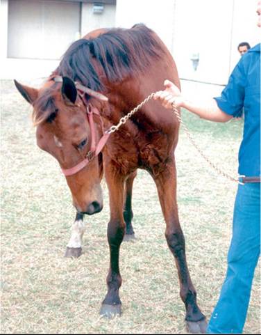
FIG. 35.18 Obtundation, right-sided head tilt neck turn, and body lean in a 3-year-old Thoroughbred colt with peracute-onset equine protozoal myeloencephalitis.
drug combination continues to perform well in wider use, it will obviously become an important treatment option.
Horses may exhibit relapse soon after the initial course of treatment is discontinued or up to several years after improvement or apparent resolution of signs. Although no data are available to guide treatment of relapses, it is common practice to change or add antiprotozoal drugs. In order to prevent future relapses, antiprotozoal therapy can be provided pro- phylactically. Protocols include FDA-approved formulations of sulfadiazine/pyrimethamine, 2 days/week at the standard dosage, or diclazuril daily at 0.5 mg/kg. A similar rationale can be used to treat horses only around the time of stressful events that may predispose to relapses.
Of the available drugs, only pyrimethamine has significant adverse side effects. Toxicity reflects both direct effects of the drug and its inhibition of mammalian dihydrofolate reductase, which induces folate deficiency. Signs include convulsions and recumbency, reproductive and neonatal disorders, oral ulcers, and bone marrow suppression, including (in decreasing frequency) leukopenia, neutropenia, anemia, and thrombocytopenia.45-47 Horses treated with sulfadiazine/pyrimethamine should be fed good-quality green forage (e.g., alfalfa hay or pasture), which is likely to be rich in folate. Supplementation with folic acid during treatment confers no advantage, and some evidence from other species indicates that this practice could increase the possibility of toxicity.46 Most cases of bone marrow suppression resolve if treatment is suspended for 1 to 2 weeks. Folic acid (100 mg daily) is appropriate for treatment of folate deficiency once pyrimethamine is discontinued. If signs are severe, treatment with folinic acid (100 mg IV or PO daily for 5 days) is indicated.
In mild acute cases of EPM (grade 1), additional therapy is unnecessary. In horses with moderate or severe signs, NSAID and antioxidant treatment is usually provided for the first 1 to 2 weeks. If the horse has grade 3 EPM or worse, a corticosteroid may be added. Vitamin E is often given throughout the period of antiprotozoal treatment for additional antioxidant effect. Oral administration of vitamin E has been shown to result in increased concentrations of α-tocopherol in serum and CSF.48 As a relatively crude way to address putative immunodeficiency and “boost” cellular immunity, immune modulators are sometimes given to horses with EPM.
■ Prognosis On the basis of drug efficacy studies and clinical experience, it is reasonable to expect that approximately 60% of moderately or severely affected horses will improve at least one grade after treatment. Only 10% to 20% of such horses completely recover, and 10% to 20% of successfully treated horses suffer at least one relapse. The outlook for mildly affected horses (grade 1) is considerably better, particularly if treatment is begun promptly. Although no data for the latter, more common class of treated horse have been published, at least 80% of such cases can be expected to improve, and at least 50% recover completely, with low possibility of recurrence.
■ Prevention An inactivated vaccine was marketed under conditional approval by the U.S. Department of Agriculture. The conditional license lapsed in March 2008, and the vaccine is no longer available. When tested in horses dosed with S. neurona and stressed by transportation, vaccination did not prevent development of clinical neurologic signs.49
Prevention of S. neurona infection may be possible by daily administration of a drug such as diclazuril or ponazuril.50,51 The disadvantage of this approach is that normal immune responses to the organism may be prevented. An alternative approach is metaphylaxis, wherein intermittent doses of drug are given to allow an initial round of protozoal proliferation and normal immune responses to take place but prevent invasion of the CNS. Ponazuril given at 20 mg/kg every 7 days but not every 14 days significantly reduced S. neurona antibody in the CSF of S. neurona-challenged horses, which suggests the potential utility of such an approach.52
Common-sense measures can be instituted to prevent contamination of feed with S. neurona sporocysts. Opossums can be trapped and removed or kept away by free-ranging dogs. Spilled grain, fallen fruit, and animal or bird feed should be removed, and horse food sources should be monitored and secured. The utility of controlling intermediate hosts in an effort to reduce environmental sporocyst load is doubtful; however, if populations of host animals on a problem premise are large, then such animals should be trapped and removed. Products useful for cleaning of areas potentially contaminated with sporocysts have been described.53