Equine Metabolic Syndrome
Nicholas Frank
Definitions
Equine metabolic syndrome (EMS) is a collection of risk factors for endocrinopathic laminitis, and the term syndrome is used because multiple factors contribute to the risk of laminitis developing in an individual horse, pony, or donkey.1 Risk factors include ID, which is the central endocrine disorder of EMS, as well as increased adiposity, hypertriglyceridemia, altered adipokine production, and cardiovascular abnormalities.
The term insulin dysregulation encompasses the different glucose and insulin alterations observed in affected horses, including fasting hyperinsulinemia, postprandial/postsugar hyperinsulinemia, and tissue insulin resistance (IR).2 It is likely that EMS has a genetic basis because the syndrome has a higher prevalence in certain breeds of horse or bloodlines. For example, Andalusians, Arabians, Morgan horses, Paso Finos, ponies, and warmbloods are more likely to be affected by EMS than Standardbreds and Thoroughbreds.If EMS susceptibility is determined by genetics, then it is essential that we identify and manage modifying factors that exacerbate the condition. Although most horses with EMS are overweight, a lean phenotype is also identified and these patients may present with the primary complaint of laminitis. It is conceivable that we will identify multiple forms of EMS in the future in the same way that researchers have recently defined different types of diabetes mellitus in humans.3 The EMS phenotype typically develops when genetically predisposed animals encounter environmental factors that promote obesity and exacerbate ID.
It is important to distinguish EMS from pituitary pars intermedia dysfunction (PPID) yet recognize that both disorders can occur concurrently and influence one another. These disorders are compared in Table 41.1.
Etiology
Genetics play a role in EMS development, and research studies provide evidence of breed differences in ID susceptibility.
■ TABLE 41.1
Comparison of Equine Metabolic Syndrome and Pituitary Pars Intermedia Dysfunction
Equine Metabolic Syndrome (EMS) Clinical Presentation
Pituitary Pars Intermedia Dysfunction (PPID) Clinical Presentation
Any mature horse
Obesity and regional adiposity; prominent fat pads along the crest of the neck, above the tailhead, or in the prepuce/ mammary gland; animals often described by owners as easy keepers
A lean EMS phenotype is also recognized with regional adiposity
Some affected horses appear normal but have excessive insulin responses to oral sugars
Infertility or abnormal cyclicity in mares
Subclinical or clinical laminitis; acute episodes occurring after exposure to pasture or feed with high sugar content
Excessive insulin responses to oral sugars
Commonly accompanied by insulin resistance
Begin evaluating horses at 10 years of age
Early PPID:
Subtle regional hypertrichosis and/or delayed shedding for a few weeks; duller, coarser hair coat
Reduced muscle mass, shift in metabolism, poor performance
Advanced PPID:
Generalized hypertrichosis (hirsutism)
Weight loss and skeletal muscle atrophy; rounded abdomen More pronounced fat deposits along the neck crest and in front of the tailhead in some cases
Recurrent or chronic infections such as sinusitis, skin infection, dental disease, or hoof abscesses
Reproductive abnormalities including infertility and persistent lactation
Chronic laminitis in some cases
Pathophysiology
Pathophysiology
Not fully established
Current theories focus on a genetic predisposition to insulin dysregulation; affected horses have excessive insulin responses to oral sugars
Hyperinsulinemia might promote increased adiposity
Obesity, lack of exercise, and PPID are modifying factors and lower insulin sensitivity, with insulin resistance exacerbating hyperinsulinemia
Laminitis results from persistent hyperinsulinemia
Loss of dopaminergic inhibition leads to hyperplasia and hypertrophy within pituitary pars intermedia
Excessive proopiomelanocortin (POMC) production with resulting increased α-melanocyte-stimulating hormone, adrenocorticotropin hormone, corticotropin-like intermediate peptide, and β-endorphin production
Management Goals
Management Goals
To reduce glucose and insulin responses to oral sugars by limiting the sugar content of the diet
Limit access to pasture
To induce weight loss if obese by restricting caloric intake
To improve insulin sensitivity through weight loss and exercise Appropriate farrier care
Improvement in clinical signs and hormone concentrations through pergolide treatment
Provision of supportive care including body clipping, appropriate farrier care, dentistry, and nutrition
Jeffcott and colleagues4 detected differences in glucose tolerance and insulin sensitivity between ponies and Standardbred horses, with fat and laminitic ponies having lower responses to exogenous insulin (and therefore lower insulin sensitivity) than Standardbreds.
Standardbred horses, mixed-breed ponies, and Andalusian cross horses also show differences in postprandial insulin responses to meals, providing further evidence of breed-related variability in glucose and insulin dynamics.5 A more detailed examination of genetics was performed in Arabian horses using genome-wide association, and an EMS locus was identified.6 Evidence has also been presented to suggest that EMS susceptibility follows familial lines in ponies associated with dominant expression of one or more genes with incomplete penetrance because of environmental or epigenetic factors.7Environmental factors, including diet and exercise, play important roles in the development of obesity and the EMS phenotype. Feeding grain to a susceptible animal is enough to induce obesity, and the grass consumed on pasture represents a significant source of calories that contributes to weight gain. Carbohydrate intake on pasture is influenced by grazing time, the area available for grazing, and the nonstructural carbohydrate (NSC) content of pasture grasses. NSC content varies by geographic location, climate, soil quality, and season. EMS affects adult horses, and there is no reported sex predilection. Age impacts insulin metabolism in horses, as it does in other species.8,9
Clinical Signs and Differential Diagnoses
The medical history may provide clues that the horse is affected by EMS because genetically predisposed animals readily gain body mass and are sometimes referred to as “easy keepers” or “good doers” by their owners. Most horses, ponies, and donkeys with EMS are obese, and a thicker than normal neck crest is noted on physical examination. This form of regional adiposity is associated with ID in affected horses, and the cresty neck score can be applied to more objectively measure the amount of fat in this region.10 Over time, regional adiposity develops in other locations such as the tail head, prepuce, or mammary glands.
Many horses with EMS suffer from generalized obesity and have body condition scores of 7 or greater on a 1 (poor) to 9 (extremely fat) scale.11 A mare suffering from EMS is shown in Fig. 41.14. Laminitis may have started to develop without the owner noticing. Horses should be turned in tight circles to detect lameness. Divergent growth rings (“founder lines”) or widening of the white line may be detected when the hooves are examined closely, and these signs provide evidence of subclinical laminitis.12,13 Radiographs are recommended as part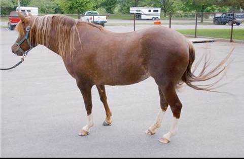
FIG. 41.14 Photograph of a 7-year-old Morgan horse crossbred mare that suffers from equine metabolic syndrome.
of the initial examination, and radiographic evidence of third phalanx displacement may be detected, even when lameness has not been reported.
Clinical Pathology
Hypertriglyceridemia and mildly increased plasma gamma glutamyl transferase (GGT) activities are detected in some horses, ponies, and donkeys with EMS, but complete blood count and other plasma biochemical analysis results are normal. Hyperglycemia is occasionally detected in affected horses and it is appropriate to use the term diabetes mellitus when this problem persists and becomes a chronic condition. Hyperglycemia and hypertriglyceridemia are more likely to occur when horses with ID are challenged by systemic disease and hospitalization, and this is sometimes referred to as metabolic decompensation. Older horses with concurrent ID and PPID are susceptible to this problem, and insulin therapy may be required to control hyperglycemia and glucosuria.14
Low resting total triiodothyronine (tT3) or total thyroxine (tT4) concentrations are detected in some horses with EMS, but the significance of this finding has been overstated in the past. It is a common misconception that obese horses suffer from hypothyroidism because obesity is associated with hypothyroidism in dogs and humans.
However, most animals have normal TRH stimulation test results, and low tT3 and tT4 concentrations, if present, should be attributed to extrathyroidal changes in thyroid hormone metabolism or low thyroid-stimulating hormone (TSH) concentrations. Phenylbutazone is an extrathyroidal factor that significantly lowers serum tT4 concentrations in horses because of protein binding, and this drug is commonly used to treat laminitis.15Thyroid-stimulating hormone concentrations vary as the body adapts to different metabolic states, and this might also explain why low thyroid hormone concentrations are sometimes detected in horses with EMS. This is referred to as secondary hypothyroidism and can be confirmed by documenting low TSH concentrations. Unfortunately, diagnostic laboratories do not measure equine TSH at present, so it is difficult to confirm the diagnosis of secondary hypothyroidism. Levothyroxine supplementation is controversial in these cases, and in the author's opinion, levothyroxine supplementation should be reserved for horses with persistently low free thyroxine concentrations and abnormal TRH stimulation test results. Levothyroxine can also be administered at higher dosages for 3 to 6 months to accelerate weight loss, and that protocol is described later.
Diagnostic Testing
Measures of adiposity should be performed, including body condition score, cresty neck score, and ultrasound measurements of abdominal or subcutaneous fat depth.16 Diagnostic testing for ID is required for all patients in high-risk breed groups, and the Equine Endocrinology Group provides current recommendations for diagnosing and managing EMS on their website (https://sites.tufts.edu/equineendogroup/). Horses are screened for ID by measuring basal (or resting) insulin concentrations under fed conditions or by performing an oral sugar test (OST).17 The assay used to measure plasma or serum insulin concentrations should be considered before either test is performed, and a diagnostic laboratory with experience handling equine samples should be selected.
Insulin concentrations have been measured by radioimmunoassay for the majority of publications on EMS, including the 2010 American College of Veterinary Internal Medicine consensus statement,1 but some diagnostic laboratories are now using chemiluminescent or enzyme-linked immunosorbent assays to measure insulin. The reference intervals provided as follows are for insulin concentrations measured by radioimmunoassay, and adjustments must be made if other insulin assays are used.Some clinicians prefer to screen horses by measuring basal insulin concentrations before performing an OST because of owner concerns (not supported by evidence) about inducing laminitis with a stimulation test. If this approach is followed, detection of high insulin concentrations confirms the diagnosis of ID, but horses with normal results must undergo dynamic testing before normal insulin status can be assumed. Insulin concentrations were previously measured under fasting conditions because this is the approach taken in human medicine,1 but this recommendation has changed and blood samples should be collected when horses are consuming hay or grazing on pasture. Grain should be withheld for 6 hours before blood collection. When measuring basal (fed) insulin concentrations, an insulin concentration greater than 50 pIU/mL provides strong evidence of ID, concentrations in the 20 to 50 pIU/mL range provide weak evidence, and there is no evidence of ID if the insulin concentration is less than 20 pIU/mL. Note that some laboratories provide insulin concentrations in pmol/L, and an approximate conversion factor of 7 is used to convert units from pIU/mL to pmol/L.
The OST is performed by fasting the horse for a minimum of 4 hours and preferably 6 hours in order to minimize the impact of gastric emptying on test results.18 When the OST is to be performed in the morning (8:00 and 9:00 a.m.), the horse should receive one flake of hay between 10:00 p.m. and 12:00 a.m.
the night before, and then all feed should be withheld until after the test is completed. Water should also be withheld during the test. Corn syrup (Karo Light, Cordova, Tenn.) is administered by mouth using 60-mL catheter-tip syringes at a dosage of 0.15 mL/kg, and blood samples are collected 60 and 90 minutes afterwards. Plasma glucose and insulin concentrations are measured, and an insulin concentration greater than 60 pIU/mL at 60 or 90 minutes provides strong evidence of ID, concentrations in the 45 to 60 pIU/mL range provide weak evidence, and there is no evidence of ID if insulin concentrations are less than 45 pIU/mL at both time points.5,10 Detection of plasma glucose concentrations greater than 125 mg/dL (6.9 mmol/L) at 60 or 90 minutes raises concerns about uncompensated insulin resistance or diabetes mellitus.19The OST can be further simplified for use in the field by instructing the owner to administer the corn syrup themselves and then the veterinarian arrives in time to collect blood samples at 60 and 90 minutes. When the OST is repeated to monitor the progress of an individual patient with ID, only dichotomous results (ID or normal insulin status) should be considered
significant because repeatability is low for absolute values.20,21 A recent study provides evidence that a higher dose of corn syrup (0.45 mL mg/kg) improves test repeatability in ponies, and a cutoff value of 40 μIUfinL at 60 or 90 minutes is applied when this dose is selected and insulin concentrations are measured by Immulite chemiluminescent assay.22
An in-feed oral glucose test (OGT) can be selected instead of the OST, and this test is performed by fasting the horse overnight and feeding a small meal containing dextrose powder (0.75 or 1.0 g/kg body weight) mixed with water and a small quantity of chaff or low-sugar chopped forage the next morning. A blood sample is collected 120 minutes later, and an insulin concentration greater than 80 μ[U7nιL is consistent with ID when the 0.75 g/kg dose of dextrose is used.23 Dextrose (1 g/kg mixed in 2 L water) can also be administered via nasogastric tube to eliminate the variability in consumption time associated with horses consuming a meal, and an insulin cutoff value of 110 μIU∕mL at 120 minutes has been proposed for diagnosing ID.24 When oral glucose test results were compared with OST results in the same group of horses and ponies, 85% agreement was detected.25
Basal glucose, insulin, leptin, and triglyceride concentrations can be measured to screen horses for ID, and referral laboratories may soon offer measurement of serum high-molecular- weight (HMW) adiponectin concentrations. Plasma leptin concentrations are positively correlated with body condition in equids, and the Cornell Animal Health Diagnostic Center uses a cutoff value of 10 mg/mL to define hyperleptinemia in horses. A reference interval has been reported for leptin concentrations in donkeys.26 Serum adiponectin concentrations are negatively correlated with insulin concentrations, and low values are therefore detected in horses with ID.21,27 Basal glucose concentrations should always be measured at the same time as insulin concentrations in case uncompensated insulin resistance or diabetes mellitus is developing.28,29
The combined glucose-insulin test (CGIT) can be used to measure tissue insulin sensitivity in horses. This test involves withholding feed overnight, collection of a baseline blood sample, and IV infusion of 150 mg/kg BW 50% dextrose solution, immediately followed by 0.10 IU/kg BW regular insulin.30 Blood samples are collected at 1, 5, 15, 25, 35, 45, 60, 75, 90, 105, 120, 135, and 150 minutes post infusion. Both glucose and insulin responses must be assessed, and insulin resistance is defined by blood glucose concentrations remaining above baseline for 45 minutes or longer. The insulin response is assessed by measuring insulin concentrations at 0 (preinjection) and 45 minutes. Insulin concentrations greater than 20 μIU/mL at baseline or greater than 100 μIU/mL at 45 minutes are considered abnormal. These values reflect the amount of insulin secreted by pancreatic beta cells in response to dextrose, as well as clearance of exogenous and endogenous insulin. This test does not, however, allow assessment of the insulin response to oral sugars, which involves secretion of the incretin hormones, glucagon-like peptide-1 (GLP-1) and gastric inhibitory peptide (GIP; also known as glucose-dependent insulino- tropic polypeptide). There is a small risk of inducing hypoglycemia when the CGIT is performed, so 60 to 120 mL of 50% dextrose solution should be administered intravenously if muscle fasciculations or profound weakness develops. Note that stress is an important cause of transient IR, and horses should remain calm before and during the procedure to avoid false-positive results.
Pathophysiology
Individual horses that are genetically predisposed to EMS readily gain weight and have high blood insulin concentrations. The specific genetic modification(s) present in equids with EMS have not been identified, but several hypotheses are being tested at present. One hypothesis is that affected horses have altered regulation of the incretin hormones, GLP-1 and GIP, which are secreted from the small intestine in response to ingested sugars and other nutrients, and stimulate the insulin secretion from pancreatic beta cells.31 Incretin hormones raise insulin concentrations and slow gastric emptying as glucose concentrations increase after feeding, and both incretin hormones are degraded by the enzyme dipeptidyl transferase 4. It is therefore hypothesized that postprandial hyperinsulinemia is more pronounced in horses with EMS because of increased secretion or slowed degradation of incretin hormones. Hyperinsulinemia might then promote lipogenesis and contribute to the development of obesity in affected animals. Active GLP-1 concentrations have been measured in horses and ponies, and research is ongoing in this area.5,21,31,32
A second potential mechanism for hyperinsulinemia is increased secretion of insulin to compensate for rising glucose concentrations. This is the concept of compensatory hyperinsulinemia, and it fits well with our understanding of diabetes mellitus in humans. Beta cells secrete more insulin to maintain euglycemia if glucose load increases, and the animal compensates for reduced insulin sensitivity using this mechanism. Detection of higher than normal basal or OST blood glucose concentrations indicates that this compensatory mechanism is failing, and the horse has entered an uncompensated state that may progress to diabetes mellitus. Horses with EMS typically maintain blood glucose concentrations within the reference interval, but decompensation can occur when stress or systemic disease is encountered, and hyperglycemia develops at these times. Evidence for compensated IR comes from research studies involving the administration of somatostatin analog octreotide to horses. Somatostatin inhibits insulin secretion from pancreatic beta cells, and horses with ID develop more pronounced hyperglycemia in response to octreotide injection, when compared with normal horses.33,34 Hyperinsulinemia therefore reflects altered glucose metabolism and ID, and it is difficult to separate the effects of these disturbances on the health of the horse. Glucose load increases with increased consumption of sugars and glucogenic amino acids in the diet, as well as with tissue insulin resistance, which occurs with obesity, pregnancy, stress, systemic inflammation, PPID, and glucocorticoid administration.2 Internal fat depots may also play a role in the pathophysiology of EMS, and the concept of internal pathologic fat must be considered. Adipose tissues 3537 secrete inflammatory cytokines as obesity progresses,35 and adipokine production shifts, with increased leptin secretion and decreased adiponectin production in obese animals.27 High- molecular-weight adiponectin concentrations are negatively correlated with insulin concentrations in horses with ID, and this may contribute to the development of insulin resistance in affected animals.27 Adipose tissue deposits in different regions of the body might also respond differently to obesity. Higher concentrations of mRNA coding for IL-1 beta and IL-6 have been detected in nuchal ligament adipose tissue, when compared with adipose tissues from other regions of the body in horses.35
Insulin secreted by pancreatic beta cells enters the portal circulation, and approximately 70% of insulin is cleared from the blood as it passes through the liver in horses.34 Decreased insulin clearance might therefore contribute to hyperinsulinemia, a hypothesis supported by the finding of low C-peptide/insulin ratios in horses with EMS.34 Potential causes of decreased insulin clearance include a decreased number of functional insulin receptors within the liver or altered hepatocyte or endothelial cell function. Lipid accumulation within the liver is a potential mechanism linking obesity with hyperinsulinemia in horses, and mildly increased plasma GGT activities are detected in some patients. Intestinal disease, endotoxemia, and systemic inflammation also compromise liver function, potentially explaining why systemic inflammation exacerbates hyperinsulinemia.38
Management
If ID is confirmed, an immediate adjustment in diet is required to lower the amount of sugar ingested. It is assumed that the risk of laminitis increases when blood insulin concentrations rise and remain increased for extended periods, so affected horses must be placed on a diet with low sugar content. Obesity, PPID, pregnancy, inadequate exercise, corticosteroid administration, and diseases inducing systemic inflammation are all modifying factors that exacerbate ID and increase the risk of laminitis. If obesity is present, the initial focus of management should be weight loss and pergolide must be prescribed if concurrent PPID is diagnosed. A thorough diet history is required, and if obesity is a factor, the diet of the horse must be adjusted to decrease energy intake. As long as hoof structures are sound, horses should be exercised to increase energy consumption and accelerate weight loss, and an exercise program consisting of daily or every other day trotting, cantering, or hillside work for 30 to 60 minutes is recommended. A more strenuous exercise regimen is required because lower intensity exercise has not been shown to improve insulin sensitivity in overweight and obese Arabian horses.39 Body fat mass decreased in ponies that were placed on a low-intensity exercise regimen, but insulin sensitivity did not improve in all animals.40 Swimming may be an effective method of exercising equids with EMS, and a recent study showed that plasma insulin concentrations decreased in healthy Thoroughbred horses subjected to this form of exercise.41
Many horses with EMS are being overfed, and removal of grain from the diet is sufficient to induce weight loss. Pasture access should be restricted in obese animals because horses with EMS often maintain the same body condition unless grass intake is limited. Recommended strategies for limiting grass consumption include (1) housing the obese horse with EMS with a companion in a small grass paddock that is one third to one half of an acre (a square with 120- to 150-ft dimensions), (2) application of a grazing muzzle during turnout, or (3) strip grazing with an electric fence. Affected horses can also be housed in a dirt paddock, but this recommendation is reserved for horses with severe hyperinsulinemia, such as those with OST insulin concentrations greater than 60 μIU/mL.
Each patient should be individually assessed before formulating a management plan. A mildly affected horse with EMS that is obese as a result of overfeeding generally responds well to energy restriction and may eventually return to pasture, as long as a grazing muzzle is used. Grazing at night or in the early morning is safer for horses with EMS, except after a hard frost when grasses rapidly accumulate sugars. In contrast, a severely hyperinsulinemic horse with recurrent laminitis should be held off pasture initially and then housing conditions can be adjusted, depending on subsequent test results.
If removing grain from the diet and limiting pasture access does not result in weight loss, the amount of hay fed should be progressively decreased. Hay should be fed in an amount equivalent to 1.5% to 2% of current body weight (e.g., 18 to 24 lb grass hay/day for a 1200-lb horse). If the horse is not losing weight after 4 weeks, the amount fed should be lowered to 1.5% of ideal body weight (15 lb hay/day for an ideal weight of 1000 lb). An obese horse or pony that is not losing weight after an additional 4 weeks can then be lowered to 1.0% of ideal body weight in hay (10 lb/day), but this is the minimum amount recommended. Trace minerals, protein, and vitamin E (1000 IU per day) should be provided to horses on a hay-only diet in order to maintain adequate micronutrient and protein intake.
Attention must also be paid to the sugar content of the diet if basal insulin concentrations are high or OST results confirm severe ID. In these cases, care should be taken to only feed hay with low NSC content because simple sugars and hydrolyzable starches raise insulin concentrations in affected horses. Mild and severe insulin dysregulation have not been defined through objective research, but the author has greater concern when OST insulin concentrations exceed 60 μ[U7mL at 60 or 90 minutes. Many commercial laboratories analyze hay samples and report water-soluble carbohydrate (WSC) and starch percentages, which are equal to the NSC content when added together. WSCs include simple sugars and fructans, whereas cellulose and lignin are structural carbohydrates. A cutoff value of 10% has been selected for defining low NSC content, but it should be noted that this value was not derived from definitive research studies. Nevertheless, hay with less than 10% NSC on a dry matter (DM) basis is recommended for horses with ID and feeds with even lower values may be required for severely affected animals. Soaking hay in cold water for 30 to 60 minutes before feeding lowers the soluble sugar content of the feed, although results vary considerably depending on the type of hay.42 A number of commercial products are also available as bagged hay or forage alternatives if low-NSC hay cannot be purchased. Most horse owners ask to feed a small amount of concentrate to encourage horses to enter their stalls and consume supplements. Half a measuring cup of low-NSC commercial pelleted feed can be fed to EMS horses twice daily when needed.
Some horses with EMS have a lean phenotype, and these patients are challenging to manage because they require sufficient energy to maintain body condition, which must be accomplished with low-NSC feeds. Many of these animals have lower body condition scores, but regional adiposity is still observed and increased internal adipose tissue depots may be detected by ultrasound examination. Measuring plasma or serum leptin concentrations provides insights into the biological activity of adipose deposits because hyperleptinemia (>10 ng/mL) is detected in affected horses, and these results can be shared with horse owners to encourage compliance with management recommendations.43 An important question to address is whether the animal is concurrently affected by PPID because this catabolic disease induces weight loss. All EMS horses older than 10 years of age should therefore undergo diagnostic testing for PPID. Other EMS horses with a lean phenotype may have been obese in the past and are lean now because of successful diet and exercise plans. Diets that provide energy in the form of fat and fiber should be selected to maintain or increase body mass in lean EMS horses, and basic feeds such as soaked molasses-free beet pulp and vegetable oil are acceptable. Commercial low-NSC pelleted feeds are also available for purchase, and vegetable oil (0.5 to 1 cup twice daily), rice bran, or a high-fat concentrate can be added to the diet if additional calories are required.
Treatment
There are two indications for pharmacotherapy to manage EMS in horses. The first is for managing obesity when other approaches have failed, and the other is for the management of hyperinsulinemia in refractory cases. Levothyroxine sodium can be prescribed for the management of obesity when this problem persists despite diet and exercise changes. Some obese horses readily lose body fat mass when placed on an energy-restricted diet, whereas others remain overweight, even when energy intake is significantly reduced. The term weight loss resistance is applied in these cases, and these are the same horses, ponies, and donkeys that are described as easy keepers.44 Ponies, miniature horses, and donkeys are particularly challenging to manage and may remain obese, even after they have spent months on an energy-restricted diet. Levothyroxine sodium (Thyro-L, Lloyd, Inc., Shenandoah, Iowa) can be administered to reduce body fat mass and improve insulin sensitivity in these horses and is administered by mouth or in the feed at a dose of 0.1 mg/kg body weight once daily.45,46 One teaspoon of Thyro-L powder contains 12 mg levothyroxine sodium, so a horse with body mass of 500 kg receives 48 mg (4 tsp) per day. A higher dosage, 0.15 mg/kg body weight, which is equivalent to 72 mg (6 tsp) per day, may be selected if there is no change in body condition after 3 months. Treatment is continued until the horse reaches ideal body condition score or until the end of the 6-month period. When treatment is discontinued, the dosage is lowered to 0.05 mg/kg (24 mg for a 500-kg horse)/day for 2 weeks and then 0.025 mg/kg (12 mg)/day for 2 weeks. Gradual discontinuation of the drug is necessary because endogenous thyroid axis activity is suppressed while it is being administered. Medical problems have not been associated with the administration of levothyroxine to healthy horses for 12 months,47 although some horses exhibit increased activity and mild hyperexcitability when treated. Pasture access must be restricted when horses are treated with levothyroxine; otherwise, effects on body mass are compensated for by increased feed intake.
Long-term administration of levothyroxine at lower dosages is controversial. Although obese horses often have low or fluctuating resting thyroid hormone concentrations, low thyroid hormone concentrations accompany many systemic conditions and are not associated with adverse health effects. There is little medical justification for thyroid hormone supplementation at lower dosages in horses with EMS, but it remains common practice.
The second indication for drug therapy is persistent hyperinsulinemia. Metformin and canagliflozin are used for the management of diabetes mellitus in humans, and they have also been administered to horses. Of the choices available, metformin is the least expensive and this drug can be used for the long-term management of postprandial hyperinsulinemia. Dosages selected for horses range from 15 to 30 mg/kg metformin hydrochloride given 2 to 3 times daily PO, and the drug should ideally be administered 30 to 60 minutes before feeding. Oral bioavailability of metformin is low in equids (7.1 ± 1.5% in unfed horses and 3.9 ± 1.0% in fed horses),48 and this may explain why responses to treatment vary among individual horses. The strength of scientific evidence supporting the use of metformin in equids is weak because only three research studies have been performed to date and just two of them included affected animals.49-51 In 2008, Durham and colleagues49 administered metformin (15 mg/kg PO q12h) to ponies and horses with ID and reported improvement in insulin concentrations and proxies for insulin sensitivity. In contrast, Tinworth and colleagues50 failed to demonstrate beneficial effects of metformin treatment (15 mg/kg PO q12h for 21 days) on insulin sensitivity in insulin-resistant ponies. Shortterm effects of metformin on glucose and insulin dynamics were examined by Rendle and colleagues51 by administering a single dose of metformin (30 mg/kg) before an oral glucose challenge test. Metformin blunted increases in plasma glucose and insulin concentrations induced by administering dextrose (0.5 g/kg body weight) with a meal. Responses to metformin appear to vary among individual horses, so one approach is to assess the effect of metformin treatment on postprandial hyperinsulinemia in the individual horse by measuring insulin concentrations measured 2 hours post feeding, before and after treatment is initiated.
Canagliflozin is another option for managing hyperinsu- linemia in equids, but this drug is expensive at present and this limits its use. At the time of writing, the safety and efficacy of canagliflozin has only been evaluated in one study,53 so this drug is only recommended for severely affected patients whose health is threatened by laminitis. Canagliflozin is available as Invokana (Janssen Pharmaceuticals, Inc., Titusville, N.J.; 100 mg tablets) for use in humans. The high cost of canagliflozin limits its use in average-sized or large-sized horses, but it can be used for short-term management of acute hyperinsulinemia if, for example, a horse with ID breaks out of its enclosure and grazes on pasture or inadvertently consumes grain. In these situations, canagliflozin may be administered for 3 to 7 days with the goal of limiting the anticipated increase in insulin concentrations after acute exacerbation of ID. Long-term treatment with canagliflozin can be considered for miniature horses and small ponies, and the author prescribes canagliflozin at a dosage of 0.4 mg/kg q24h PO. Care should be taken to assess liver function and plasma triglyceride concentrations before prescribing canagliflozin. The drug should not be administered to patients with liver disease and care should be taken to monitor plasma triglyceride concentrations after treatment is initiated because canagliflozin may exacerbate hyperlipidemia in equids. Hypoglycemia, ketosis, and urinary tract infections are complications observed in humans receiving canagliflozin,54 but these problems were not encountered in the preliminary study.53 It should be noted, however, that canagliflozin has only been administered to a small number of horses to date, so it is possible that additional complications will be discovered if the drug gains wider use. For this reason, it is important to explain to clients that Invokana is designed for use in humans and is being used off-label in horses. Potential complications must be explained, and a good client-veterinarian relationship should exist before this new medical treatment is selected. Canagliflozin is a sodium-glucose co-transporter-2 (SGLT2) inhibitor, and inhibition of this transporter within the proximal tubule of the kidney increases glucose loss in the urine.54 Blood glucose concentrations decrease in response to treatment, and the amount of insulin secreted to maintain euglycemia decreases as a result.
PPID may develop in middle-aged or older horses with EMS, and this endocrine disorder exacerbates ID. Pergolide is a dopamine receptor agonist used for the treatment of PPID in horses. It is available as Prascend (Boehringer Ingelheim Vetmedica, Inc., Ingelheim, Germany). The starting dosage is 0.002 mg/kg PO daily, and higher doses are selected for horses with more advanced PPID. Several studies provide evidence of an association between PPID and ID, but it is important to note that PPID horses can have normal insulin status, so diagnostic tests for ID must be performed to evaluate the individual animal.55-57 Three groups are identified: equids with ID alone, those with PPID and ID, and equids with PPID alone. Medical management of PPID with pergolide typically improves insulin status in horses affected by both endocrine disorders, and this suggests that PPID exacerbates ID by increasing insulin secretion, decreasing insulin clearance, or lowering insulin sensitivity. It has been proposed that pergolide exerts an independent effect on insulin dynamics in horses, but only one study has been performed to test this hypothesis. Horses with clinical signs of EMS were placed on 1 mg pergolide/day and OST insulin concentrations decreased over time (McFarlane D.; personal communication).
Wellness Evaluations
Horses belonging to breed groups with a high risk of EMS should undergo wellness evaluations every 6 to 12 months. The minimum database for each visit includes an updated diet and exercise history; physical examination and body condition scoring; visual inspection of the hooves for signs of laminitis; basal glucose, insulin, leptin, and adiponectin concentrations; and/or OST results.