Equine Motor Neuron Disease
Robert J. MacKay
EMND is an acquired neurodegenerative disorder of the ventral horns of the gray matter of the spinal cord and selected brainstem nuclei.
■ History and Epidemiology EMND was first reported in 1990 in 11 horses from four northeastern states.1 Hundreds more cases occurred in the northeast section of the United States over the subsequent 20 years; fewer cases were scattered elsewhere across the country.2,3 Additional solitary cases or outbreaks of EMND have been reported in many other countries, including Great Britain, Canada, Ireland, Switzerland, Korea, Belgium, Japan, Brazil, and The Netherlands.2-6 The annual incidence of EMND cases in the United States peaked in 1997 and has since declined to low levels, with less than one new admission annually to Cornell University College of Veterinary Medicine’s Equine Animal Hospital.7
The disease usually occurs sporadically, with only one case on a given premise at any time; however, one or more additional cases have occurred on the same premise within 2 years of the original diagnosis.2 An unusual cluster of more than 80 cases occurred among horses in a city police cavalry in Brazil during the 1990s.2 The age range of animals in reported cases is 2 to 27 years3,8; the mean age is 9 years, and the age at estimated maximal risk is 16 years.9,10
In the United States, Quarter Horse breed and age,9 duration of residence, lack of recent rabies vaccination, exercising in a dirt paddock, a history of cribbing or coprophagia, use of pelleted feed (alone or with sweet feed), and frequent supplementation with vitamin/mineral supplements lacking vitamin E/selenium were significant nutritional risk factors in multivariate logistic regression models.10 Of most importance is that after other significant risk factors were controlled, a strong negative association was demonstrated between serum/plasma α-tocopherol concentration and EMND risk.11 In Europe, 13 of 32 horses with EMND had part- or full-time exposure to pasture, and yet all had low plasma levels of α-tocopherol, which suggests that bioavailability of ingested vitamin E was low in these horses.8 A horse in Finland developed EMND despite supplementation with 300 mg α-tocopherol daily for the previous 10 years.12 In that case, daily supplementation with excessive amounts of iron was suggested as a possible cause.
In the United States, horses with EMND that had normal access to pasture had enteric or hepatic disease, which presumably resulted in malabsorption of vitamin E.3,13 Eosinophilic enteritis associated with the presence of giant ciliated protozoa was described in five young Andalusian horses with EMND, which led the authors to speculate that these lesions either caused α-tocopherol deficiency or were the result of α-tocopherol deficiency.14Although epidemiologic analyses of EMND data do not suggest a familial basis for the condition, histologic evidence of both NAD/EDM and EMND were found in two young Thoroughbreds and a Paint horse.15 One horse had the NAD/ EDM phenotype, one had the EMND phenotype, and one had clinical signs of both conditions.
These horses had a common Thoroughbred ancestor within five to six generations.■ Cause and Pathogenesis EMND is a spontaneous, progressive, sporadic, even solitary, condition that closely resembles progressive spinal muscular atrophy, a variant of amyotrophic lateral sclerosis (Lou Gehrig disease) of humans.9 Although sporadic amyotrophic lateral sclerosis is probably multifactorial, it is widely believed that oxidative stress is involved in the final common pathways of neuronal injury. Evidence of systemic oxidant stress in horses with EMND includes the dominant involvement of oxidatively active type 1 myofibers in atrophied skeletal muscles and the abundant deposits of ceroid lipofuscin found in the retinal pigmented epithelium and in the endothelium of spinal cord capillaries.3,16 Such deposits are thought to be the end products of peroxidation of membrane polyunsaturated fatty acids.17
In addition to the epidemiologic evidence of an association between hypovitaminosis E and EMND, the disease has been reproduced experimentally by prolonged feeding of vitamin E-deficient diets. After 21 to 28 months on a diet deficient in vitamin E but high in the pro-oxidant transition elements iron and copper, 4 of 8 horses developed clinical signs of EMND that were confirmed histologically.18 In a separate experiment, 4 of 10 horses on the vitamin E-deficient diet but with normal iron and copper levels developed clinical signs of EMND after 18 to 37 months, and in the remaining horses, the disease was confirmed histologically via peripheral nerve biopsy or necropsy.19 Together, the results of these experiments and epidemiologic studies confirm that a diet low in vitamin E is a strong risk factor for EMND. Left unresolved is whether vitamin E intake is the only responsible nutritional or management factor influencing the development of EMND in these experimental horses and in other affected horses around the world.
■ Clinical Signs The clinical signs of EMND reflect denervation of skeletal muscles. Signs of muscle weakness and atrophy dominate the clinical presentation.2-4,13,18 Weakness commonly occurs acutely in affected horses with noticeable muscle wasting; it is uncommon in affected horses without such wasting. In four horses that developed EMND when fed a vitamin E-deficient diet, the mean weight loss was 92 kg at the time signs of limb weakness occurred.2 In rare cases, the atrophy may advance insidiously without sudden weakness. Typically, there is acute onset of trembling in antigravity muscles and generalized sweating. Episodes of recumbency are frequent. When standing in one place, horses with EMND adopt a characteristic “horse-on-a-ball” stance with the limbs gathered close together under the body (Fig. 35.22).20 Affected horses lack the strength necessary to engage the stay apparatus of the limbs when standing still; thus they constantly shift weight between limbs and have great difficulty standing in a confined area such as stocks. Acutely affected horses are more comfortable walking than standing, although even brief walking exercise may cause signs of distress such as sweating, tachypnea, and
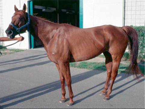
FIG. 35.22 Equine motor neuron disease in a 13-year-old Quarter Horse mare whose feet are placed close together under the body in a characteristic “horse on a ball” stance. Also note the generalized muscle atrophy, extended tail, and flared nostrils (indicative of severe exercise intolerance).
nasal flaring. Typically, at the walk, protraction phases appear somewhat short and hypometric. Horses with EMND do not have signs of ataxia such as interference between limbs or circumduction of the pelvic limbs.
In most horses with EMND, the head is carried below the shoulders, usually reflecting obvious wasting of the neck muscles.2 The tail head is elevated in many cases because of atrophy and contracture of the dorsal coccygeal muscles.2 Widespread muscle atrophy, if not present at presentation, develops quickly (days to weeks) and is most obvious in the quadriceps, triceps, and gluteal areas.
Despite the frequent occurrence of pathologic changes in the motor nuclei of cranial nerves V and XII, noticeable atrophy of the masseters or tongue is rarely reported.21-23
In approximately 40% of cases, fundoscopic examination reveals a horizontal band of pigment above the optic disk at the tapetal-nontapetal junction.2,17 The pigment may be brownblack to yellow-brown and has a reticulated or mosaic appearance. Surprisingly, problems with vision have not been reported even in EMND horses with severe pigment retinopathy.17
■ Laboratory Findings Typical cases have below-range plasma/serum α-tocopherol concentrations. However, as a stand-alone test, α-tocopherol concentration is quite nonspecific20: Normal pasturemates of horses with experimentally induced18 or naturally occurring13 EMND often also have low α-tocopherol concentrations. In healthy pasture-fed horses, α-tocopherol concentrations are variable during the day and often drop transiently below the normal range.24 For this reason, it is recommended that plasma (or serum) from serial blood collections throughout the day be combined for the purpose of α-tocopherol assay.24 Although CSF α-tocopherol concentrations in horses with EMND have not been published, they are presumed to be lower than the published normal range of 4.1 to 13.5 ng/mL.25,26
Mild to moderate increases in plasma or serum CK and AST activity are found during the initial rapid progression of clinical signs, but muscle enzyme activities may be normal in chronic stable cases.
Cytologic changes in CSF are not observed in EMND; however, there may be increased total protein concentration and CK activity.3,18 The IgG index is high in many cases, which suggests the presence of intrathecally produced IgG.
■ Ancillary Tests
NEEDLE ELECTROMYOGRAPHY. Needle EMG can be performed in a standing or recumbent, anesthetized horse.
Horses with EMND often have abnormal EMG findings, including prolonged insertional activity and pathologic spontaneous activity such as fibrillation potentials and trains of positive sharp waves.20 Signs of denervation are found in facial muscles, as well as appendicular and epaxial muscles.3 Standing EMG is preferred in order to avoid anesthesia in weak, compromised animals; however, interpretation of these studies is difficult because of movement artifacts associated with the reluctance of affected horses to stand in one place. The use of sedation and caudal epidural anesthesia (lidocaine, 0.2 mg/ kg) was reported to facilitate the procedure in a horse with EMND.21 Techniques for quantitative analysis of motor action potentials have been published, and results are abnormal in horses with EMND.20,27,28 Regardless of the EMG technique used, abnormal results are nonspecific as to cause; they suggest only denervation or myopathy.20GLUCOSE ABSORPTION/METABOLISM TESTING. Oral glucose tolerance tests in horses with EMND yielded abnormal results in approximately 50% of reported cases, whereas results of oral xylose absorption tests were normal or slightly low.2,13,28 It appears that increased glucose metabolism, possibly in association with increased glucose sensitivity, rather than reduced intestinal absorption, is the cause of abnormal oral glucose tolerance in horses with EMND. Euglycemic clamp results from one horse showed a 5.4-fold increase in insulin sensitivity in comparison with control horses.29
NERVE BIOPSY. Biopsy of the ventral branch of the spinal accessory nerve allows reliable antemortem diagnosis of EMND, at least in subacute and chronic cases.3,30 A 5-cm section of the nerve is excised as it courses over and into the medial belly of the sternocephalicus muscle. The procedure can be performed when an anesthetized horse is either standing or recumbent and does not induce visible muscle atrophy at the surgical site.
A positive histologic result is evidence of mild to severe wallerian degeneration of axons and Schwann cell proliferation. Chronic cases are marked by loss of myelinated fibers, presence of compact Bungner bands, and increased endoneurial collagen. The positive predictive value of nerve histologic findings for EMNd was 77.4%, and the negative predictive value was 90%.30 False-negative results appeared to be most likely to occur in acute cases (90%) but relatively low specificity.3 In chronic EMND cases, histologic study of nerve biopsy samples may have higher sensitivity for diagnosis than that of muscle samples.3■ Diagnosis Antemortem diagnosis of EMND is suspected on the basis of typical clinical signs, patient's age of 2 years or older, and diet low in green forage and high in carbohydrates. Needle EMG findings of denervation, especially in the deep limb muscles, are technically difficult to obtain because of movement artifact in weak horses, but they do support the diagnosis, as do mild to moderate increases in plasma CK and AST activity and low plasma α-tocopherol concentration. Evidence of denervation in muscle or nerve biopsies in the context of the preceding clinical and laboratory findings is tantamount to a definitive diagnosis of EMND. In occasional cases of insidiously progressive EMND, especially in nonendemic areas, diagnosis is made only post mortem.
■ Necropsy Findings Although horses with EMND exhibit obvious weight loss, fat deposits noted post mortem are usually within normal limits.1,4,20,31 Acute cases are characterized by widespread degeneration and loss of somatic motor neurons in the ventral horns of the spinal cord, accompanied by degenerative axonal changes in the ventral roots and peripheral nerves. All brainstem cranial nerve somatic motor nuclei, except those of cranial nerves III, IV, and VI, are variably involved. Most affected neurons are swollen, markedly chromatolytic, and diffusely argyrophilic; severely affected neurons are shrunken or vacuolated. Horses with cases of disease burnout (described later) demonstrate glial scars consisting of astrocytes and lipofuscin-laden microglia. Additional deposits of ceroid lipofuscin are found in the retinal epithelium and occasionally in the liver and intestine. Relatively minor neurodegenerative changes may be found in dorsal root ganglia. In contrast with grass sickness, minimal or no lesions are present in the autonomic nervous system. In skeletal muscles, angular atrophy affects all myofiber types, with some selectivity for type I fibers. Atrophied fibers are intermingled among normal fibers and fascicles. Involvement of the deeper muscles of the limbs may be grossly evident as pale discoloration. Histologic signs of EMND concurrent with NAD/EDM were identified in three young horses.15 A genetic basis for these cases is suspected.
■ Treatment The only treatment recommended for horses with EMND is vitamin E. This can be supplied in good-quality grass and alfalfa hay or by vitamin E supplementation (at 10 to 20 IU/kg/day). The water-dispersed form of natural vitamin E (RRR-α-tocopherol) is five to six times more bioavailable than synthetic vitamin E (all-rac-α-tocopherol acetate or d,l-α-tocopherol), and its administration results in rapid rises in plasma and CSF concentrations of α-tocopherol. After an initial period of at least 2 weeks' supplementation with water- dispersed RRR-α-tocopherol designed to raise plasma and CSF α-tocopherol concentrations into the normal range, treatment can be transitioned to less expensive synthetic RRR-α-tocopherol acetate for subsequent maintenance therapy.26
The frequent clinical observation that many horses with EMND have increased appetite may be explained by an estimate that daily caloric requirement of horses could be doubled.20 Thus as part of the convalescent program of surviving horses, high-quality pasture, hay, and concentrate are recommended in high quantities.
■ Clinical Course Within 6 weeks after onset of signs, approximately 40% of horses progressively deteriorate and must be euthanized; a similar number undergo marked improvement in clinical signs (usually after relocation to another premise or administration of antioxidants).2 Some of these “recovered” horses look normal but may show relapse under the pressure of intensive training or competition.1 In the remaining 20%, the disease appears to “burn out”; such horses survive with permanent and obvious muscle wasting and emaciation.