Esophageal diverticula
An esophageal diverticulum is a pouch-like dilation of the esophageal wall. Diverticula may be congenital or acquired and are of two types, pulsion or traction diverticula. Pulsion diverticula represent an outpouching of the esophageal mucosa through a defect in the muscular and adventitial layers of the esophagus.37,45 It is usually acquired secondary to increased intraluminal pressure and deep esophageal inflammation.
Predisposing disorders include esophagitis, esophageal stricture or foreign body, or hiatal hernia.10 Traction diverticula are generally acquired as a result of inflammation in the thoracic cavity in close proximity to the esophagus. This leads to the formation of fibrous tissue, which then contracts pulling the esophageal wall outwards.10 This type of diverticulum affects all four layers of the esophageal wall and most commonly occurs secondary to a foreign body perforation.10 Both types have been reported in the dog while only one case of an esophageal diverticulum has been reported in a cat.45-47 Clinical signs of small diverticula may not be apparent. Large diverticula allow food trapping, leading to postprandial dyspnea, regurgitation, odynophagia, and anorexia.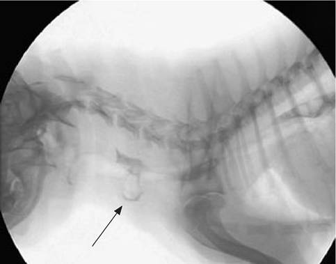
Figure 3.10:
Esophageal diverticulum. Contrast Videofluoroscopic image of a 1-year-old castrated male Shih Tzu with a small esophageal diverticulum. The study was performed 1 month after endoscopic removal of a bone foreign body as a follow-up. The dog was asymptomatic for the diverticulum.
The results of a minimum diagnostic database are usually unremarkable. Survey thoracic radiographs usually reveal an air-, fluid-, or food-filled outpouching of the esophagus. Small diverticula must be differentiated from normal esophageal redundancy that occurs in some animals, especially Chinese Shar Peis, young animals, and brachycephalic breeds.10,17 Contrast radiography can help delineate the diverticulum and differentiate it from other soft tissue structures in the thorax (Figure 3.10). Endoscopy can be used to confirm the diagnosis and will allow identification of any ulceration or scarring.16 Small diverticula may be managed with a bland, soft diet fed with the patient in an upright position, and generally carry a favorable prognosis. Large diverticula require surgical excision, justifying a less favorable prognosis, but successful surgical management has been reported.45,46
a
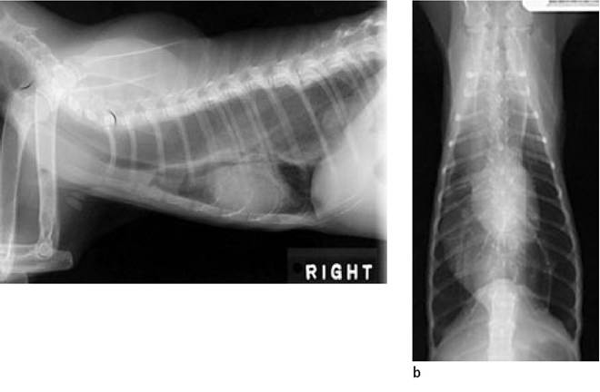
Figure 3.11 a,b:
Megaesophagus. Lateral and VD radiographs of a 2- year-old spayed female DSH with chronic regurgitation and weight loss. A complete work-up did not identify a cause for the megaesophagus.
3.3.7