Esophageal strictures
Esophageal strictures occur secondary to damage of the submucosal and muscular layers of the esophagus. Damage to these layers stimulates the production of fibrous connective tissue.38 Damage to the mucosa alone generally does not result in stricture formation.
Benign esophageal strictures have been reported in the dog and cat, and are usually secondary to damage from foreign bodies, ingestion of caustic substances, or most commonly, secondary to GER during an anesthetic pro- cedure.18,23,32,39,40 Esophageal strictures have also been reported in cats secondary to oral doxycycline administration.41The clinical signs of patients with an esophageal stricture include regurgitation, ptyalism, dysphagia, odynophagia, inappetence, and weight loss.18,23,38 The physical examination is usually unremarkable, unless aspiration pneumonia has developed, in which case fever and coughing may be seen.
The results of a minimum diagnostic database, including thoracic radiographs, are often normal. Occasionally, a dilated segment of the esophagus may be seen cranial to the stricture on survey thoracic radiographs (Figure 3.6). A definitive diagnosis is made via contrast videofluoroscopy (Figure 3.7) and/or esophagoscopy (Figure 3.8).23,40 Esophagoscopy allows the clinician to fully evaluate the esophageal mucosa to aid in treatment decisions.
Currently, the treatment of choice for esophageal strictures is balloon dilation (Figure 3.9).12,18,23,39,40 This is most commonly done under endoscopic guidance, but may also be done under fluoroscopic guidance.18,23,39,40 Strictures can vary in number, location, and severity.18,23,39,40 The balloon dilation procedure typically must be repeated an average of 2-4 times per animal.18,23,39,40 The interval between procedures also varies, but is generally 4-7 days.18,23 Complications associated with balloon dilation include esophageal perforation and esophageal tears.18,23,40,42 Placement of a gastrostomy tube has been recommended in cases where the esophageal damage is severe, but this may not be necessary in all cases.17,18,39 Therapy for esophagitis should be instituted after the balloon dilation procedure. The use of corticosteroids has been advocated to prevent reformation of the stricture.10 However, the efficacy of such corticosteroid therapy has not been proven in a clinical setting, although it has been reported as an adjunct therapy by some authors.18,23 Bougienage is also a reported technique for dilation of esophageal strictures, but is not as commonly used as balloon catheters are more readily available and used quite successfully.12 Surgical removal of strictures and other surgical techniques have been reported, but surgery is generally reserved for refractory cases or those in which perforation has occurred.43,44 In clinical studies, 70-88% of patients with esophageal strictures had a good to excellent outcome with balloon dilation of the esophageal stricture, returning to eating canned or dry dog food with minimal to no regurgita- tion.18,23,39,40
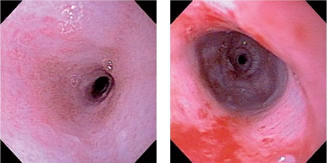
Figure 3.8 (left):
Esophageal stricture.
Endoscopic view of the esophageal stricture of the dog in Figures 3.6 and 3.7. Note the pale to white fibrous tissue. The diameter of the stricture was approximately 3 mm.Figure 3.9 (right):
Esophageal stricture after balloon dilation. Endoscopic view of the esophageal stricture in the same dog shown in Figures 3.6-3.8 after balloon dilation. The diameter of the strictured area is now approximately 15 mm. Note some hemorrhage and mucosal tearing, which can often be observed after balloon dilation. The dog underwent a total of three balloon dilation procedures and returned to being able to eat canned dog food without regurgitation.
3.3.6