False Pregnancy (Pseudopregnancy) and Hydrometra
Goats occasionally exhibit prolonged luteal function and absence of estrus even though they are not pregnant. Some of these does were bred and may have conceived at the time that the persistent corpus luteum first developed.
One study summarized by Chemineau et al. (1999) documented that approximately half of the pseudopregnancies identified by ultrasonography at 45 days post breeding were preceded by an embryo that lived long enough to produce PSPB. Other affected does have never even been exposed to a buck.Historical Perspectives and Clinical Signs
Until the availability of real-time ultrasound, it was very difficult to diagnose or study this phenomenon. Results of progesterone analysis (Holdsworth and Davies 1979) and amplitude depth ultrasound were suggestive of pregnancy. The rectal Doppler examination indicated increased blood flow to the uterus. Radiographic evaluation (absence of fetal skeleton) or laparotomy was required to make the diagnosis. Owners noticed that some apparently pregnant does (no return to estrus, with or without a gradual enlargement of the abdomen) delivered fluid, but no placenta or fetus. The lay term “cloud burst” has been applied to the natural termination of a false pregnancy accompanied by discharge of large volumes of fluid. When practitioners examined goats with marked abdominal distension that had not delivered at the calculated due date, hydrometra was sometimes recognized. Surgical drainage of the uterus was soon replaced by treatment with prostaglandin to induce resorption of the corpus luteum followed by voiding of the retained fluid. A few animals that had been included in various reproductive research trials spontaneously developed false pregnancy, thereby providing retrospective hormonal profiles.
Etiology
The etiology of the condition is not yet known. It seems likely that the prevalence is greater in herds trying to delay breeding of some does (for winter milk production) than in those in which all does are bred on the first estrus of the breeding season.
Certain infectious diseases - trypanoso- mosis if controlled with trypanocidal drugs (Llewelyn et al. 1987), toxoplasmosis (Debenedetti et al. 1989), border disease (Loken 1987) - may possibly increase the prevalence of false pregnancy. Involvement of phytoestrogens in the forage has been proposed as contributing to the occurrence of false pregnancies in certain herds (Malher and Ben Younes 1987). Others have suggested that treating short cycles with hCG or GnRH may predispose the goat to development of false pregnancy (East 1983).Prolonged luteal function and hydrometra have been recreated experimentally by immunizing goats against prostaglandin F2 alpha (Taverne et al. 1995; Kornalijnslijper et al. 1997). Fluid accumulation is first detectable between days 29 and 38 of the luteal phase. Prolactin does not appear to be involved in the etiology (Hesselink et al. 1995), although administration of bromocriptine to suppress prolactin will empty the uterus (Taverne et al. 1988). Possible suppression of testosterone secretion has not been investigated in relation to false pregnancy. Testosterone is usually required for luteolysis in the goat (Cooke 1989). The possibility of a genetic predisposition is supported by a study in which the daughters of goats with hydrometra had a 38% frequency of hydrometra, compared with 9% of the daughters of unaffected goats (Hesselink and Elving 1996). In another study, 20% of 125 daughters of 5 sires developed pseudopregnancy, compared with 0% of the 326 daughters of 12 other sires, all in the same herd (Chemineau et al. 1999).
Epidemiology
The prevalence of the condition in privately owned herds is uncertain, but this information should be easy to estimate when pregnancy diagnosis by real-time ultrasound becomes routine. In one French herd studied in 1986, the buck was introduced to 124 adult goats in early April. Ultrasound at day 54 revealed that 92 goats were pregnant, but 21 other goats (17%), all of which had experienced a normal gestation the past year, had hydrometra (Malher and Ben Younes 1987).
In another study in France (Mialot et al. 1991b; Duquesnel et al. 1992), ultrasound was performed more than 5000 times on 68 farms in 1989 and on 71 farms in 1990, and pseudopregnancy was diagnosed in 2.1% and 2.9% of the examinations. In 10% of the herds, the prevalence was more than 5%. The condition was most commonly diagnosed in adult goats after a fall (November to December) kidding without rebreeding or after an out- of-season synchronization program that did not include prostaglandin. A Dutch study found a mean incidence of 9%, with the condition being more common in older goats (Hesselink 1993a), whereas in a more recent Dutch study the incidence was 17% (Van den Brom et al. 2019). In a German study, 143 of 2434 dairy goats were affected, as diagnosed by ultrasound, with an average incidence of 5.78% (Wittek et al. 1997, 1998). From Brazil, 18.6% of 80 goats found non-pregnant by ultrasound after natural mating displayed hydrometra or mucometra (Moraes et al. 2007).Diagnosis
With the development of real-time ultrasound, it has become possible to easily differentiate a false from a true pregnancy and to institute early treatment and monitor the success of therapy (Pieterse and Taverne 1986). When a goat with advanced hydrometra is scanned from either flank, large fluid-filled compartments are seen (Figure 13.4 and Menzies 2019). Thin tissue walls separate the compartments and undulate when the goat moves or succussion of
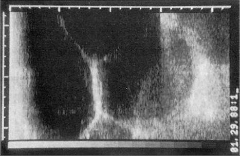
Figure 13.4 Real-time ultrasound image of hydrometra. Source: Courtesy of Dr. M.C. Smith.
the abdomen is performed. These walls represent sections of curved horn overlapping each other rather than internal trabeculae (Hesselink and Taverne 1994). Fetus and caruncles are absent. White flecks may be seen in the fluid and settle down like snow after succussion. Diagnosis of an early stage of hydrometra is more difficult. Before 40 days, when caruncles may not yet be visible by ultrasound, hydrometra is difficult to distinguish from normal pregnancy because it is not always possible to visualize the fetus when scanning an early pregnancy.
In these goats, and in those when the breeding date is unknown, reevaluation several weeks later verifies the existence of hydrometra if fetus or caruncles still cannot be found.Diagnosis can also be confirmed by laparotomy or necropsy. The volume of fluid in the uterus is quite variable, 1-7.2 L in one study (Mialot et al. 1991b), and 0.25-8.3 L in another (Wittek et al. 1997). In the goat that spontaneously voids the fluid (mucoid discharge on tail, decreased abdominal size), the diagnosis is presumptive. Some does that correct spontaneously early in the course of a pseudopregnancy have a bloody discharge. The condition in these animals cannot be distinguished from early embryonic loss (unless the goat was not bred) without a prior ultrasound diagnosis.
Treatment and Prevention
Prostaglandin is the treatment of hydrometra as confirmed by ultrasound or of false pregnancy as suspected in an anestrous doe that cannot possibly have been exposed to a buck. Both natural (5-10 mg prostaglandin F2 alpha) and synthetic (125-250 μg cloprostenol) hormones cause regression of the corpus luteum and emptying of the uterus. Repeated doses of oxytocin (50 IU twice a day for four days) also induce emptying of the uterus, and might be attempted if scanning several days after prostaglandin therapy indicates continued presence of fluid in the uterus. The rare animal that fails to respond to therapy may have segmental aplasia of the uterus or cervix (Webb 1985; Batista et al. 2006). Many goats return to estrus after treatment and can become pregnant within two months of resolution of the hydrometra (Pieterse and Taverne 1986; Duquesnel et al. 1992; Moraes et al. 2007). Some of these develop a new pseudopregnancy the following year. Recurrence shortly after treatment has also been observed (Batista et al. 2001), but the risk of this decreases if the goat receives a second dose of prostaglandin 12 days after the initial discharge of fluid (Hesselink 1993b). Other authors report very poor conception rates after two doses of prostaglandin (only 10 of 19 showed estrus and only two of these became pregnant; Souza et al.
2013).In intensively managed herds that practice out-of-season breeding, ultrasound should be performed on all bred does before drying off (Duquesnel et al. 1992). Those that are pseudopregnant should then remain in the milking herd and be treated with prostaglandin, and their milk production will increase. Likewise, goats to be synchronized out of season should be examined by ultrasound first, so that affected does that would be expected to have poor fertility at the induced estrus can be treated with prostaglandin and synchronization can be delayed until resolution of the hydrometra is achieved.
Preventing hydrometra in pet goats that will not be bred, or even just preventing estrus and associated noisy behavior in these pets, can be achieved by ovariectomy. The surgery is done under general anesthesia (Tibary et al. 2017) or heavy sedation and a local block. The animal is placed in dorsal recumbency and each ovary is removed after ligation of the pedicle by laparoscopy or via a ventral midline incision just cranial to the udder. Temporary elevation of the rear end of the doe by 25 or 30° simplifies location of the ovaries, but prolonged weight of the rumen on the diaphragm is not desirable (Wolfe and Baird 1997). Exposure of the contralateral ovary is difficult from a flank incision, but this approach is preferred for unilateral ovariectomy, as for an ovarian tumor. Laparoscopic ovariohysterectomy has also been described, usually for pet goats (Daniel et al. 2019).
A case report of mucometra in a mature West African pygmy goat in Germany documented persistent accumulation of fluid in the uterus in the absence of a functional corpus luteum. This doe did not respond to prostaglandin, but was successfully managed by ovariohysterectomy performed from a ventral midline incision caudal to the umbilicus (Trasch et al. 2002).