Pregnancy Diagnosis
An excellent general discussion of pregnancy diagnosis has been provided by Williams (1986a) and updated to include B-mode ultrasonography by Matsas (2007). Table 13.1 outlines many different methods of pregnancy diagnosis and the sensitivity and specificity of the tests for which published information is available from early studies.
In fact, the apparent accuracy of most of these methods varies from farm to farm, according to the prevalence of infectious abortions, nutritional deficiencies causing early embryonic loss, and hydrometra. Experience of the examiner also affects accuracy.Table 13.1 Pregnancy diagnosis techniques for goats.
Accuracy (%)
Stage of
| Technique | pregnancy | Pregnant | Non-pregnant | Potential errors | Comment |
| Estrus detection | 18-24dor until term | Varies with detection method, may stand when pregnant | End of breeding season, buck not “acceptable” | Observe tail wag; use buck, testosterone- treated doe, intersex | |
| Serum progesterone (Thibier et al. 1982) | 19-24 d | 87 | 97 | Irregular cycle, false pregnancy, early abortion | Best used as nonpregnancy test |
| Milk progesterone (Pennington et al. 1982; Dionysius 1991) | 19-24 d | Varies with each 71-98 | kit/antiserum 80-100 | False negative unless antiserum detects five-pregnanedione; false positive may occur during estrus (Holdsworth and Davies 1979; Bretzlaff et al. 1989) | Concentrations more variable and higher than serum progesterone (Murray and Newstead 1988) |
| Urinary or serum estrone sulfate (Williams 1986a; Sardjana et al. 1988a) | After 50-60 d, some possible by 30 d | About 100 | About 100 | Testing milk unreliable unless in full production | Negative in false pregnancy; secretion decreases rapidly if abortion |
| Pregnancy-specific protein B (PSPB) | After 25 d | High | High | False positive if recent fetal death | |
| Ballottement | After 100 d | Goat fat or tense | Swing entire abdomen or push in with fist | ||
| Radiography (Barker and Cawley 1967) | After 70-90 d | About 100 | About 100 | Diagnosing by uterine enlargement rather than skeleton | Hold off feed; can count fetuses |
| Amplitude depth ultrasound | 60 (80)-120d | False pregnant does positive; late-pregnant does may be negative | Clip hair right side above udder | ||
| Doppler (Lindahl 1969; Fraser et al. 1971; Ott et al. 1981) | >35 d (rectal) | 95 | 25-75 | Increased blood flow in false pregnancy | Fetal heart two times maternal rate |
| Real-time ultrasound (Buckrell 1988; Baronet and Vaillancourt 1989) | >25 d (rectal) >35 d (abdominal) | About 100 | 87 | False pregnancy (fluid, no fetus or caruncles); late pregnancy more difficult to visualize | Transabdominal test earlier in older does |
Diagnostic Techniques
The most accurate tests are those that measure or detect something that is only produced by a viable fetus and that is always present when the goat has reached a certain stage of pregnancy.
Hormonal Assays
Hormones that have been used as predictors of pregnancy include progesterone, estrone sulfate, placental lactogen, and pregnancy-associated glycoproteins.
Progesterone, because it is produced only by the ovaries and not by the placenta, is best used as a non-pregnancy test (Dionysius 1991; Gonzalez et al. 2004), although the probability that a goat with elevated progesterone is actually pregnant is increased during the anestrous season (Fleming et al. 1990). Goats with low progesterone in serum or milk five days or more after breeding are judged to be not pregnant. The exact cutoff varies with test method, and most commercial kits have not been validated for goats. Goats with elevated progesterone at 21 days after breeding could be pregnant or could have a different cycle length or be pseudopregnant. One study found that using milk progesterone kits at 20 days after breeding was no more accurate for pregnancy diagnosis than using a buck to detect return to estrus at 18-24 days (Engeland et al. 1997a).
Estrogens are produced by both the ovaries and placenta, but estrone sulfate is thought to be a product of steroid conjugation by fetal or placental tissues (Refsal et al. 1991). Estrone sulfate in whole milk or whey can be used for pregnancy diagnosis (Chaplin and Holdsworth 1982; McArthur and Geary 1986), and total urinary estrogen has also been used by a commercial laboratory (B.E.T. Reproductive Laboratories, Lexington, KY, USA), beginning at about 50 days of gestation. Occasional false-negative results in the B.E.T. test were reported in pregnant does well beyond 50 days of gestation, and the laboratory no longer offers the test. Currently US producers are using urine test strips that detect estrone sulfate and are marketed by Emlab Genetics (Riverside, IL, USA) under the trade name P-Test™ for pregnancy diagnosis of goats after 50 days.
Placental lactogen is a hormone produced only by the placenta (Hayden et al. 1980; Byatt et al.
1992). Concentrations in plasma or milk can be assayed after 60 days to diagnose pregnancy. The hormone concentration is also correlated with the total weight of kids produced (Sardjana et al. 1988b). Commercial assays for the caprine hormone are not currently available. Several pregnancy-associated glycoproteins (PAGs) are produced by chorionic binucleate cells from the placenta and have been detected in plasma as early as 21 days of gestation by radioimmunoassay (Gonzalez et al. 1999, 2000, 2004) and in milk by 32 days (Gonzalez et al. 2001). No PAG is detected if a pseudopregnancy develops early, and sequential tests show a decrease in PAG concentration when one fetus in a litter dies (Zarrouk et al. 1999a). An enzyme-linked immunosorbent assay (ELISA) for PAG has been positive in plasma of Boer goats by 28 days (Shahin et al. 2013) and an ELISA for PAG in goat milk (Alertys®, available from IDEXX in many countries) is used to detect pregnancy as early as 28 days after breeding. Currently, an ELISA test for the closely related pregnancy-specific protein B (PSPB) is available from Bio Tracking (Moscow, ID, USA). This is a placental hormone that has been used as an indicator of pregnancy in goats (Humblot et al. 1990), cattle, and sheep (Ruder et al. 1988). Detection of pregnancy is possible using PSPB by 26-30 days in goats. It is not known how long the test remains positive in a pseudopregnant goat when an embryo reached the stage of hormone production before dying. It is also not yet known if concentrations of placental hormones vary according to previous selection pressures on a given breed for milk or meat production.Amplitude Depth Ultrasound
The first ultrasound machines on the market detected the interface between a fluid-filled organ (the uterus) and the abdominal viscera. These machines are still commonly used for pregnancy diagnosis in swine and are relatively inexpensive. They are very unsatisfactory for use in dairy goats because of low accuracy.
Non-pregnant animals with fluid in the uterus (false pregnancy) are classified as pregnant, thus escaping detection. Also, a false-negative diagnosis is delivered if the ultrasound beam encounters fetus rather than large pockets of fluid.Doppler
When the limitations of the amplitude depth machines were recognized, interest switched to Doppler techniques. Doppler machines permit the operator to hear the blood flow in maternal and fetal vessels. When used rectally (after 35 days), the external iliac vessels are first located by directing the probe laterally within the pelvic canal. When the maternal pulse rate is ascertained, the probe is advanced and directed ventrad to search for a fetal heartbeat or blood flow in the umbilical artery, at approximately twice the maternal rate. The fetal heart rate is negatively correlated with fetal age (Fraser et al. 1971). Loud whooshing sounds at the maternal rate but ventrally located indicate increased blood flow to the uterus; this should not be taken as conclusive evidence of pregnancy, because it is also heard in goats with hydrometra and might be detected in does that have recently kidded (Wani et al. 1998). After 45 days, detection of the fetal heartbeat can be accomplished transabdominally. This technique may also be used to assess fetal viability in goats with pregnancy toxemia. The equipment is more affordable than many real-time ultrasound machines, but the method is no longer in vogue, as it provides less information about fetal numbers and stage of pregnancy.
Real-Time Ultrasound
A real-time ultrasound machine eliminates worries about false positives if the diagnosis is always based on visualizing either fetus or caruncles (Haibel 1990b). False negatives are still possible if later, unrecorded matings have occurred. Administering prostaglandin, then, should generally be delayed until the doe has been rechecked, unless later breedings are absolutely impossible. Accuracy also improves with operator experience (Bretzlaff et al.
1993). Sector scanners generally have superior resolution to linear array systems and make counting fetuses easier, but the equipment is more expensive. Portable generators permit the use of real-time technology even under range conditions. With some machines, it is important not to disconnect the probe or let the generator run out of fuel while the power is on. Smaller, battery-operated units are now available.Transrectal and Transvaginal Scanning
Equine and bovine linear array units with 5 MHz transducers can be used rectally for early diagnosis (20-30 days) or transabdominally later in pregnancy (day 35 and beyond) (Buckrell 1988). Withholding food and water for 12 hours improves visualization of the uterus, but is usually not necessary and might lead to pregnancy toxemia (Bretzlaff et al. 1993). Taping an insemination rod or a longitudinally split section of plastic pipe to the head and cord of the transducer simplifies manipulations within the rectum. Although more expensive, a human prostate probe also works well. A small amount of lubricant is introduced into the rectum before inserting the lubricated probe. The pregnant uterus is usually located anterior and ventral to the urinary bladder. Elevating the abdomen manually or by placing the goat across a hay bale makes transrec- tal diagnosis reliable even after 35 days (Baronet and Vaillancourt 1989). The earliest that the embryo is reliably detected by transrectal exam is 25 or 26 days after breeding (Gonzalez et al. 2004). In another small study, at least one embryo of each pregnant doe had a detectable heartbeat by day 23 (Martinez et al. 1998), while transrectal evaluation of Boer does using a 7.5 MHz probe disclosed a fetal heartbeat by day 20-26 (Padilla-Rivas et al. 2005). Transvaginal scanning has been performed less frequently, but with an appropriate 7.5 MHz annular sector probe pregnancies can best be identified in the standing doe at five to six weeks after mating, though fetal counting is not accurate (Koker et al. 2012).
Transabdominal Scanning
For transabdominal examination, does are generally scanned standing, from the right side, and clipping hair immediately lateral and dorsal to the udder improves contact between transducer and skin. Otherwise (show does, for instance), much contact gel (such as methyl-cellulose rectal lubricant), vegetable oil, alcohol, or a mixture of alcohol and contact gel is needed to wet the hair. Examination in dorsal recumbency (in a padded trough) has been used in early research trials (Tainturier et al. 1983), but stresses the doe to an unnecessary degree.
Early in pregnancy (30-45 days), the ultrasound beam should be directed toward the pelvic inlet. Subsequently, the uterus is usually positioned against the right ventral abdominal wall (Haibel 1986a). Late in pregnancy, the 5 MHz transducer may not penetrate as far as the fetus (caruncles are visible, but not well outlined by fluid), or part of a fetus may fill the screen and be overlooked unless bones or beating fetal heart (non-echogenic because of the blood in it) is noted. A 3.5 MHz transducer is preferred, but not imperative in late gestation.
A sector scanner is preferred for determining fetal numbers. The best time for counting fetuses is between 40 and 70 days of gestation (Lavoir and Taverne 1989; Hesselink and Taverne 1994). The number of triplet litters is underestimated before 50 days (Dawson et al. 1994).
In early pregnancy, numerous fluid-filled cross-sections of uterus are visualized. This phenomenon can be explained by circumferential folds of endometrium that protrude into the lumen (Kahn 2004). If a fetus is clearly visualized, its heartbeat can be detected by 25-30 days. It has been stated that caruncles are routinely found by day 30 (Buckrell et al. 1986; Baronet and Vaillancourt 1989) or day 35 (Doize et al. 1997), but others feel that day 40 or 50 is a safer date if a goat is to be called open because caruncles are not visualized. This allows for variations in resolution of the machine and position of the uterus. The caruncles appear to be doughnut- or C-shaped by 45-50 days (Haibel 1986a) and are outlined by fluid (Figure 13.2). There are approximately 120-125 caruncles in a goat's uterus, arranged in four rows in each horn (Lyngset 1968c).
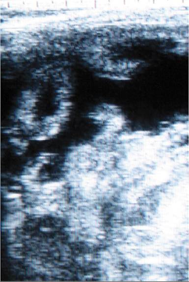
Figure 13.2 Transabdominal ultrasound of a pregnant doe showing a doughnut-shaped caruncle clearly outlined by black uterine fluid. Source: Courtesy of Dr. M.C. Smith.
Fetal Age Determination
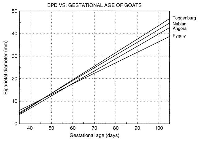
With practice, the size of the fetus can be used to estimate stage of gestation. In Saanens and Alpines, the crown-rump length of the fetus is approximately 40 mm at 45 days, 100 mm at 60 days, and 250 mm at 90 days (Mialot et al. 1991a). Fetal age in dairy goats also can be estimated by measuring the width of the fetal head during real-time examination (Figure 13.3). Between 40 and 100 days’ gestation, the biparietal diameter (measured from a symmetrical, maximum-width image) correlates very closely with age (Haibel 1988; Haibel et al. 1989). A similar correlation has been reported for second-trimester Pygmy goat fetuses (Reichle and Haibel 1991). Later in pregnancy, there is too much difference in size of the fetuses and it is no longer possible to date pregnancies accurately enough to permit safe induction of parturition based on the estimated fetal age.
Caruncular size is also quite variable and not very useful for estimating age, except early in pregnancy. The caruncles increase in size until a maximum is reached at about 90 days of gestation. Before this time the diameter of the caruncles in the uterine body as imaged transrectally can be used as a predictor of stage of pregnancy, but less precision might be expected from transabdominal measurements of caruncles in random parts of the uterus (Doize et al. 1997).
Fetal Sex Determination
Real-time ultrasound permits visualization of the genital tubercle of the developing fetus, an embryologic structure that is initially located between the hindlimbs, but moves forward to near the umbilicus in males and back toward the tail in females. Transrectal scanning using an adaptor to manipulate the probe gives excellent results for single kids between 55 and 75 days of gestation, but it is not always possible to visualize the genital tubercle or external genitalia in twins or triplets (Santos et al. 2007). Amer (2010) reported an accuracy of transrectal fetal sexing of 93% at 40-60 days and 82% at 61-70 days of pregnancy, whereas transabdominal fetal sexing at 90-109 days was 57% successful. Some authors prefer to use the presence of two thin teats between the hind legs and absence of a triangular- appearing scrotum in this region to diagnose a female fetus, because the tail may obscure the female genital tubercle (Burstel et al. 2002). The penis/prepuce is located immediately behind the non-echogenic, circular umbilical cord.
Figure 13.3 Correlation of biparietal diameter (BPD) and fetal age. Source: Haibel 1990b / With permission of Elsevier.
Other Techniques
Where Doppler or real-time ultrasound is not available and hormonal assays are prohibitively expensive, other, less sophisticated techniques may be used. For instance, rapidfield laparotomy has been used to diagnose pregnancy in goats. Hulet’s rod for recto-abdominal palpation has also been used in goats, but American practitioners will probably find this technique less than satisfactory. Goats do not lie quietly on their backs for this examination, as is typical for sheep to do, and rectal lacerations and abortions may occur (Ott et al. 1981). In a relaxed doe in late pregnancy, it may be possible to palpate or ballot a fetus in the right ventral abdomen or to observe fetal movements.
A bimanual technique has been developed for detecting early pregnancy in goats and sheep (Kutty 1999). After overnight fasting of the doe, the gloved index finger of the left hand is introduced into the rectum of the standing animal and used to evacuate fecal pellets and express the bladder. Then the examiner’s right hand is pushed vertically in the caudal abdomen, to displace intestines forward and the uterus caudally into the pelvic canal, where it can be palpated by the left index finger. Ovaries could also be trapped between the two index fingers and palpated. A fluid distension of the uterine horns is reported to be palpable at 30 and 45 days, but cannot be distinguished from hydrometra. By 90 days, retroversion of the uterus is no longer possible, but placentomes and softening of the cervix are palpable.
For the answer to the in-passing “do you think she’s pregnant?” question, the sacrotuberous ligament and other ligaments near the base of the tail should be evaluated for softening. This is easiest when comparison with a nonpregnant doe is possible. In practiced hands and in the second half of pregnancy, this is a free version of the urinary estrone sulfate test. Increased elasticity of the skin around the vulva and pinbones has also been used by laypeople as an indicator of pregnancy (DeArmond 1990). Development of an udder, even in yearlings, should not be taken as proof of pregnancy (see inappropriate lactation syndrome, discussed in Chapter 14).
Use of Test Results
Pregnancy diagnosis is performed for a variety of reasons, depending on the management of the goat herd. It may be desirable to cull non-pregnant does after the end of a breeding season or in times of feed shortage. Verifying pregnancy before drying off a dairy doe is becoming routine. It is also very important to monitor the success of out-ofseason breeding programs, because of extremely variable results and the relatively high risk of hydrometra in these does.
What one does with the results of a pregnancy test depends on one’s faith in the test results. If a goat is thought to be pregnant, usually nothing is done unless mismating has occurred or nutritional resources are so short that only well-grown, pregnant goats can be fed adequately to maintain pregnancy. Animals carrying multiple fetuses might be fed more than those with singles, to decrease the risks of pregnancy toxemia in one group and obesity in the other (see Chapter 19). When the diagnosis is that of nonpregnancy, the owner may choose to restrict feed or continue milking the goat, or stop staying up nights, waiting for it to kid. If rebreeding is desired, it is very likely that the use of prostaglandins to induce estrus will come under discussion. At this point, the practitioner must be very certain of his or her diagnosis of “open.” Goats with negative progesterone test results 21 days after synchronized breeding could be given a new vaginal sponge or ear implant (de Montigny 1988), but even these techniques frequently include prostaglandin in the synchronization protocol.