FECAL INCONTINENCE
Fecal incontinence denotes uncontrolled release of rectal contents. Although it is not a common disorder in dogs and only rarely occurs in cats, the ramifications of this problem for a household pet and its owner are highly significant.
Pets with a fecal incontinence problem that cannot be reasonably controlled are often euthanized because of the impracticality of maintaining the animal on a long-term basis in terms of the problems associated with fecal soiling.There are many potential causes of fecal incontinence (Box 1-12). Most incontinence disorders can be classified as neurogenic or nonneurogenic. Causes include anatomic disruption of the anal sphincters or pudendal nerve trauma resulting from surgery (e.g., perineal hernia repair, perianal fistula repair, anal sac removal, tumor removal), obstetric trauma or other injuries (e.g., lacerations, bite wound trauma with subsequent ascending bacterial neuritis as may occur from a cat fight injury), and various non—surgery-related neurologic problems. Neurologic problems may include peripheral neuropathies, cauda equina syndrome, and congenital defects of the caudal vertebral column and spinal cord (e.g., sacrocaudal agenesis of Manx cats). Incontinence may be related to aging in some patients. Also, any disease that causes rapid transit of large volumes of diarrhea (e.g., severe enteritis) may produce transient fecal incontinence in patients with healthy continence mechanisms.
The mechanisms of anal continence are complex, and a detailed description is beyond the scope of this discussion. In the dog and cat the internal and external anal sphincter muscles and the puborectalis muscle (the caudal portion of the levator ani muscle) play major roles in maintaining continence. The most important muscle in maintaining the sphincter component of the continence mechanism may be the puborectalis muscle.
The external anal sphincter is innervated by the caudal rectal branch of the pudendal nerve, originating from the sacral spinal cord segments (S1 to S3).Bilateral transection of the pudendal nerve or sacral cord lesions will result in fecal incontinence. However, unilateral transection usually does not lead to major dysfunction because the remainder of the innervated external anal sphincter muscle can compensate for the denervation. Surgical procedures involving full-thickness circumferential resection at the anorectal area always carry the risk of ensuing fecal incontinence. The internal anal sphincter is innervated by branches of the pelvic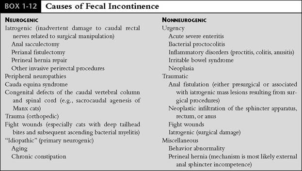
nerve (afferent and efferent) and pudendal and hypogastric nerves (efferent).
The colon also plays an important role in helping to maintain fecal continence through its reservoir function. Reflex activity in the colon appears to allow the external anal sphincter to retain fecal material while the internal anal sphincter relaxes, thereby allowing the colon to dilate and accommodate increases in fecal mass. Simultaneously there is a brief (several minutes) decrease in propulsive contractions in the colon, which also helps facilitate the accommodation process. The colon continues to readapt with subsequent peristaltic delivery of fecal material until a time when defecation is appropriate. If the colon is presented with large volumes of watery fecal material in a short period of time, as may occur in patients with severe viral or bacterial enteritis, this reservoir function can become overwhelmed and transient incontinence (urge incontinence) may result. Urge incontinence can also be associated with moderate to severe proctitis or colitis, in which the patient experiences significant discomfort (perhaps a “burning” sensation) with a resultant sense of urgency to defecate and overriding of the continence mechanism.
The internal and external anal sphincter muscles and the puborectalis muscle are primarily responsible for maintaining a high-pressure zone in the terminal rectum that maintains continence at rest.
Studies have shown that the internal anal sphincter contributes 50% to 80% of the resting tone in the high-pressure zone. The primary function of the external anal sphincter is to actively contract over short periods of time to resist the action of peristaltic waves.Diagnosis
Important factors in diagnosis include obtaining a detailed history so that any potential causative factors (e.g., trauma, difficult whelping, history of significant constipation problems) can be elucidated, physical examination (including neurologic assessment), and completion of any indicated diagnostic tests.
The signalment is very important in evaluating a patient with fecal incontinence. Manx and other tailless cats and Old English sheepdogs, bulldogs, and Boston terriers may be affected with an agenesis of the sacrocaudal vertebrae and spinal cord. The neurologic deficit is present from birth but is often first noted at weaning. Clinical signs include both urinary and fecal soiling and irritation around the perineal and abdominal skin. Occasionally there is complete paralysis of the pelvic limbs as well. Aging patients with gradual onset of incontinence are most likely to have some type of neurologic problem.
Once the existence of incontinence has been established, the clinical evaluation begins with an assessment of the frequency, severity, and circumstances surrounding the incontinent episodes. How acute are the symptoms? Are the episodes associated with urgency, or is there no prior warning? The owner should be asked whether or not the dog still assumes an appropriate posture for defecation, and, if so, does this take place at an appropriate time and place? Some dogs with mild incontinence still do this on a fairly routine basis but on occasion will inappropriately release stool when asleep, during periods of relaxation, or while on a walk. These episodes may occur in response to increased rectal pressure related to the presence of stool that overrides a now compromised continence mechanism.
Excitement may also cause spontaneous evacuation of stool. In some patients incontinence episodes become much more frequent (i.e., major incontinence versus partial), and this suggests a severe anorectal sensory disorder. Unconscious anal dribbling of small amounts of fluid and residue may become common, especially during periods of increased abdominal or rectal pressure (e.g., associated with coughing, excitement, or exercise).
The owner should also be asked if the patient can urinate normally. Because micturition relies on nerve pathways similar to those involved in fecal continence, abnormalities involving both functions suggest that the fecal incontinence is of neurologic origin.
If the fecal incontinence has been a very recent development, questions regarding trauma are asked. Lumbosacral and sacrocaudal fracture, subluxation, and luxation can cause fecal incontinence (distended bladder and atonic tail often result as well). In cats these injuries are commonly associated with getting their tails caught by something. Cats with bite wounds around the tailhead may develop abscessation and ascending bacterial neuritis and meningomyelitis of the caudal spinal cord. Any history of anorectal surgery is reviewed. Generally symptoms develop and are reported shortly after any surgery in which nerve damage occurs. Any incidence of significant straining episodes should also be discussed. Severe proctitis, for example, may cause so much irritation that urge incontinence occurs.
In my experience one of the most common presentations for the complaint of fecal incontinence is an aging dog with no obvious predisposing history of major trauma, anorectal disease, or neurologic disease. Careful studies using electromyography and other neurophysiologic techniques in human patients with “idiopathic” incontinence have identified striated muscle denervation damage in the majority. Detailed studies have not been done in animals, but it is suspected that a similar mechanism exists.
Physical Examination
The most important aspects of the physical examination in a patient with fecal incontinence include close perianal inspection and digital examination of the anorectum. The skin in the perianal and perineal regions may show evidence of irritation from fecal or urinary soiling. Perianal fistulas, which may be accompanied by mild leakage of residue from the rectum, are readily identified on visual inspection. Rectal examination may reveal evidence of proctitis (e.g., increased sensitivity on palpation, irregular rectal mucosa) or a perineal hernia. Fecal incontinence has been documented as a potential complication of perineal herniation, most likely occurring secondary to external anal sphincter incompetence. Anal tone is assessed during digital palpation, but it should be noted that this provides only a crude assessment (unless there is loss of tone altogether) of true anal sphincter function. The presence of fecal impaction and tumors can also be determined. Abdominal palpation is done to assess bladder tone (a large, distended, easily expressed bladder found in conjunction with fecal incontinence and a dilated anus supports diagnosis of a neurogenic disorder), colon content, and any sensitivity of the colon that might suggest colitis. Signs of hindlimb paresthesia and hyperesthesia suggest the possibility of cauda equina syndrome.
Neurologic examination includes evaluation of gait, pelvic limb postural reactions and proprioception, and spinal reflexes of the pelvic limbs, anus, tail, and bladder. The anal reflex is elicited by pinching or touching the anus and observing contracture of the anal sphincter. If the S1 to S3 spinal segments or nerve roots are damaged, the anus is dilated and unresponsive.
Diagnostic Testing
Baseline analysis of patients with fecal incontinence without a readily explainable cause includes survey radiography of the lumbar spine and lumbosacral and tailhead areas and proctoscopy with examination and biopsy of the rectum and colon.
Myelography or epidurography can be done to help diagnose spinal cord and cauda equina disorders. Electrodiagnostic evaluation by electromyography (EMG) of the muscles of continence is very useful and may reveal denervation or myopathy. In patients with incontinence associated with an acute diarrheal illness, diagnostic evaluation usually requires no specific anorectal evaluation other than evaluation of the diarrhea itself.Treatment
The treatment of fecal incontinence is directed toward the underlying cause if one can be identified. The prognosis for resolution or adequate control of incontinence also varies with the type and extent of involvement, but, in general, it is somewhat worse when neurogenic disorders (especially those resulting from surgical damage) are present.
General treatment principles include dietary manipulation, pharmacologic therapy to increase anal tone and decrease colonic transit rate (opiate derivatives), and proper supportive care for any injuries that may have been incurred.
The goal of dietary therapy is to minimize fecal volume. This is best accomplished through feeding a low-residue, highly digestible diet. Homemade diets consisting of cottage cheese and rice or tofu and rice often work well. Commercial diets such as Eukanuba Low-Residue (Iams), IVD Select Care Sensitive and Canine Vegetarian Formula diets, or Prescription Diet Canine i/d (Hill's) may also be effective. Two to three small meals rather than one large meal should be fed each day.
When fecal incontinence is associated with chronic diarrhea, urgency, or decreased anal tone as may be seen in aging patients, symptomatic improvement can often be effected with opiate derivatives (diphenoxylate [Lomotil], loperamide [Imodium]). Solid stools are much easier to retain than liquid stools, and thus antidiarrheal therapy alone may eliminate all symptoms in patients with loose stools. Several human studies have suggested that loperamide is superior to diphenoxylate in reducing soiling and improving continence of rectally infused saline. Pharmacologic actions include increased anal tone, increased fluid absorption in the colon, decreased secretions, and increased rhythmic segmentation in the bowel, thereby decreasing colonic transit rate. These drugs can be safely administered on a long-term basis if necessary. I have had good success in managing geriatric patients with age-related incontinence with low- residue diets used in conjunction with loperamide. Dosage recommendations for these drugs are presented in Chapter 8. It may also be useful to manage geriatric patients with incontinence problems with periodic enemas in an effort to decrease fecal volume in the rectum and colon, as well as to help decrease episodes of fecal soiling.
Inflammatory disorders (colitis, proctitis, anusi- tis) are treated with antiinflammatory medications used either singly or in combination. The most commonly used drugs include sulfasalazine (Azulfidine), metronidazole (Flagyl), and prednisone. Use of these drugs for large bowel disorders is discussed in detail in Chapter 8. Loperamide or diphenoxylate may be used in conjunction with any of these drugs as well. Dietary therapy in the form of a highly digestible low- residue diet, as discussed previously, is also used.
Surgical intervention is indicated for patients with incontinence related to perineal herniation or anal or rectal neoplasia and for some with diseases of the lumbosacral spine. The prognosis is guarded when profound urinary and anal sphincter disturbances exist. Attempts have been made to correct complete neurogenic fecal incontinence surgically with such techniques as fascia slings or silicone elastomer slings. However, results have not been encouraging, and these procedures are not commonly recommended.