FUNCTIONAL ANATOMY
The stomach is a pouch-shaped organ positioned transversely between the lower esophageal sphincter (LES) and the pylorus. The stomach has four functionally distinct anatomic and functional regions (Figure 5-1).
The cardia, fundus, and body are located to the left of midline, whereas the antrum lies mostly in a transverse position to the right of midline. convergence of muscles of the esophagus and stomach forms the gastric inlet or cardia. The main function of the LEs and cardia is to allow entry of ingesta into the stomach while preventing reflux of gastric contents into the esophagus. The fundus is the dome-shaped portion of the stomach located left and dorsal to the cardia. The fundus dilates during gastric filling to accommodate a volume of ingesta without increasing intragastric pressure. The body is the large middle portion of the stomach extending from the cardia and fundus to the antrum. The body stores ingesta and secretes hydrochloric acid, pepsin, and lipase for the initial phase of digestion. The distal third of the stomach is the tubularshaped antrum, which extends from the incisura angularis of the lesser curvature to the pylorus. The pylorus is the most distal and narrowly tubular part of the stomach. The pylorus is composed of a thick muscular wall to form the small-lumen pyloric sphincter, which connects to the duodenum. The primary function of the antrum is to grind food into smaller particles, whereas the pylorus limits the size of food particles that pass into the duodenum and prevents reflux of duodenal contents into the stomach.The stomach wall has three layers: the mucosa, the muscularis, and the serosa. The mucosa consists of the surface epithelium, the glandular lamina propria, and the muscularis layer. columnar surface epithelial cells of the mucosa secrete copious amounts of mucus and bicarbonate that protect the underlying tissue from the damaging effects of luminal acid and proteolytic pepsin. The lamina propria contains glands composed of columnar epithelial cells that are functionally different in each part of the stomach.
Glands in the cardia secrete mucus and pepsinogens, whereas glands in the fundus and body have parietal cells that secrete hydrochloric acid and chief cells that secrete pepsinogens. These chemicals hydrolyze dietary proteins and inactivate ingested microbes. Glands in the antrum also secrete pepsinogens and mucus and contain specialized G cells that secrete gastrin, which is a potent secretagogue for acid production. The muscularis consists of an inner circular and an outer longitudinal layer of smooth muscle, with a thin oblique muscle layer in between. The thickness of the muscular coat increases distally through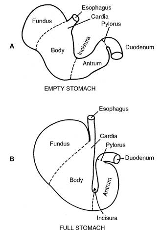
Figure 5-1 Diagram of cross section of stomach showing anatomic and functional regions. A, Empty stomach. B, Full stomach, showing that increase in size is due to changes in the proximal part of the stomach. (From Guilford WG, Strombeck DR: Gastric structure and function. In Guilford WG et al., eds: StrombeckS Small Animal Gastroenterology, ed 3, Philadelphia, 1996, WB Saunders.)
the stomach, reaching maximum thickness at the pylorus. The serosa is the outer layer of the stomach.
The blood supply to the stomach is from the celiac, hepatic, and splenic arteries. Venous return to the hepatic portal vein is through the gastrosplenic and gastroduodenal veins. Gastric lymph empties into the cisterna chyli via the hepatic and mesenteric lymph nodes. Extrinsic autonomic innervation consists of sympathetic afferent and efferent fibers from the celiac plexus and parasympathetic efferent fibers from the vagus nerves. The intrinsic myenteric plexus innervates the muscular and submucosal layers and functions to control mucosal secretion.