Glucagonoma
Introduction
In 1974, the first series of nine human patients with a gluca- gon-producing pancreatic tumor was reported. To date, only seven cases of canine glucagonoma have been conclusively diagnosed, and this syndrome has not yet been reported in a cat.2,29
Clinical presentation
Glucagonomas in humans are associated with typical skin lesions characterized by marked erythema, destruction of the superficial epidermis, and a tendency to heal and progress to other areas of the body.
These skin lesions are descriptively termed necrolytic migratory erythema (NME).30 Similar skin lesions have been reported in several dogs and have been descriptively termed superficial necrolytic dermatitis (SND).31 However, approximately 90% of canine patients with SND are diagnosed with hepatic disease, diabetes mellitus or, less commonly, other conditions.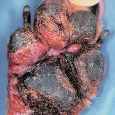
Figure 9.23:
SND foot pad. This dog shows severe crusting lesions on his foot pads with significant interdigital ulceration. A biopsy must be collected to confirm the presence of SND. Most dogs with SND do not have a glucagonoma, but have hepatic disease instead. (Courtesy of Dr. Sandy Merchant, Louisiana State University, Baton Rouge, LA.)
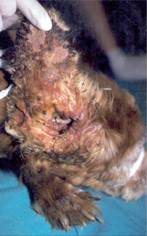
Figure 9.24:
SND ear. This figure shows severe crusting and ulceration on the concave pinna of a dog. A biopsy must be collected to confirm the presence of SND. (Courtesy of Dr. Sandy Merchant, Louisiana State University, Baton Rouge, LA.)
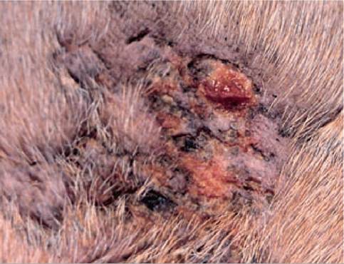
Figure 9.25:
SND skin. This image is a close-up of crusting with ulceration on a pressure point area of a dog.
A biopsy must be collected to confirm the presence of SND. (Courtesy of Dr. Sandy Merchant, Louisiana State University, Baton Rouge, LA.)Human patients with glucagonoma present with NME, weight loss, diabetes mellitus, glossitis, stomatitis, cheilitis, and a tendency for thromboembolic disease. The small number of dogs diagnosed with a glucagonoma have had crusting and scaling skin lesions, most commonly of the footpads, hocks, abdomen, elbows, perineum, nose, and mucocutaneous junctions (Figures 9.23, 9.24, and 9.25). These canine patients also exhibited depression, peripheral lymphadenopathy, and anorexia. Hyperglycemia was reported in 4 of 7 dogs, but was mild in most cases. Also, in some dogs, diabetes mellitus was diagnosed after the diagnosis of the glucagonoma. It is, therefore, appropriate to suspect a glucagonoma in dogs presenting with SND that do not show evidence of hepatic disease, even if they do not concurrently present with diabetes mellitus.
Diagnosis
Most dogs with a glucagonoma are hypoaminoacidemic. Measurement of serum or plasma glucagon concentrations may be helpful in confirming a diagnosis. While an assay has been validated for use in domestic animals, to the author’s knowledge, no veterinary endocrine laboratory currently offers this assay. However, samples can be submitted to a human laboratory for assay. In such cases, the laboratory must be contacted for special submission instructions. Caution is advised in interpreting high serum or plasma glucagon concentrations, as they have also been reported in human patients with other conditions, such as chronic renal failure, diabetic ketoacidosis, starvation, acute pancreatitis, hyperadrenocorti- cism, and sepsis.30
If a glucagon assay is not available, the diagnosis of SND is confirmed by histopathology and other potential causes are ruled out. An exploratory laparotomy should then be considered for definitive diagnosis and treatment.
Treatment
The same treatment guidelines for localization and surgical exploration discussed for other NETs also apply for glucagonomas.
As for gastrinomas, metastatic disease at the time of diagnosis is common in dogs with a glucagonoma. Since most patients with a glucagonoma have hypoaminoacidemia preop- eratively, total or partial parenteral nutrition may improve the overall condition of the patient. An exploratory laparotomy was performed in four of the seven dogs diagnosed with a glucagonoma and a mass was removed in three, two of which died or were euthanized within 3 days after surgery for pancreatitis. One dog survived for 9 months before the skin lesions returned and the patient was euthanized.32Medical management should be considered if abdominal exploration is not an option in cases of a metastatic glucagonoma or in recurrent cases. Medical therapy can include exogenous insulin, intravenous infusion of essential amino acids and fatty acids, zinc supplementation, and octreotide therapy.2 However, more clinical information is needed before more specific treatment recommendations can be given.
9.4.5