Hands-on Examination
PHYSICAL APPEARANCE, BODY WEIGHT, AND BODY CONDITION. It has been increasingly recognized that genetics influences the risk of perinatal mortality, with the heritability for perinatal mortality ranging from 0.03 to 0.13.285 Congenital disorders may present with subtle changes in physical appearance and behavior.
For example, Salers calves with beta-mannosidosis have moderately domed heads, mild superior brachygnathism, and a slight head tremor.286 Similarly, in utero infections that lead to growth retardation may be reflected by low birth weight and poor body condition. Low birth weight, contracted tendons, and/or abnormal mentation should prompt consideration of testing for persistent infection with bovine viral diarrhea (BVD). Head. Observation of facial expression provides an opportunity to assess cranial nerve function. Normal eyeball position and movement is consistent with normal function of cranial nerves III, IV, VI, and VIII. Normal facial expression and ear and eyelid position suggest normal functioning of cranial nerve VII. Vision (cranial nerve II) is assessed by watching the neonate navigate its environment. Because the menace response is learned, it is not particularly useful in very young animals. Normal sucking behavior is consistent with normal functioning of cranial nerves V, VII, IX, and XII. Absence of atrophy of the neck is consistent with normal functioning of cranial nerve XI. Functioning of cranial nerves V, VI, and VII are further assessed by the palpebral and corneal reflexes, and cranial nerves II and III are assessed by the pupillary light response. Eyeball position and skin tent duration are used to assess dehydration and estimate base deficit in calves with diarrhea (Table 19.5).
Depressed mentation is a feature of a number of neonatal diseases. Sepsis and diarrhea are both commonly associated with depressed mentation secondary to toxemia and/or metabolic derangements.
Bacterial meningitis is common in neonates following bacteremia; diarrhea, septic arthritis, omphalophlebitis, and uveitis are frequent concurrent clinical problems. The clinical signs of meningitis in neonates (lethargy, anorexia, recumbency) are nonspecific. Concurrent metabolic derangements may appear to provide an adequate explanation for the observed depressed mentation. In a retrospective study of 32 cases of bacterial meningitis in calves, concurrent metabolic derangements included hyperkalemia (15 of 25, 60%), respiratory acidosis (11 of 24, 46%), hypernatremia (3 of 20, 15%), and hypoglycemia (3 of 7, 43%).287 The more classical clinical signs described for bacterial meningitis in older animals (fever, opisthotonus, extension of the head, convulsions, hyperesthesia, signs of neck pain) may not be evident or perceived in neonates. Seizures in calves are often subtle, manifesting as facial twitches or jaw champing. Common causes of cerebral disease in lambs and kids include polioencephalomalacia and focal symmetric encephalomalacia. Affected animals are typically depressed and blind but have normal pupillary light reflexes. Mouth. Examination of the mouth includes assessment of the mucous membranes, moisture, color, and capillary refill time. Mucous membranes should be moist and pale pink with a capillary refill time of less than 2 seconds. Gray or cyanotic mucous membranes are associated with severe hypoxia, circulatory collapse, hypovolemic shock, cardiovascular anomalies causing a right-to-left shunt, and/or severe pulmonary disease. Absence of cyanosis is not a reliable indicator of adequate oxygenation in the neonate since the partial pressure of oxygen may reach very low levels (12
>20 | Adapted from Ravary-Plumioen B: Resuscitation procedures and life support of the newborn calf. Rev Med Vet 160:410, 2009; Constable PD, Walker PG, Morin DE, Foreman JH: Clinical and laboratory assessment of hydration status of neonatal calves with diarrhea.
J Am Vet Med Assoc 212:991, 1998; Smith GW: Treatment of calf diarrhea: oral fluid therapy. Vet Clin North Am Food Anim Pract 25:55, 2009. ![]()
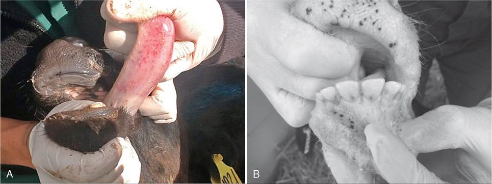
FIG. 19.1 A, Petechial hemorrhages on the underside of the tongue of a calf with thrombocytopenia. B, Petechial hemorrhage on gums.
may be corrected by placing vertical or horizontal mattress sutures in the affected eyelid. Treatment following correction of entropion includes topical administration of 1% atropine to dilate the pupil and relieve ciliary body spasm and topical antibiotics to prevent bacterial infection.
Examination of the sclera is useful to detect icterus, scleral injection, and scleral hemorrhage. Icterus may be observed secondary to liver or hemolytic diseases; both tend to be uncommon in neonatal milk-fed ruminants. Conversely, scleral injection is a common finding and suggests sepsis. Observation of fibrin in the anterior chamber of the eye of a neonate is highly indicative of sepsis. Ecchymotic hemorrhages may be observed on the sclera with type II BVD infections, bovine neonatal pancytopenia, or disseminated intravascular coagulopathies, or following birth trauma.
Ears. Bilateral drooped ear carriage is a common observation in sick calves and should prompt a closer examination. Unilateral ear droop may be observed with otitis and facial nerve paralysis. Mycoplasma spp., Pasteurella spp., and Haemophilus spp. are reported to cause otitis in calves. Clinical signs may include facial and vestibular nerve deficits and a purulent discharge from the ear. Calves infected with Mycoplasma spp. may also present with tenosynovitis and respiratory disease. Poor perfusion is reflected by cold extremities. A high incidence of pinna abscesses usually reflects contamination of equipment used to place ear tags. Ears should be warm and mobile.
Neck and Back. There are a number of congenital anomalies that affect the alignment of the vertebral column.
These include atlantoaxial malformations, scoliosis, kyphosis, lordosis, and combined anomalies, such as kyphoscoliosis. Atlantoaxial malformations may occur with or without cervical scoliosis or signs of spinal cord compression. Holstein calves affected by the hereditary condition complex vertebral malformation may present with a range of abnormalities that include retarded growth, malformation of the head (dysplasia or palatoschisis), bilateral symmetric flexion of the carpal and metacarpophalangeal joints, posterior arthrogryposis, and interventricular septal defects. Of these, growth retardation, vertebral malformation, and symmetric arthrogryposis are the most consistent findings.288 The neck and brisket should be examined to evaluate jugular fill and to detect brisket edema. Distention of the jugular veins and brisket edema are consistent with right-sided heart failure but may be absent with left-sided heart failure. Examination of the skin on the side of the neck is a good place to detect lice and fleas, and a skin fold test is performed to assess hydration. Parotid and prescapular lymph nodes should also be palpated to assess size and symmetry. Enlargement of all peripheral lymph nodes may be observed with juvenile lymphoma. Enlargement of specific nodes suggests a localized inflammatory response.
Thorax. Heart rate and respiratory rate vary with activity, excitement, and feeding. Tachypnea can be attributed to elevated temperature humidity index, fever, thermoregulation dysfunction, excitement, pain, stress, respiratory disease, hypoxia (cardiac disease, pulmonary disease, shock), dehydration, shock, toxemia, or metabolic acidosis. Periodic apnea and abnormally slow respirations are often the result of metabolic disturbances (hypoglycemia, hypocalcemia), hypothermia, prematurity, or hypoxia-induced suppression of the respiratory center. Tachycardia may be present with excitement, pain, injury, inflammation, toxemia, dehydration, shock, infectious disease, metabolic acidosis, hypokalemia, hypocalcemia, or heart disease.
Bradycardia of neonatal ruminants may be associated with hyperkalemia, central nervous system dysfunction, hypothermia, or starvation. Cardiovascular disease in neonates may be primary or secondary to other disease processes.
Congenital cardiac anomalies may be asymptomatic. Dyspnea and coughing are often the predominant clinical signs of congestive heart failure in calves. Cardiac auscultation should be carried out systematically with the aim of evaluating cardiac rhythm and detecting normal heart sounds and cardiac murmurs. In most calves the first and second heart sounds are easily audible. The third heart sound is not normally heard in calves, and the fourth heart sound is heard infrequently.289 Cardiac murmurs are not uncommon during the newborn period. Various congenital anomalies of the heart occur in cattle as the result of defective septa and formation of the cardiac chambers.290 The prevalence of congenital heart disease in cattle has been reported to be approximately 0.7%,291 with ventricular septal defects representing the most common type.292 Other frequent defects include noncyanotic anomalies (atrial septal defect, patent ductus arteriosus without pulmonary hypertension) and cyanotic anomalies (tetralogy or pentalogy of Fallot, Eisenmenger’s complex associated with reversion of a previously left-to-right shunt secondary to pulmonary hypertension).290 The prevalence of the different congenital heart diseases in calves is shown in Table 19.6. Serious heart anomalies may not be accompanied by any murmur, whereas a closing ductus arteriosus may cause a murmur that is very loud. This normal ductal murmur is best ausculted on the left
■ TABLE 19.6
Prevalence of the Most Common Congenital Heart Diseases in Calves, According to Ohwada and Murakami (2000) 290327
| Congenital Heart Diseases | No. of Cases |
| Ventricular septal defects | 198 |
| Atrial septal defects | 148 |
| Double outlet right ventricle | 66 |
| Aortic stenosis | 53 |
| Double cranial vena cava | 50 |
| Patent ductus arteriosus | 45 |
| Anomaly of the caudal vena cava | 42 |
| Tetralogy of Fallot | 32 |
| Hypoplastic left ventricle | 25 |
| Transposition of the great arteries | 24 |
| Abnormal pulmonary vein connection | 21 |
| Total | 704 |
aA total of 469 cases of congenital heart diseases were studied.
side of the chest at the third intercostal space at the level of the shoulder. In some instances, however, it is described as a continuous machinery-type murmur, but far more commonly a grade II to V/VI holosystolic murmur is ausculted. Cardiac auscultation will determine heart rate, rhythm, and the presence of murmurs. Abnormal radiation of heart sounds caudal to the fifth intercostal space or dorsal to the level of the scapulohumeral joint on either side of the thorax should be noted. Thoracic percussion may be used to identify the presence of thoracic fluid, cranioventral consolidation, or atelectasis in neonatal ruminants.
Cardiac arrhythmias are observed sporadically in neonates, often associated with diarrhea. Metabolic acidosis secondary to losses of electrolytes and water causes a transcellular shift of potassium ions into the extracellular fluid in exchange for hydrogen ions.293 Hyperkalemia decreases the membrane potential and slows down impulse conduction by inactivating sodium (Na) channels.294 As serum potassium increases (>5.5 mEq/L), aberrations in cardiac excitability occur and are manifested as progressive atrial standstill, progressing to ventricular fibrillation and asystole.295 The response of the myocardial cells to catecholamines decreases with the reduction of blood pH.294 Influxed H+ ions repress ventricle contraction by competing with Ca2+ ions in binding to myocardial troponins.294 Tachyarrhythmias may be observed in calves with cardiomyopathies, ionophore toxicosis,296 or hypomagnesemia.
Cardiomyopathy secondary to selenium deficiency, gossypol, monensin, or lasalocid toxicity may present as a syndrome of sudden death during periods of excitement precipitated by feeding or moving calves out of hutches into group pens.297
Inspiratory stridor is often a feature of extrathoracic airway obstruction, and increased abdominal effort on expiration is often an indication of pulmonary disease causing reduced lung compliance. Lung disease in the newborn is usually diffuse and is the result of infection acquired in utero or postpartum and/ or lung atelectasis associated with immaturity, recumbency, or surfactant dysfunction. Signs of lung disease include increased work of breathing characterized by nostril flare, rib retractions, and increased abdominal effort. It is useful to palpate the ribs carefully to check for rib fractures, as these may also lead to tachypnea and dyspnea. A cough and nasal discharge, salient features of respiratory disease in older neonates, are infrequent findings in newborns with lung disease. Animals with few or no audible thoracic abnormalities may have severe respiratory disease.
Auscultation of the trachea, thorax, and occasionally the nasal passage are essential elements of the respiratory examination. Tracheal compression should not induce multiple coughs. Lung sounds of neonates are typically easier to hear than those of adults, but lung sounds do not always correlate well with the severity of respiratory condition that is present. Interpretation of the intensity of thoracic breath sounds can be validated by comparison to tracheal breath sounds. The intensity of tracheal breath sounds is gradually attenuated as the stethoscope is moved from trachea to the lung hilus, again to the anteroventral thorax, and finally to the caudal thorax in normal animals. Loud tracheal breath sounds that are not attenuated over the thoracic area may be indicative of atelectasis, lung consolidation, or cardiac enlargement. Added breath sounds such as crackles or wheezes should be noted but may not be heard without augmenting the depth and frequency of the breathing effort with the use of a rebreathing bag.
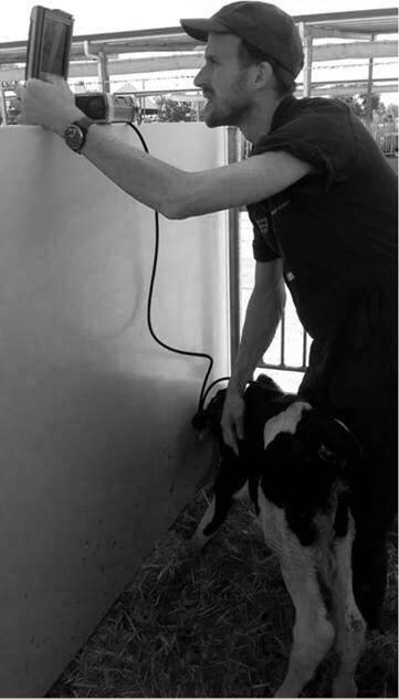
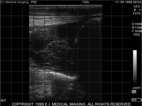
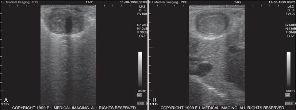
The advent of portable ultrasound machines commonly used on-farm for reproductive work provides a useful tool for assessing the thorax of calves. The pleural cavity and lungs can be examined through the intercostal spaces. The 5.0 to 7.5 MHz rectal linear transducers used for reproductive work are suitable for this task. Thoracic ultrasound has similar sensitivity (76.5%, 60.2% to 88.8%) for diagnosis of bronchopneumonia as auscultation but a higher specificity (92.9%; 86.5% to 97.1%).298 In young calves, the lung fields extend cranioventrally from the tenth to the first and second intercostal spaces on the right and left sides, respectively.299 When standing, it is easiest to scan reaching over the dorsum to the opposite side (Fig. 19.2). The transducing agent of choice is 70% isopropyl alcohol, and the hair is not clipped. Bronchopneumonia most commonly localizes to the cranial aspect of the right cranial lung lobe, followed by the right middle lung lobe and the caudal aspect of the left cranial lung lobe. The cranial aspect of the right cranial lung lobe is imaged from the right first and second intercostal spaces. The right middle lung lobe is imaged from the right fifth intercostal space, whereas the caudal aspect of the left cranial lung lobe can be imaged between the left fourth and fifth intercostal spaces.299 It is important to scan both sides of the thorax because consolidation may occur unilaterally in up to 1 in 3 dairy calves. When imaging normal lungs the sound waves are reflected at the pleural surface, precluding visualization of the lung. Normal scans reveal a thin, white echogenic, moving pleural line. An increase in pleural fluid is reflected by separation of the visceral and parietal pleura; fibrin may be observed moving in the pleural fluid with the respiratory cycle (Fig. 19.3). Ultrasound is useful for identifying the optimal site for thoracentesis. The inundation of fluid in a consolidated lung provides an opportunity to visualize pulmonary architecture. Similarly, thoracic and/or pulmonary abscesses located adjacent to the thoracic wall may be imaged.
Abdomen. Much can be learned about gastrointestinal function by observing abdominal contour, appetite, and fecal consistency and volume. A normal abdominal contour and vigorous appetite associated with the passage of an appropriate volume of pasty stool suggests normal gastrointestinal function. Examination of the neonatal ruminant should include assessment of abdominal size and contour.
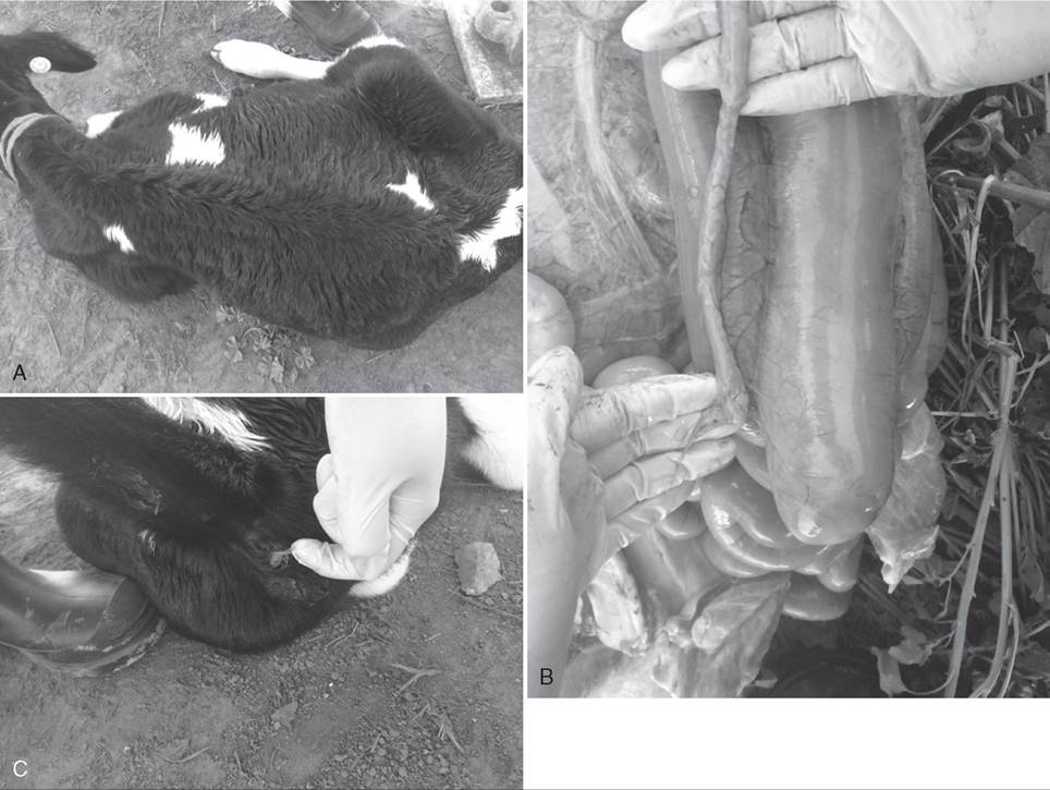
Gradual abdominal distention may be observed with congenital atresia of the gastrointestinal tract (Fig. 19.4, A). Atresia may be observed in the colon (coli) (Fig. 19.4, B), rectum (recti), or anus (ani); the spiral loop of the ascending colon (spiral colon) is the most commonly affected segment of intestine in calves.300 Neonates presenting with atresia often have a distended abdomen and a history of declining appetite. Astute owners may notice the lack or reduced volume of feces (Fig. 19.4, C).
External palpation of the abdomen can be rewarding, depending on the cooperation of the animal and the tenseness of the abdominal musculature. In calves it is usually possible to palpate
![]()
FIG. 19.2 Scanning calf lungs.
![]()
FIG. 19.3 Pleural effusion containing fibrin in cranial ventral thorax.
enlargement of the umbilical vein and arteries. The inguinal rings and umbilical area should also be palpated for hernias. Abdominal auscultation, while important, is not used for the forestomach and intestinal motility parameters typically evaluated in the adult ruminant; along with simultaneous auscultation and succession; however, ballottement, or percussion may show evidence of gas, fluid, or disordered motility. Abnormal forestomach function in neonatal ruminants is often reflected by altered abdominal contour. Left and right abomasal displacement and abomasal torsion are observed sporadically in calves. Succussion (simultaneous auscultation and percussion) is useful for delimiting the boundaries of distended visci. Passage of a stomach tube helps distinguish rumen and abomasal distention and facilitates collection of a rumen fluid sample. A putrid odor to neonatal rumen fluid is common with putrefactive indigestion when milk is delivered to the rumen in greater quantities than normal by escaping the esophageal groove or via excessive backflow from the abomasum. Reflux of abomasal contents into the reticulum and rumen, independent of feeding, occurs in connection with abomasal inflammation and obstructions.301 Evaluation of rumen fluid pH and renin activity are useful for distinguishing abomasal reflux from esophageal groove overflow. Rumen fluid pH is usually 7 or higher with rumen putrefaction and low to normal with abomasal reflux.301 Chymosin (renin) is normally present in abomasal juice, and renin activity in rumen fluid suggests abomasal reflux.302 Renin activity is measured by adding 2 mL rumen juice to 2 mL whole milk on a California mastitis test (CMT) plate. Presence of renin in the ruminal fluid causes coagulation of the casein in the milk. Chloride ion concentration in rumen fluid from calves is higher than in adults (55 to 102 mmol/L calves303 vs 25 mmol/L adults301), possibly reflecting the high chloride content of milk (45 mmol/L); as such, the chloride concentration of rumen fluid is not useful for identifying abomasal reflux in calves.
Abomasal ulceration may lead to localized or generalized peritonitis. Normal peritoneal fluid from calves has a higher nucleated cell count than that of adult cattle (3350 cells/jL vs 1371 cells/jL). Total protein concentration in the peritoneal fluid of calves is similar to that in adults (2.5 g/dL vs 3.1 g/dL).304
Umbilicus. After rupture of the umbilical cord, the two umbilical arteries retract actively, and the urachus is pulled back passively by these vessels into the abdomen. Smooth muscle contraction brings about luminal closure. The umbilical arteries ultimately become the lateral ligaments of the urinary bladder.
The umbilical vein does not retract but rather collapses in association with some minor smooth muscle contraction. The umbilical sheath shrinks and dries within 3 days postpartum. The body wall closes completely around the umbilical structures within days to a few weeks after birth.305
During the physical examination the navel is assessed for diameter, discharge, and pain while the neonatal ruminant is standing and again while it is in lateral recumbency. Abdominal palpation, using both hands pressing together, is useful for evaluating the umbilicus of standing calves. Enlargement of the umbilical arteries can be palpated coursing caudally toward the bladder and enlargement of the umbilical vein coursing cranially to the liver. Application of pressure caudal to the xiphoid often elicits a soft grunt from calves with a septic umbilicus and associated peritonitis. Extensive adhesion of bowel to inflamed umbilical structures produces a large, easily palpable intraabdominal mass. The uterine vein and artery may also be traced within the abdomen with the calf in recumbency. Common abnormalities of the calf umbilicus include umbilical hernias, omphalophlebitis, external umbilical abscess, urachal abscess, and omphaloarteritis. Patent urachus is uncommon in calves. Disorders of the urachus or the umbilical arteries are often associated with pollakiuria and a prolonged stance of urination. Umbilical hernias in cattle do not tend to close spontaneously and are believed to be hereditary.306
Ultrasound examination of the calf is performed with the calf standing; occasionally the umbilical vein is easier to identify with the patient in left lateral recumbency.307 The umbilical vein courses from the umbilicus to the liver, which in the calf is located on the right side. The lumen is generally larger near the body wall, with a diameter ranging from 10 to 25 mm. It appears as a round-to-oval anechoic to hypoechogenic structure. Within 3 weeks after birth, ultrasonographic identification of the intraabdominal umbilical vein becomes impossible in up to 50% of healthy calves. If still visible, the vein appears
![]()
FIG. 19.4 A, Atresia coli progressive abdominal distention. B, Atresia coli dilated proximal section of spiral colon and nondistended distal colon. C, Atresia coli—mucus with the absence of feces.
hypoechogenic to the surrounding tissue and indistinctly marginated.305 In cattle the umbilical arteries and urachus retract into the abdominal cavity when the cord ruptures and thus cannot be identified in the external umbilical stalk in normal calves.307 The umbilical vein of calves is scanned from the umbilical stalk to the liver along the right abdominal wall. The umbilical vein enters the liver caudoventral to the gallbladder. The umbilical arteries are most easily located adjacent to the urinary bladder and cannot normally be identified much beyond the apex of the urinary bladder, unless enlarged and abnormal. Identification of a urachal remnant in calves is abnormal.307 Umbilical ultrasound is useful for evaluating the need for umbilical surgery and identifying other organs involved that may influence the complexity of the procedure (Fig. 19.5).
MUSCULOSKELETAL SYSTEM. The musculoskeletal system should be examined for evidence of birth trauma, including fractured ribs, long bones, and mandibles; brachial plexus injuries; and soft tissue trauma, which includes an edematous head and tongue from excessive traction or compression in the pelvic canal. The vertebral column, ribs, and limbs should be palpated carefully to elucidate swelling, heat, or painful areas. Strenuous manipulation during dystocia can result in Salter Harris type 1 fractures characterized by disruption of the distal physis in one or more limbs. Femoral nerve paralysis occurs as a sporadic complication of dystocia associated with “hip lock” in calves.308
Dwarfism is a relatively common inherited problem in most cattle breeds. Osteopetrosis has been reported in Angus, Hereford, and Simmental breeds of cattle. Syndactyly is considered an inherited disorder in Holstein Friesan cattle, and affected animals are also predisposed to hyperthermia. Growth retardation, vertebral malformation, and bilateral symmetric arthrogryposis affecting the carpal and metacarpophalangeal joints are features of complex vertebral malformation in Hol- steins, a genetic condition associated with a mutation in the gene SLC35A3 coding a uridine diphosphate V-acetylglucosamine transporter.288
Peripheral pulses in the middle coccygeal, brachial, and auricular arteries should be strong and regular, and the peripheral extremities should be warm. Assessment of pulse quality includes consideration of pulse strength and frequency. Concurrent cardiac auscultation is useful to verify that heart rate and rhythm matches the pulse. With cardiovascular disease the pulse may be weak and/or uneven and may not correlate with the heart rate and rhythm.
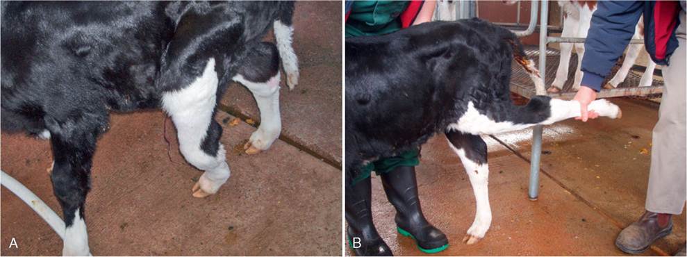
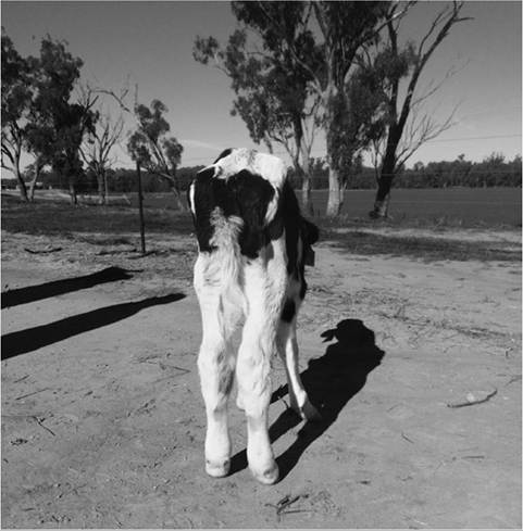
The limbs should be examined for laxity, flexural, and angular limb deformities. Flexor tendon laxity occurs more commonly in calves born prematurely and in calves that are small for their gestational age. Unilateral or bilateral hyperextension of the tarsus can be encountered in newborn calves following forced extraction. A rupture of the peroneus tertius causes the hyperextension of the tarsus (Fig. 19.6). The gait is abnormal but does not appear to be painful.309 Congenital contracted
![]()
FIG. 19.5 A, Enlarged umbilical vein cranial to external umbilical stump. B, Enlarged umbilical vein just before entering the liver.
![]()
FIG. 19.6 Peroneus tertius rupture. A, Standing. B, Extended, showing hyperextension of the tarsus with concurrent flexion of the stifle.
flexor tendon is a common defect in cattle; etiologic origins include inherited factors, in utero malpositioning, and overcrowding caused by the size of the fetus relative to the dam. Contracted tendons may occur with other congenital abnormalities, such as cleft palate, dwarfism, and arthrogryposis.309 Arthrogryposis, abnormal fixation of joints because of damage to or contracture of soft tissues, may be hereditary, secondary to infection in fetal life with teratogenic viruses, or may result from ingestion of teratogenic plants. Following teratogenic viral infections, the clinical presentation is secondary to mild to extensive loss of ventral horn motor neurons in the spinal cord gray matter and Wallerian degeneration of ventral spinal motor nerves.310 Hereditary congenital deformity in Hereford cattle is characterized by arthrogryposis, kyphosis, torticollis, scoliosis, and cleft palate.311 Ingestion of lupines by pregnant cows between 40 and 70 days' gestation has also caused various degrees of arthrogryposis with other associated congenital defects.312
Skeletal muscle myopathy is an important differential for lame neonatal ruminants. Affected neonates may present with a history of falling behind the mob when mustered and dropping into sternal recumbency. Close examination reveals firmness of the triceps and quadriceps. Deep palpation of affected muscles elicits a pain response. Lambs and kids with selenium and/or vitamin E deficiency are often mentally bright, reluctant to stand, walk with a stiff gait, and cry when forced to move. Evaluation of serum creatinine phosphokinase, blood selenium, and plasma vitamin E concentrations is useful to confirm the diagnosis.
Any heat or cold, swelling, edema, pain around the joints or physes, or lameness should be noted (Fig. 19.7). Due to the propensity of neonates to succumb to sepsis, a swollen joint should be considered infected until proven otherwise. When sepsis is present it is not uncommon to observe enlargement of the corresponding prescapular or prefemoral lymph node in the affected limb. When multiple animals on the same property are affected, this may reflect an infectious etiology such as salmonellosis or mycoplasma; alternatively, poor colostral management may contribute to a high incidence of opportunistic infections of immunologically compromised neonates leading to joint ill. An inflammatory leukogram provides supportive evidence of a septic process, and aseptic collection of joint fluid for cytology and culture provides confirmatory evidence. When interpreting neonatal hematology and biochemistry results, it is important to be aware of age-associated normal values (Table 19.7).
In older neonates metabolic bone disease should be considered as a differential diagnosis for lameness associated with flaring of the physis. Diets high in energy and protein that have low calcium and high phosphorus promote rapid weight gain, increasing the physical load on metabolically compromised
![]()
FIG. 19.7 Calf with cold, swollen hind limbs and serum exuding through the skin on the left hock. The calf was developing gangrenous necrosis secondary to Salmonella enterica serotype Dublin infection.
growing bones. Damage to the growing physis may result in the subsequent development of angular limb deformities. Calcium deficiency in calves leads to reduced mineralization of bone. The transverse processes of the lumbar vertebrae become soft and bend when palpated. Copper deficiency causes metabolic bone disease that is manifested by physitis and brittle bones, reflected by a propensity to spontaneous fractures.
Posterior paresis is common in neonates; causes include border disease, enzootic ataxia, vertebral body abscesses, caprine arthritis encephalomyelitis virus, and vertebral body fractures associated with dietary copper deficiency (or secondary copper deficiency due to molybdenosis) or calcium phosphorous imbalance. These neurologic problems are discussed in Chapter 33.