Helminth Diseases
Nematode Gastroenteritis
Nematode infection of the gastrointestinal tract is one of the most significant causes of wastage and decreased productivity in goats worldwide, especially under grazing conditions.
The natural history of caprine nematodiasis is in many ways similar to that seen in cattle and sheep. Studies in the goat, however, suggest some important species differences in terms of parasite susceptibility, age- related immunity, and the pharmacokinetics of anthelmintic therapy. These differences must be understood and taken into account for effective control of nematode parasites in goats.Etiology
A multitude of nematode parasites are at home in the gastrointestinal tract of goats. The major genera are first presented here in taxonomic family groups to facilitate discussion of common and distinct aspects of their life cycles. Subsequently, the individual species are discussed according to their locations in the caprine alimentary tract, so that their pathophysiologic effects can be better understood. More detailed information about the biology of the various nematodes is available in standard veterinary parasitology texts (Soulsby 1982; Bowman 2019).
Trichostrongylidae The trichostrongyles are responsible for most of the disease and economic loss associated with nematode parasites in goats. Genera in this family include Haemonchus, Trichostrongylus, Cooperia, Nematodirus, Marshallagia, Mecistocirrus, Ostertagia, Teladorsagia, and Camelostrongylus. All have a direct life cycle. Adult nematodes in the alimentary tract of the host produce ova that are passed in the feces. First-stage larvae develop in ova within one day of passage to the external environment. For most species, first-stage larvae then break out of ova and molt to second-stage larvae. These molt one more time and become infective third-stage larvae.
Development to the infective stage usually takes 7-10 days under favorable conditions, but can vary according to environmental factors, mainly temperature and moisture.Nematodirus spp. are distinguished from the other genera in that development to infective third-stage larvae occurs within the ova during the three-week period after passage in the feces. This adaptation enhances survivability over periods of adverse weather. In Marshallagia spp., larvae develop to the second stage before hatching from the ova.
Infective larvae of all genera are ingested by the host during consumption of contaminated forage. Infective larvae migrate upward toward the tops of grasses during mornings and evenings, enhancing ingestion by grazing hosts. After ingestion, the larva travels to its predilection site in the alimentary tract. The larva then burrows into mucosal folds or digestive glands, and molts within one to two days to a fourth-stage larva. This larva may remain in place for as long as 10 days, and then returns to the mucosal surface to finally molt into an adult capable of producing ova to complete the life cycle. The average prepatent period is between three and four weeks.
Cases of human trichostrongylosis have been reported in two suburban goat keepers in Australia who presented with abdominal pain and diarrhea. Eggs of Trichostrongylus spp. were identified from the patients and larvae of Trichostrongylus Colubriformis from the goats. One patient had used goat manure as fertilizer on his vegetable garden (Ralph et al. 2006).
Trichuridae These parasites also have a direct life cycle. In Trichuris spp. of this family, known as whipworms, development to the third larval stage occurs within the egg and release of the larva does not happen until after ingestion by the host. Embryonation within the egg occurs after passage in the feces, taking three weeks or longer, depending on temperature and moisture conditions. Ingested eggs release larvae that penetrate the small intestine.
Larvae develop for 2-10 days before moving to the cecum for maturity to the adult stage. The prepatent period is 7-9 weeks.Oxyuridae The members of this family that affect goats are Skrjabinema spp., referred to as pinworms. The life cycle is direct. Eggs ingested by the host hatch in the small intestine and larvae migrate to the large intestine to become adults within 25 days of infection. Eggs are fully embryonated and are deposited by the adult female pinworm on the perianal skin of the host.
Strongylidae Oesophagostomum spp. in the family Strongylidae are referred to as nodular worms. They have a direct life cycle similar to the Trichostrongylidae, except that infective larvae penetrate deep to the submucosa of the alimentary tract, reaching the lamina propria for development to fourth-stage larvae. This can occur anywhere from the pylorus to the rectum. After the molt, fourth-stage larvae return to the mucosal surface and migrate to the colon to develop into adults. The prepatent period is approximately six weeks. The characteristic nodules that occur in the intestine of affected animals due to Oesophagostomum columbianum are associated with a host reaction to the deeply burrowed infective larvae, not the surface-feeding adults. The third-stage larvae of Chabertia spp. develop deep in the intestinal wall, but nodule formation is not a characteristic of chabertiosis. The prepatent period is approximately seven weeks.
Ancyclostomidae This family includes the hookworms of goats, in the genera Bunostomum and Gaigeria. These nematodes have a direct life cycle, but a different route of transmission than the Trichostrongylidae. Infective larvae penetrate the skin or oral mucosa of the host. Such larvae enter the bloodstream via capillaries and end up in the lungs. They disrupt pulmonary capillaries and enter the alveoli, where they molt to fourth-stage larvae. These larvae are coughed up and swallowed. Migration to the small intestine and development of adults then occur.
The prepatent period is approximately 9-10 weeks, but adult worms may be present and feeding in the goat colon by four weeks after infection (Arantes et al. 1983).Strongyloididae Strongyloides papillosus is the sole caprine pathogen in this family. The life cycle is unique among the gastrointestinal nematodes. S. papillosus is parthenogenic, so free-living or parasitic development can occur. Under adverse environmental conditions, ova produced by parthenogenic females are passed in the host feces and develop only to infective third-stage larvae. Under more favorable environmental conditions, the free-living cycle is more common.
Ova are passed in the host feces again, but develop rapidly into sexually mature free-living males and females. After copulation, the females produce a single generation of infective larvae. In both cases, the resulting infective larvae must enter a suitable host to complete a subsequent life cycle. Infection of the host may occur by penetration of the skin, or the oral or esophageal mucosa. Penetrating larvae enter the bloodstream and localize in the lungs, where they enter the alveoli, migrate up the airways, and are swallowed. They mature to adults in the small intestine. The prepatent period is six to seven days. In addition, transmission of infective larvae to neonates via the milk of infected dams has been documented to occur in goats, though transplacental transmission was not demonstrated (Moncol and Grice 1974; Yvore and Esnault 1986). Very young kids infected via milk or colostrum can pass ova in the feces, and S. papillosus may be the only gastrointestinal nematode found in preweaned kids.
Gongylonematidae Gongylonema spp. of the family Gongylonematidae (superfamily Spiruroidea) have an indirect life cycle. Eggs passed in feces of primary hosts such as the goat hatch after ingestion by coprophagous beetles. Infective larvae develop within the beetles for about 30 days. Goats are infected by eating beetles containing infective larvae.
Location of Nematodes in the Host
Nematodes of the Esophagus and Rumen Nematodes of the genus Gongylonema, Gongylonema pulchrum, Gongylonema verrucosum, and Gongylonema monnigi exist in the esophagus and forestomachs of the goat. Adult worms may be visible embedded in the mucosa and submucosa of the esophagus and rumen, but these parasites are essentially non-pathogenic and are of little clinical significance. Trematode parasites of the rumen, mainly Paramphistomum spp. and Cotylophoron spp., are more pathogenic, and are discussed later in this chapter.
Nematodes of the Abomasum The abomasal worms of goats regularly associated with morbidity, mortality, and production losses on a worldwide basis are Haemonchus contortus, Teladorsagia circumcincta, and Trichostrongylus axei. H. contortus is generally considered the most seriously pathogenic in goats. It is distinguished from the others in that the fourth-stage larvae and adults are voracious blood suckers. Mecistocirrus digitatus, like H. contortus, is an aggressive blood feeder and is a serious pathogen of goats in Central America and Southeast Asia.
Certain other abomasal worms affecting goats are less pathogenic or have a more limited geographic distribution. Haemonchus longistipes is a stomach worm of camelids in North Africa and India that occurs naturally in goats commingled with camels (Hussein et al. 1985). Pathogenicity for goats has been demonstrated experimentally (Arzoun et al. 1983). Haemonchus placei, usually associated with cattle and sheep, is found in the abomasum of goats in the Philippines (Tongson et al. 1981). Teladorsagia (Ostertagia) trifurcata often occurs in conjunction with T circumcincta. Several species of Ostertagia, which are mainly associated with cattle, show variable infectivity for goats (Bisset 1980). Ostertagia lyrata has been found to occur naturally in New Zealand feral goats (Andrews 1973). Angora goats in Australia are readily infected with Ostertagia ostertagi when grazing contaminated paddocks (Le Jambre 1978).
This parasite is also known to occur in goats in Chile, Cyprus, and the Ukraine.Teladorsagia davtiani occurs primarily in goats in temperate regions. Marshallagia marshalli occurs in tropical and subtropical regions. Marshallagia mongolica infects goats, sheep, and camels in central Asia. Camelostrongylus mentulatus is a common, non-pathogenic abomasal worm primarily of camels in the Middle East and Australia that also can infect goats. All four species are morphologically similar to the Ostertagia and are considered to be minor pathogens.
Trichostrongylus axei is one of several important Trichostrongylus spp. to infect goats, but it is the only one found principally in the abomasum. It may also be found infrequently in the small intestine of the goat as well (Tongson et al. 1981; Akkaya 1998).
Nematodes of the Small Intestine The major pathogens of the small intestine of goats recognized worldwide are the black scour worms T colubriformis and Trichostrongylus vitrinus, the small intestinal worm Cooperia curticei, the thin-necked worms Nematodirus filicollis and Nematodirus spathiger, and the hookworm Bunostomum trigonocephalum. This hookworm is an active blood sucker and may contribute significantly to the development of anemia. This is also true of the hookworm Gaigeria pachyscelis, which occurs in goats in Indonesia, India, and Africa. As few as two dozen
G. pachyscelis feeding in the proximal small intestine can lead to acute death from blood loss. S. papillosus, the threadworm of the small intestine, is moderately to markedly pathogenic in goats. Field observations in Namibia followed by experimental studies in South Africa indicated that S. papillosus may be associated with severe clinical disease in young goats up to 12 months of age. Some of the signs observed were typical of gastrointestinal parasite infection, such as abnormal feces or diarrhea, anorexia, cachexia, and dehydration, but other more dramatic and unexpected signs were also present, including ataxia, stupor, nystagmus, and head pressing, as well as rupture of the liver, with the latter presenting as sudden death (Pienaar et al. 1999).
Other nematodes of the caprine small intestine are geographically restricted or of limited pathogenicity. Nematodirus oiratianus and Nematodirus abnormalis are common caprine pathogens in cold regions of central Asia (Neiman 1977). Nematodirus battus has emerged as an important pathogen of lambs in the United Kingdom and North America. It is reported to occur in goats in northern Europe but, unlike in lambs, is not a significant cause of disease (Holm et al. 2014) N. battus was found in 5.8% of goat alimentary tracts examined at an abattoir in Nigeria (Nwosu et al. 1996). Trichostrongylus falculatus and Trichostrongylus rugatus infect goats in South Africa and Australia. Trichostrongylus longispicularis is primarily a parasite of cattle, but has been reported from a goat in Brazil (Lima and Guimaraes 1985).
Nematodes of the Cecum The whipworm Trichuris ovis occurs worldwide in goats, but is not considered a primary cause of disease or production loss. It occurs commonly in mixed infections and may contribute to poor condition. Injfectionis of goats with T. ovis and/or other Trichuris spp. have been reported to be more common during dry seasons in Birrzil and Nigeria (Travassos et al. 1974; Okon 1974). Trichuris spp. burrow into the cecal mucosa and pierce vessels with their styletted mouth parts, subsequently feeding on the pools of blood created. The cecal worm Skrjabinema ovis occurs worldwide in goats, but is generally considered non- pathogenic. A separate species, Skrjabinema caprae, occurs in the United States and elsewhere. Adult forms of these small, non-pathogenic pinworms are sometimes present around the anus of goats, where they deposit their eggs. If observed, they may cause concern to owners.
Nematodes of the Colon Adult O. columbianum reside in the colon, but the nodular lesions produced by infective larvae occur throughout the intestines. The parasite occurs worldwide. The remaining Oesophagostomum spp. that infect goats are not known to produce the characteristic intestinal nodule and are considered non-pathogenic. Experimental infection of goats with Oesophagostomum venulosum was reported to produce small nodular lesions in goats, but this is generally not observed in natural infections (Goldberg 1952; Chhabra 1965). Chabertia ovina can contribute to clinical parasitism in goats worldwide. Morbidity caused by Chabertia infection alone is uncommon. However, in experimental challenge studies, C. ovina worm burdens of more than 800 were fatal to 4-6-month-old kids (Kostov 1982).
Epidemiology
The successful infection of goats by gastrointestinal nematodes and the completion of the parasitic life cycle depend on a variety of environmental, parasitic, and host-related factors and interactions.
Environment-Host Interactions The feeding behavior of ruminant species is a major factor in the development of parasitism. Animals such as sheep and cattle that graie close to the ground are exposed to massive numbers of infective larvae. Free-ranging goats are less exposed to infective larvae, because their feeding behavior includes a large component of browsing at levels well above the ground. In surveys of infection intensity in sheep and goats, where the animals are allowed to follow their natural feeding behaviors, sheep have been shown to carry heavier worm burdens than goats (Le Riche et al. 1973). Domesticated goats, however, are often managed in situations where access to browse is restricted and pasture graiing is obligatory. Under these conditions, goats may have equal or greater risk of nematode parasitism; this has been demonstrated experimentally in Australia (Le Jambre and Royal 1976).
Elimination characteristics also play a role. Splattering of naturally fluid cow feces when it hits the ground facilitates the dissemination of nematode ova on pasture. The fecal pellets of goats and sheep are not as accommodating, but natural disintegration of the feces with spreading of ova or larvae by heavy rain, melting snow, trampling by hooves, and the action of coprophagous beetles can achieve the same result, albeit more slowly. In addition, goats on lush pasture in spring, a time of increased ova production, often develop a more fluid manure that loses its pelleted character. Overt diarrhea caused by clinical parasitism also facilitates the dissemination of ova on herbage.
Lush, dense pasture in turn provides a protective umbrella for developing larvae, screening out direct sun to reduce desiccation. Direct sunlight has been shown to reduce the survival time of nematode larvae contained in goat fecal pellets (Tongson and Dimaculangan 1983). Survival time of infective larvae may also decrease in very hot weather due to the increased metabolic rate experienced by the larvae.
Overstocking and/or overgraiing of pastures generally promote increased parasitism. While intensive, rapid consumption of herbage may reduce the survivability of ova and larvae by eliminating the protective plant growth, the total number of ova produced and deposited on the pasture each day increases directly with the number of animals present. Wild ruminants using pastures may also transmit nematodes to the goat, as demonstrated with C. mentulatus and Trichostrongylus probolurus transmitted to goats from blackbuck antelope (Thornton et al. 1973).
Management systems also play a role in the type and intensity of caprine nematodiasis, and a well-recogniied advantage of confined housing systems is the marked reduction in nematode parasite loads. In a survey of 49 dairy goat farms in France, indoor housing was associated with a low incidence of clinical parasitism, attributable mainly to Chabertia and Oesophagostomum. In goats with access to yards ostertagiasis was more common, and in pastured goats haemonchosis was the predominant problem (Cabaret et al. 1986).
Environment-Parasite Interactions Nematodes have evolved a number of adaptive strategies for surviving severe environmental stresses such as freeiing, overheating, and desiccation. These include deep burrowing of larvae into soil during adverse seasons; delay of ova hatching until optimal conditions of temperature, moisture, and season are met; development of the infective larvae within the protective shell of the ova, as is seen in Nematodirus spp.; and the production of huge numbers of ova by a single female, as is seen in H. contortus, which produces up to 10 000 eggs per day.
The most dramatic adaptation to hostile environments Is hypobiosis, or arrested development. In hypobiosis, infective larvae consumed by the host during periods of environmental adversity remain voluntarily dormant and progress to adulthood only when environmental conditions favor development and survival of larvae outside the host. Host factors may also trigger renewed development, as discussed below. In temperate regions, decreasing temperatures may signal the larva's commitment to hypobiosis, while in tropical regions with distinct seasons, hypobiosis is triggered by the onset of hot, arid conditions (Chiejina et al. 1988). Hypobiosis of H. contortus in goats in association with the hot, dry season has been reported from Kenya (Gatongi et al. 1998) and Togo (Bonfoh et al. 1995). Even in tropical regions where temperature and humidity conditions can support free-living larvae year-round, some degree of hypobiosis may occur in response to increasing moisture content of the soil (Ikeme et al. 1987).
Synchronous resumption of larval development in the host can lead to clinical disease, referred to as type II disease. Type II ostertagiasis has been reported in goats in Israel, with a significant increase in fecal egg counts occurring at the end of the hot, dry summer and continuing through the subsequent cooler rainy season (Shimshony 1974). Type II infection also has been reported in goats in Spain, with clinical signs occurring in January and February (Tarazona et al. 1982).
Environmental temperature is an important factor in the survival of nematode ova and free-living larvae. Various nematode ova in the feces of goats all died within six days at a temperature of 40 °C (104 °F). Ova survived and hatched optimally within eight to nine days at temperatures between 30 and 35 °C (86 and 95 °F). Hatching was delayed for 14 days at temperatures between 20 and 25 °C (68 and 77 °F). At 0 °C (32 °F), ova remained alive, but still did not hatch after 30 days (Tripathi 1980).
Some nematodes are best suited to tropical and subtropical conditions, notably Haemonchus spp., M. digitatus, and O. columbianum. H. contortus is representative of this group. No hatching of ova or larval development occurs when temperatures are 10 °C (50 °F) or below. The optimal conditions for development of H. contortus from ova to infective third- stage larvae are reported to be 28°C (82.4 °F), with humidity greater than 70% (Rossanigo and Gruner 1995).
The ova are highly susceptible to desiccation and do not survive in regions where hot, dry summer follows winter rainfall, or where winters are intensely cold. Infective larvae, when developed, are more resistant to weather and can survive repeated periods of desiccation. Warm, humid regions of the world with summer rainfall and temperate regions with mild winters are conducive to the development of infective larvae on pasture.
For H. contortus, hypobiosis largely replaces overwintering of free larvae as a means of survival for the species. This is true not only for tropical countries, but also for cold, temperate regions. A report from Sweden indicated that H. contortus in sheep showed almost 100% arrested development in the early fourth larval stage as early as mid-summer, thus evolving a strategy to survive the long, cold winters entirely within the host as the arrested larval stage, and relying on the ewe to complete its life cycle with the periparturient resumption of egg-laying in the spring (Waller et al. 2004). The pattern of development of O. columbianum is similar to that of H. contortus.
B. trigonocephalum and G. pachyscelis are best suited to humid subtropical and warm temperate regions. They thrive under management conditions where housing and bedding are allowed to remain continuously damp, because early larval stages are particularly susceptible to desiccation. Percutaneous penetration of larvae on the feet and legs of livestock is facilitated by grazing in wet herbage or housing in damp conditions.
Nematodes better adapted to cooler, temperate climates include Teladorsagia (Ostertagia) spp., Trichostrongylus spp., and C. ovina. T. circumcincta is the prototype of this group. Trichostrongylus spp. are more resistant to cold and desiccation than are H. contortus, and are capable of overwintering. Ova accumulate on pasture until suitable conditions of moisture and temperature occur, at which time large numbers of infective larvae develop. Development occurs within four to six days at 27 °C (80.6 °F), but can take as long as one month when temperature and humidity are unfavorable. Trichostrongylus fare poorly in very hot, dry summer conditions. They are capable of hypobiosis as a survival mechanism.
Teladorsagia (Ostertagia) spp. are broadly adaptable, tolerating both colder winters and hotter, drier summers than Trichostrongylus spp. Overwintering of Teladorsagia (Ostertagia) larvae is successful as long as winters are not excessively dry. Survival is enhanced by slow release of infective larvae from disintegrating fecal pellets. This permits some larvae to persist on pasture for as long as one year. Nevertheless, the Teladorsagia (Ostertagia) readily undergo hypobiosis when necessary. In regions of cold winter, larvae are conditioned to arrested development in the late autumn. In regions of hot, dry summer, larvae are conditioned in the spring.
Nematodirus spp. are well adapted to cold climates. They produce low numbers of ova, but survival rate is enhanced by the adaptation of larval development within the protective shell of the egg. They are extremely resistant to cold and dryness, and survive drought conditions. Hatching of ova of N. filicollis can begin in late autumn and continue through spring (Boag and Thomas 1975). N. spathiger does not show delayed hatching and behaves on pasture more like Trichostrongylus spp.
Host-Parasite Interactions Two phenomena in the hostparasite relationship favor success for the parasite. These are hypobiosis and the periparturient egg rise phenomenon. The goat is not totally defenseless, however, and some host mechanisms are known to limit parasite infection. These are immunity, the phenomenon of self-cure, and genetic resistance.
With regard to hypobiosis, environmental cues can trigger arrested development, as discussed in the preceding section. However, host factors may also trigger arrested development of ingested larvae. Host factors include immunity acquired from previous nematode infections, ingestion of large numbers of infective larvae, or a sizable preexisting adult worm burden. Resumption of larval development also may be triggered by host factors, including depression of host immunity, removal of adult worm burdens by anthelmintic therapy, pregnancy-induced changes in hormone levels, and increased prolactin levels related to lactation. The latter two signals also are related to the phenomenon of periparturient egg rise.
The periparturient egg rise commonly observed in sheep results from maturation and ova production by previously arrested larvae, particularly of H. contortus and T. circumci- ncta. In temperate regions, when lambing occurs in the spring, ova deposited during the periparturient egg rise are largely responsible for the infections of grazing lambs that occur in summer. Overwintered larvae are usually killed off by the time lambs or kids are actively grazing. Lambs and kids in turn are more susceptible to infection than their older, possibly resistant dams. Lactation after pregnancy is a strong stimulus for renewed development of arrested larvae. Periparturient egg rise in goats has been documented. Fecal ova counts were highest in does one week after parturition and remained elevated for four weeks. This was independent of the season when they kidded, and no changes in fecal ova counts were observed in infected male goats sampled concurrently (Okon 1980). Increasing prolactin levels in goats have been reported to be associated with the periparturient egg rise (Chartier et al. 1998a). It has also been postulated that a reduction in parasite-specific immunoglobulin (Ig)A antibodies in the dam associated with the transfer of maternal antibody to the colostrum around parturition may also facilitate the periparturient egg rise (Jeffcoate et al. 1992).
Immunity is an important host defense. While the neonate is immunologically naive to parasites and colostral antibody does not appear to be protective, immunologic resistance to nematode parasites can develop over time and is enhanced by the continuous exposure to parasites. The development of resistance to infection with T. colubriformis in goats exposed to decreased weekly doses of infective larvae has been demonstrated experimentally (Pomroy and Charleston 1989).
The intensity of immunity with advancing age and parasite experience varies among domestic ruminant species. Goats show the weakest degree of immunity, sheep somewhat more, and cattle the most resistance to infection as adults. It is postulated that selection pressure on goats to develop parasite resistance is not as strong as in cattle and sheep, because the grazing behavior of sheep and cattle subjects them to intense contact with infective parasite larva on herbage, which selects for the development of immune mechanisms for survival. In contrast, the browsing behavior of goats in natural settings or under extensive range management systems does not expose them to high concentrations of infective larvae and the selection pressure to develop immunity to parasite infection is less. Therefore, when goats are required to graze under commercial farming conditions, their susceptibility to parasite infestation is pronounced (Hoste et al. 2010)
The greater susceptibility of goats is manifest in numerous studies. Significant worm burdens have been observed much earlier in kids than in lambs, with worm burdens of more than 17 000 noted in kids between 3 and 4 weeks of age (McKenna 1984). When Merino sheep and Angora goats grazed the same contaminated pasture for four months, the goats had higher worm burdens of all species of gastrointestinal nematodes present except Nematodirus spp. (Le Jambre and Royal 1976). Adult goats carried mixed worm burdens similar in intensity to kids and yearlings in a survey of 47 dairy goat farms in New Zealand (Kettle et al. 1983). No difference in intensity of worm burdens or fecal egg counts was apparent between years one and two of pasturing when feral goats in New Zealand were pastured. In addition, when compared with sheep infections, worms infecting goats were more fecund, producing increased numbers of eggs per worm, and the return of high fecal egg counts was much quicker after anthelmintic therapy in goats than in sheep (Brunsdon 1986). These findings suggest that direct extrapolation of sheep parasite control measures to goats may often be ineffective because of species differences in response to parasite infection. One practical consideration of these differences between goats and sheep is that adult goats represent a significant risk for the contamination of pastures and that goat parasite control programs, to be effective, must account for this risk (Hoste and Chartier 1998b).
Immunity to parasites is not absolute. It predictably declines in the periparturient period, and may also be impaired by concurrent illness or malnutrition. It has been demonstrated by experimental challenge with H. contortus that goats on a low plane of nutrition subsequently pass more ova in the feces than do better-fed goats (Preston and Allonby 1978). Goats with paratuberculosis are predisposed to increased worm burdens, and in Africa goats with Trypanosoma congolense show increased susceptibility to
H. contortus infection (Griffin et al. 1981).
The phenomenon of “self-cure” is known in sheep that ingest large numbers of infective larvae of H. contortus and subsequently expel their existing adult worm populations. Because these animals usually become immediately reinfected, the adaptive significance in terms of parasite control is unclear. Nevertheless, it does serve as an indicator of the potency of the immune response of the host to infection. There are two reports suggesting that self-cure is observed in goats (Fabiyi 1973; Preston and Allonby 1978). However, more recent and extensive field observations, as well as experimental studies, suggest that if self-cure does occur naturally in goats, the response is weaker and less reliable than that seen in sheep (Kettle et al. 1983; Brunsdon 1986; Watson and Hosking 1989). The failure of self-cure in pastured goats may lead to increased levels of sustained fecal egg counts and a resulting increased intensity of pasture contamination compared with sheep.
A final host defense against parasites is genetic resistance, the existence of which has been studied extensively in sheep (Courtney 1986; Gruner and Cabaret 1988). Evidence for genetic resistance in goats to helminths has been reported as well. In East Africa, imported Saanen goats showed more resistance to challenge with H. contor- tus than either of two indigenous breeds, the Galla and East African (Preston and Allonby 1978). It was postulated that the European dairy breed has been selected for resistance to H. contortus through generations of grazing behavior, while the indigenous breeds, as innate browsers, have not. In a later study, the Small East African goat breed was shown to be more resistant than the Galla breed, based on significantly lower fecal egg counts in the postweaning period (Baker et al. 2001). In India, during a natural outbreak of haemonchosis after unexpected heavy rains, small grazing or pen-fed breeds such as the Black Bengal goat were far less affected than large browsing goats such as the Beetal and Jamunapari (Yadav and Sengar 1982). Thai native goats demonstrated greater resistance to H. contor- tus infection based on fecal egg counts and worm counts on necropsy than did Thai native (50%)-Anglo Nubian (50%) crosses (Pralomkarn et al. 1997).
In addition to breed differences, selection within breeds can also lead to greater resistance, as indicated by longterm studies involving large numbers of Creole goats in Guadeloupe (Mandonnet et al. 2001, 2006). Another extended study with cashmere goats in Scotland naturally exposed to mainly T. circumcincta concluded that selection for reduced fecal egg counts was possible in breeding programs (Vagenas et al. 2002). Breeding for helminth resistance in small ruminants has been reviewed (Gruner 1991; Baker 1998).
Hemoglobin type in sheep has been associated with resistance to helminth infection, and this has also been studied in goats. Five hemoglobin phenotypes were identified in Red Sokoto goats in Nigeria and helminth egg counts on feces during the rainy season were correlated with these types. Significant differences in infection rates were observed, and hemoglobin types associated with high infection rates were less frequent in the older goat population than in the kid population, suggesting increased mortality among the more parasite-susceptible phenotypes (Buvanendran et al. 1981).
In summary, the development of gastrointestinal parasitism in goats depends on a complex interrelationship of parasite, host, and environmental factors, many known and some unknown. In general, the types and degree of parasitism that develop in goat populations may be predicted on the basis of geographic and climatic location, management system, and prevailing weather conditions. Nematodes, by evolving diverse strategies and rates of development, different mechanisms for feeding, and different predilection sites in the host alimentary tract, appear to have maximized their exploitation of the host, while minimizing competition among each other. This diversity encourages development of multiple nematode infections, which are the rule in goats.
The outcome of infection depends on host resistance, level of infection, variety and types of parasites involved, the development of parasite resistance to anthelmintics, and the extent of appropriate therapeutic intervention. Acute death, clinical disease, or subclinical infection with adverse effects on growth and lowered productivity are all possible outcomes. Worldwide, numerous studies on the causes of wastage in goats, particularly young goats, have confirmed that clinical gastrointestinal nematodiasis is a major cause of morbidity and mortality. However, specific studies concerning the impact of subclinical gastrointestinal nematodiasis on production parameters in dairy, fiber, and meat goats are comparatively scarce.
The impact of nematode parasites on dairy goat production is beginning to become clearer. One French study indicates that elimination of gastrointestinal nematodes in lactating does by treatment with thiabendazole resulted in a 17.6% increase in milk production compared with untreated control goats (Farizy and Taranchon 1970). Later studies have revealed a number of interesting findings. Subclinical parasitism with H. contortus and T. colubri- formis in lactating does resulted in a decrease in body condition score, as well as a persistent decrease in milk yield ranging from 2.5 to 10% compared to uninfected controls. However, when the highest producers were assessed separately from other infected does, reductions in milk output ranged between 13 and 25.1% and the milk had lower fat content. It was concluded that high-producing goats had less resistance and/or resilience to parasite infection than did lower-producing goats, leading to more severe depression of milk output (Hoste and Chartier 1993). Further studies reinforced these observations that resistance to parasite infection differed according to level of milk yield in lactating does (Chartier and Hoste 1997; Hoste and Chartier 1998a).
Pathogenesis
Various pathogenic mechanisms are involved in gastrointestinal nematodiasis, depending on the genera involved. The principal effect of hematophagous worms on the host is a progressive debilitating anemia. Blood feeders include H. contortus and M. digitatus in the abomasum, B. trigono- cephalum and G. pachyscelis in the intestine, and Trichuris spp. in the cecum. Each H. contortus adult in the abomasum may be responsible for the loss of 0.05 mL of blood per day, either by active feeding or by moving to new feeding sites and leaving old sites to continue hemorrhaging. Death caused by acute blood loss is possible when infection rates are high (more than 10 000 adults per host) and development of large adult populations is synchronous, as can happen after arrested development.
In less severe infections, three stages of anemia may be recognized in infected hosts (Dargie and Allonby 1975). In the initial stage of blood loss, PCV may decrease markedly, because intraluminal blood loss in the alimentary tract is not a strong trigger for hematopoiesis. In experimental haemonchosis of goats, the PCV decreased from a mean of 29% to 16% within 19 days of infection with 9000-12 000 larvae (Al-Quaisy et al. 1987). In the second stage, regenerative erythropoiesis begins and the PCV stabilizes, albeit at less than the normal level, for as long as 6-14 weeks. During this time, however, iron stores in the host are reduced by loss in the feces. In the final stage, the PCV begins to decrease again as erythropoiesis is impaired by the progressive iron deficiency. Concurrently, a steady loss of serum proteins occurs as a result of parasite feeding. Serum albumin levels may be maintained initially due to replacement by tissue catabolism, but hypoalbuminemia eventually develops, accompanied by cachexia and clinical evidence of hypoproteinemia such as intermandibu- lar edema.
The remainder of the nematode parasites of the caprine alimentary tract are not primarily blood feeders, though blood constituents are gradually lost in the process of feeding during chronic or large-scale infection. In these cases, anemia does not occur acutely or reach the severity seen with the blood feeders. In the field, however, this distinction may not be evident, because mixed infections of hematophagous and non-hematophagous parasites often occur.
Infective larvae of T. axei develop to adults within the abomasal mucosa; the adults feed there and lead to erosion of the mucosal epithelium, catarrhal inflammation, hyperemia, edema, and diarrhea. Plasma loss from the mucosal damage contributes to the hypoproteinemia seen with this infection.
Infective larvae of T. (Ostertagia) circumcincta enter the gastric glands of the abomasum to undergo the third and fourth molts to adulthood. When synchronous maturation of large numbers of arrested larvae occurs (type II osterta- giasis), a severe gastritis results. Gastric glands become hyperplastic, intercellular tight junctions are weakened, hydrochloric acid secretion diminishes, and the pH of stomach content increases. One result of the pH change is that pepsinogen is not converted to pepsin and pepsinogen may leak across the abomasal mucosa to the circulating blood. Increases in blood pepsinogen support a diagnosis of type II ostertagiasis. Other plasma proteins also are leaked from the abomasum. Hypoproteinemia and diarrhea are cardinal signs of ostertagiasis.
The Trichostrongylus spp. that infect the intestine tunnel under the mucosal epithelium to feed. This results in a protein-losing enteropathy accompanied by diarrhea. Larval stages can be as destructive as adult worms. Over time, marked villous atrophy occurs. The pathogenesis of Nematodirus spp. is similar.
O. columbianum presents a unique situation in small ruminants. Infective larvae burrow into the submucosa of the small intestine, encyst for the third molt, and then return to the intestinal lumen to migrate to the colon for the final molt. In first-time infections, this process occurs unremarkably. In previously exposed, sensitized hosts, however, encysted third-stage larvae produce a dramatic local inflammatory response around the cyst, leading to the formation of caseating nodules. The larvae inside may die or resume migration at a much later time. Nodules may occasionally rupture serosally, causing peritonitis, adhesions, and partial or complete obstructions of the intestine. Even without rupture, widespread nodular formation may impair digestion, absorption, and passage of excreta. In addition, adult nodular worms can produce a severe catarrhal colitis with much mucus production. Diarrhea with mucus, weight loss, and hypoproteinemia occur in severe infections.
Anorexia with reduced feed intake is the most consistent finding in all forms of intestinal nematodiasis, and is accompanied by poor growth, decreased productivity, and weight loss. The causes of these host responses are complex and not fully understood. Failure of muscle growth in young animals results from a decrease in skeletal muscle synthesis, with a shift to increased albumin production by the liver to counteract ongoing protein loss. Muscle wasting in severely affected animals may be associated with muscle catabolism. In fiber-producing animals, fiber growth is also impaired by the shift in protein synthesis. In growing goats, bone growth is compromised too, possibly as a result of decreased intake of calcium and phosphorus and depletion from bones (Fitzsimmons 1966). The pathophysiologic adaptations of the host to gastrointestinal parasitism have been reviewed (Hoste 2001).
Clinical Findings
Mixed parasitic infections are common and often it is not possible to attribute clinical signs to a single parasite. In general, infections with Trichostrongylus, Teladorsagia (Ostertagia), Cooperia, and Nematodirus spp. produce a similar clinical picture. Young, grazing animals are most likely to be affected, particularly after weaning. A gradual, progressive loss of condition, poor growth, a dull attitude, and a decrease in feed intake are the most consistent findings. In more severe infections, a dark green to black diarrhea is evident, with staining of the hair and skin of the tail and perineal region. When the course is prolonged, inter- mandibular edema may develop secondary to hypoproteinemia. Chronically infected animals also develop a pot-bellied appearance, a rough dry haircoat, and flaky skin. Evidence of anemia is usually not pronounced. Deaths may be reported, and are usually spread out over a period of days or weeks. A more acute presentation may also be seen with type II disease when maturation of parasites resumes after arrested development.
Nodular worm (O. columbianum) infection may result in signs of abdominal pain such as hunched back and reluctance to move when nodule formation occurs and localized peritonitis results. Affected animals may be febrile. Occasionally, nodules may abscess and rupture. Pus may be passed per rectum if the rupture is intraluminal. Diffuse peritonitis may result if the rupture is intra-abdominal. In sheep, intussusceptions are reported sometimes in association with intestinal nodules, but this has not been reported in goats. When nodule formation is minimal, as in firsttime exposures, signs of O. columbianum infection may be limited to diarrhea in young kids, or in older animals intermittent passage of soft, mucus-laden feces flecked with blood and progressive loss of condition. Excessively mucoid feces with occasional blood is also associated with C. ovina infection. Anemia is uncommon.
When hematophagous parasites such as H. contortus infect goats, clinical evidence of anemia predominates. When infections are massive, peracute haemonchosis can
Figure 10.11 Submandibular edema (bottle jaw) associated with hypoproteinemia in a goat with severe haemonchosis. Note also the associated poor body condition and rough haircoat. Source: Reproduced by permission of Dr. Jaroslaw Kaba, Faculty of Veterinary Medicine, Warsaw University of Life Sciences, Warsaw, Poland.
occur, with animals dying of gastric hemorrhage. Acute and chronic forms are more common. Affected animals exhibit marked pallor of mucous membranes and conjunctivae, and respiratory and heart rates may be increased. Hemic murmurs may occasionally be heard. Submandibular edema, or “bottle jaw,” is common (Figure 10.11). Weakness, reluctance to move, and exercise intolerance are observed. Constipation is more common than diarrhea in uncomplicated haemonchosis. In prolonged disease, weight loss is also a common finding.
Diarrhea may be seen after constipation in hookworm infections. Restlessness and pruritus, particularly on the legs, may accompany skin penetration and migration of hookworm larvae in the host.
Clinical Pathology and Necropsy
In goats with clinical gastrointestinal nematodiasis, serum albumin is consistently below 2.5 g/dL and often less than
1. 5 g/dL. Total serum or plasma protein is also usually low, but in chronic cases may be normal due to concurrent hypergammaglobulinemia. Anemia is a variable but important finding. PCVs below 9% can occur in severe haemonchosis. In less severe or chronic infections, PCVs in the range of 15-25% are likely and red cells may be hypochromic due to iron deficiency. A mild to moderate anemia may occur during severe or prolonged infections with nonblood-feeding nematodes.
When larvae of abomasal nematodes develop in the gastric glands and cause gastric inflammation, serum pepsinogen levels may be increased. Serum pepsinogen levels between 400 and 3500 mU tyrosine (tyr) were measured in naturally infected goats with mixed trichostrongylid burdens, including some with type II ostertagiasis (Tarazona Vilas 1984). In experimental ostertagiasis, serum pepsinogen in non-infected goats remained below 800 mU, while levels between 1000 and 1500 mU were seen beginning 15 days after infection. In experimental haemonchosis, serum pepsinogen in non-infected control goats also remained less than 800 mU, while infection produced levels between 1000 and 3500 mU three days after larval challenge (Kerboeuf and Godu 1981). Because clinical disease may occur in type II infections before adult nematodes pass eggs in the feces, the estimation of pepsinogen may be a helpful diagnostic tool. These elevations can be highly variable in affected individuals, so several suspected animals should be tested to establish the presence of type II disease in the affected population. A slaughterhouse study in Sri Lanka demonstrated a strong correlation between increasing H. contortus abomasal worm burdens and increasing serum pepsinogen concentration in goats (Paranagama et al. 1999).
Baseline values for serum pepsinogen from normal, strongyle-free French Alpine and Saanen dairy goats have been reported (Chartier et al. 1993). Mean baseline serum pepsinogen for French Alpine goats under 6 months of age was 490 ± 175 mU tyr, and for adults (more than 12 months of age) 825 ± 414 mU tyr. For Saanen goats it was 397 ± 135 mU tyr in young goats and 709 ± 274 mU tyr in adults. In addition to age and breed, farm and duration of lactation were identified as sources of variation. In general terms, serum pepsinogen levels above 1000 mU tyr are indicative of significant abomasal strongylosis in goats.
Microscopic examination of feces for parasite ova by direct smear, flotation techniques, or quantitative methods can aid in diagnosis. Specific identification of H. contortus eggs following fecal flotation can be achieved using fluorescent tagged peanut agglutinin that specifically binds to Haemonchus eggs. When viewed under a fluorescent microscope, Haemonchus eggs will fluoresce while other strongyle eggs will not (Palmer and McCombe 1996).
Fecal specimens should be fresh or refrigerated. Most of the gastrointestinal nematodes have ova of approximately equal size (60-90 μm long) and morphology, making specific etiologic diagnosis difficult. This requires in vitro cultivation of larvae and morphologic identification. Some infections, however, can be distinguished by ova structure. Nematodirus and Marshallagia spp. ova are distinctly larger than the rest, with average length of 160-180 μm. Trichuris ova are barrel shaped, with obvious bipolar caps or plugs. S. ovis and S. papillosus ova are smaller than average and contain fully developed embryos.
Ova may be counted by methods such as the McMaster technique, but direct correlations between egg counts and severity of infection do not always exist. The obvious example is type II disease, in which serious damage to the abomasum may occur before adults even develop to produce ova. In severe T. colubriformis infection, marked clinical illness can result from larval feeding before patency of infection occurs (Fitzsimmons 1966). There also may be wide variation in the number of eggs produced by different species, and some prolific egg producers are not always the most serious pathogens. Precise parameters for ascribing significance to ova counts from goats or using them as a basis for triggering therapeutic intervention are not established, but a generally accepted guideline is that 0-500 eggs per gram (epg) represents a low parasite burden, 500-2000 epg a moderate burden, and more than 2000 epg a heavy burden. In a study from New Zealand, there was reasonable correlation between fecal egg counts and worm burdens in individual lambs within these three categories (McKenna 1981). In a Venezuelan study, a 10-30% mortality rate in goats was associated with H. contortus infection when fecal egg counts were in the range of 650-4100 epg (Contreras et al. 1976).
At necropsy, with the exception of haemonchosis, where affected animals may die acutely and still be in good body condition, nematodiasis in general is suggested by emaciation with reduced fat reserves or serous atrophy of fat around the heart and kidneys. Subcutaneous edema may also be observed, especially in the intermandibular space, when hypoproteinemia is marked. In haemonchosis, the characteristic red- and white-striped females can be seen on the abomasal mucosa with careful inspection or by passing the abomasal contents through a sieve, although they may be absent in the extremely anemic goat at the time of death. Multiple sites of hemorrhage, ulceration, or both may be present. In Teladorsagia (Ostertagia) infection, the wall of the abomasum is edematous, and the mucosal surface has a grainy, “Moroccan leather” appearance, caused by distension of gastric glands with developing larvae. An increase above the normal pH range of the abo- masal content supports a diagnosis of severe type II disease.
For most of the intestinal worms, gross findings at necropsy are non-specific, consisting only of catarrhal inflammation. The worms themselves are very difficult to see, especially in the small intestine, but may be larger in the large intestine. Preparation of mucosal impression smears and staining with aqueous iodine solution help to establish the presence and intensity of the worm burden. A catarrhal enteritis and soft, unformed, dark feces in the colon are consistent with most forms of nematodiasis. Transmural nodular lesions anywhere along the intestinal tract support the diagnosis of O. columbianum infection. Blood staining of intestinal content, particularly in the proximal small intestine, is suggestive of hookworm infection. Petechiation in the colon with edema and thickening of the colon wall are suggestive of C. ovina infection. Extensive mucus covering the colonic mucosa is associated with adult Oesophagostomum spp. infection.
Diagnosis
Any combination of clinical signs of anemia, edema, poor body condition, and diarrhea should suggest gastrointestinal nematodiasis in goats. Evidence of increased fecal egg counts, heavy worm burdens in necropsied herd mates, or, in the case of type II disease, characteristic Moroccan leather-type lesions of the abomasal mucosa, a preponderance of arrested fourth-stage larvae, and/or increased serum pepsinogen levels supports the diagnosis. When anemia is a predominant sign, various hemoparasites, hepatic fascioliasis, and cobalt or copper deficiency must also be considered in the differential diagnosis. Causes of anemia in goats are discussed in Chapter 7. In young goats, coccidiosis is the most important cause of diarrhea that must be differentiated from nematodiasis, but giardiasis and cryptosporidiosis should also be ruled out.
In certain regions, particularly Africa and Southeast Asia, concurrent hemoparasite, gastrointestinal nematode, and liver trematode infections commonly occur in goats, so the clinician must look beyond gastrointestinal nematodia- sis as the sole explanation for the clinical signs.
Subclinical parasitism often presents as poor growth in young animals or prolonged weight loss in adults, with few additional clinical findings except in lactating goats, where a drop in milk production may be apparent. The differential diagnosis of progressive weight loss in goats is complex and is discussed separately in Chapter 15.
Treatment
Clinically affected individuals require supportive care to reverse the process of parasitic debilitation, as well as anthelmintic therapy to eliminate existing infections. In severely debilitated goats, anthelmintics of low toxicity such as thiabendazole, fenbendazole, or ivermectin should be used, because animals may be more susceptible to the adverse effects of drugs with a narrower margin of safety, such as levamisole or organophosphate compounds.
Even when PCVs are less than 10%, blood transfusions may not be a necessary aspect of therapy as long as animals are kept in quiet conditions of confinement with food and water provided. Perhaps more significant than anemia is the hypoalbuminemia. If serum albumin is below 1.5 g/dL, development of edema and anasarca will progress unchecked unless plasma or whole blood transfusions are administered to increase total serum protein. A goodquality, digestible hay or forage of high protein content should be fed during convalescence, with a gradual supplementation of concentrate when available to restore body condition. Parenteral administration of iron as iron dextran may promote erythropoiesis, because iron deficiency often results from prolonged parasitism.
A wide variety of anthelmintics have been used for treatment and prevention of gastrointestinal nematodiasis in goats. Some of the older drugs, notably the chlorinated hydrocarbons and organophosphates, are now rarely used because of toxicity and environmental safety issues. Other drugs are no longer available because markets for the products were limited and their distribution discontinued. Table 10.8 presents the anthelmintics that are currently in general use for goats, grouped according to anthelmintic class. Dosages appropriate for oral treatment of goats are given, because this is the preferred route of administration to delay the development of resistance. Many of these anthelmintics, notably the benzimidazoles, also are useful in the treatment of cestode and trematode infections, nematode lungworm infections, and, in the case of the macrocyclic lactones, arthropod parasites of the skin. The dosages and indications for these other infections are discussed in more detail in the pertinent sections of this book.
In most countries, the approved use of various anthelmintics in food-producing animals is regulated by relevant government agencies. The list of drugs approved for use in goats varies considerably between countries, as do the restrictions regarding slaughter withholding times and milk discard times after use. Where goats are considered a minor species, drug manufacturers may not invest the resources to seek regulatory approval for the use of their products specifically in goats, even though those products may be useful in goats. Countries may address this by allowing veterinarians to prescribe specific products for extralabel use in goats under clearly defined conditions. For instance, this practice is permitted in the United States under the Animal Medicinal Drug Use Clarification Act (AMDUCA) of 1994, and in the European Union under Directive 2004/28/EC, which outlines a prescribing cascade for extralabel use.
In the United States, for example, only four anthelmintics, morantel tartrate, fenbendazole, albendazole, and thiabendazole have been approved for use in goats, and thiabendazole is no longer marketed in the country. Morantel tartrate can be used in lactating goats with no milk withholding time. Thiabendazole, when available, can be used in lactating goats with a 96-hour milk withholding time. Albendazole and fenbendazole are not approved for use in lactating goats. In France, fenbenda- zole, febantel, and oxfendazole are approved for use in lactating goats with required milk discard times of 8.5, 9.5, and 14 days and slaughter withholding times of 16, 17, and 28 days, respectively. Eprinomectin is also now approved
Table 10.8 Dosages for various anthelmintics used orally in goats to treat gastrointestinal nematodiasis.
| Anthelmintic | Goat oral dose (mg/kg) | Comments |
| Benzimidazoles | ||
| Thiabendazole | 44 | No longer marketed in USA but approved for use in goats |
| Fenbendazole | 10 | Two days in a row preferred |
| Oxfendazole | 10 | Basically, the same drug as fenbendazole |
| Albendazole | 20 | The recommended dose for nematodes is 20 mg/kg split into two equal doses (10 mg/kg each) given 12 hours apart. This is more effective than a single dose. Should not be used in the first 30 days of pregnancy |
| Netobimin | 20 | |
| Macrocyclic lactones | ||
| Ivermectin | 0.4 | |
| Doramectin | 0.4 | Doramectin has little efficacy advantage over ivermectin, but has much longer persistence and can therefore promote resistance, so its use in goats is discouraged |
| Topical use only | Where approved for use in goats, it is available as a pour-on for use at 1 mg/kg with no milk withholding time | |
| Moxidectin | 0.4 | |
| Imidazothiazoles | ||
| Levamisole | 12 | |
| Tetrahydropyrimidines | ||
| Morantel tartrate | 10 | |
| Pyrantel tartrate | 25 | |
| Monepantel | 5 | This is two times the sheep dose |
for use in goats in France as a pour-on at a dose of 1 mg/kg, with no milk withholding time and a one-day withholding time for slaughter.
Benzimidazoles andPro-benzimidazoles The benzimidazoles are a useful group of broad-spectrum anthelmintics, with the parent compound being thiabendazole. The probenzimidazoles, febantel and thiophanate, are broken down metabolically to benzimidazoles by the host. Both fenbendazole and the pro-benzimidazole febantel are metabolized in the goat to oxfendazole. In general, where resistance has not developed, these drugs are highly effective against adult and active immature stages of Haemonchus, Teladorsagia (Ostertagia), Trichostrongylus, Cooperia, and Chabertia spp., moderately effective against Oesophagostomum, Nematodirus, Bunostomum, Gaigeria, and Strongyloides spp., and less effective against Trichuris spp. (Bali and Singh 1977; Kirsch 1979; Sathianesan and Sundaram 1983).
The newer compounds oxfendazole, febantel, fenbenda- zole, and albendazole are highly effective against arrested larvae of Teladorsagia (Ostertagia) and are useful in the control of type II disease. The benzimidazoles are ovicidal as well. The benzimidazoles, often referred to as “white drenches” due to their color, are only available for oral use, but a number of oral formulations are available, including boluses, pastes, drenches, and feed or salt supplements. The spectrum of activity of these drugs can be a function of dose. Fenbendazole is effective against tapeworms in goats at 15 mg/kg, but not at the usual sheep and cattle nematode dose of 5 mg/kg. Parbendazole is effective against Oesophagostomum and Trichostrongylus spp. at 10 mg/kg, Teladorsagia (Ostertagia) and Strongyloides spp. at 20 mg/ kg, and Nematodirus spp. at 30 mg/kg (Theodorides et al. 1969). Whipworms that are not commonly killed at recommended doses of benzimidazoles often may be removed by doubling the recommended doses. Albendazole administered either in a single dose of 7.6 mg/kg or two daily doses of 3.8 mg/kg was highly effective against Teladorsagia (Ostertagia) and Trichostrongylus spp., but the latter dose regimen was less effective against O. venulosum and neither regimen was effective against Nematodirus spp. (Pomroy et al. 1988). A slow-release capsule formulation of albendazole has been evaluated in dairy goats (Chartier et al. 1996b). The capsule contains 3.85 g of the drug and is designed to release 36.7 mg/day for 105 days.
This would provide at least 0.5 m/kg of albendazole for goats under 70 kg, which is sufficient to control T. circumci- ncta, the dose-limiting species. For non-resistant parasite strains, the treatment eliminated 92-99% of existing infections and prevented new infections for 85-91 days post treatment.
As a group, these drugs are quite safe. Thiabendazole has been used safely in goats at doses up to 100 mg/kg (Bell et al. 1962). In toxicity studies of Angora goats, a death loss of 20% was observed at doses of 815 mg/kg (Snijders 1962). It has been suggested that the benzimidazoles can enhance the activity of thiaminase in the gut, thereby increasing the risk of polioencephalomalacia, but field confirmation is limited (Roberts and Boyd 1974). There is concern that overdosing with oxfendazole and its precursors, albendazole, fenbendazole, or febantel, can produce teratogenic effects, particularly if given in the first 30 days of gestation in goats; fetal defects have been observed in rats. Goats given either febantel or fenbendazole at 50 mg/kg during early pregnancy, however, had no embryotoxic or teratogenic effects (Savitskii 1984). Cambendazole and parben- dazole have produced teratogenic effects in sheep in early gestation. This has not been substantiated in goats. However, cambendazole may be toxic when administered to goats on a high grain ration. Grain should be withheld for 24 hours before treatment (Howe 1984). Thiabendazole has antimycotic properties and is partially cleared in the milk. It can inhibit inoculation molds used in cheese making.
The pharmacokinetics of at least some benzimidazoles and thus the dose rates are different in goats than in sheep. At an oral dose of 5 mg/kg fenbendazole is absorbed relatively poorly from the gut of goats, with 43% of the dose excreted unchanged in the feces. Peak mean plasma concentration was 0.13 μg∕mL, compared with 0.40 μg∕mL in sheep (Short et al. 1987). At a dose of 5 mg/kg, fenbenda- zole was undetectable in the milk of lactating does by 48 hours, and by 72 hours at a dose of 25 mg/kg (Waldhalm et al. 1989).
Macrocyclic Lactones The macrocyclic lactone anthelmintic class is comprised of avermectins and milbemycins, all of which are derived from soil microorganisms of the genus Streptomyces. They are often referred to as the “clear drenches.” The commercially available products with use reported in goats include the avermectins ivermectin, doramectin, and eprinomectin, and the milbemycin moxidectin. Despite a comparatively high cost, avermectins are very popular with livestock owners because they have a very broad spectrum of action against gastrointestinal and pulmonary nematodes, including adult worms, infective larvae, and arrested or hypobiotic larvae, as well as activity against some ectoparasites, including mange mites and sucking lice. They also have a persistent effect, continuing to control new infections of gastrointestinal nematodes for up to several weeks following administration.
Ivermectin is the oldest compound in this class, the most studied and most widely used. The drug is available for oral, subcutaneous, or topical use. Pharmacokinetic studies indicate that the bioavailability of ivermectin in goats is less than that of sheep and cattle (Alvinerie et al. 1993; Lanusse et al. 1997; Gonzalez et al. 2006). Accordingly, the current recommended dose for goats is 300-400 μg^g bw, which is 1.5-2 times the cattle and sheep dose (200 μg^g). A double sheep dose requires an extension of the meat withdrawal time to 14 days and a milk withdrawal of 9 days (Baynes et al. 2000). The drug has a wide margin of safety. There are anecdotal reports of goat owners giving an entire tube of ivermectin paste formulated for an adult horse to a goat with no ill effect. Ivermectin, however, may be highly irritating to some individual goats when given subcutaneously. These goats may run around frantically after injection and attempt to rub the injection site vigorously against available objects. If the injection is given in the neck, they may throw their heads back, giving the suggestion of opisthotonos. However, the reaction invariably subsides within several minutes and no lasting local or systemic effects have been reported. The drug is highly lipophilic and, as such, it concentrates in milk. Therefore, it cannot be used in lactating animals, and if given subcutaneously in error, a meat withdrawal time of 35 days and milk withdrawal of 40 days are recommended (Baynes et al. 2000).
Eprinomectin is the least lipophilic of the macrocyclic lactones. Because of its partitioning profile between serum and milk, only 0.1% of the total topical dose is eliminated in the milk of cows, and it is approved for use lactating dairy cattle with no milk withholding time worldwide. By contrast, 2.9% of the total subcutaneous dose of doramec- tin was recovered from milk, while 5.7% and 22.5% of an oral or subcutaneous dose of moxidectin, respectively, was recovered from milk in goats (Carceles et al. 2001). Pharmacokinetic studies indicate that the systemic availability of eprinomectin is significantly lower in goats than in cattle (Alvinerie et al. 1999). Also, it has been noted that the mean residence time for the presence of eprinomectin in the lactating goat is markedly less (2.67 days) than in non-lactating goats (9.42 days), and that 0.3-0.5% of the total drug dose given is recovered in the milk, with residues never exceeding the maximum acceptable limit set for cattle (Dupuy et al. 2001). The effective dose of eprinomectin in goats is 1 mg/kg, which is twice the established cattle dose of 0.5 mg/kg (Hamel et al. 2015).
Moxidectin, though not approved in goats, has been reported to be effective at a dose of 0.2 mg/kg bw (Pomroy et al. 1992; Praslicka et al. 1994). At that dose, the subcutaneous route is preferred to the oral route in goats, due to the superior pharmacokinetic profile of the parenteral route. If given orally to goats, the dose should be doubled to 0.4 mg/kg (Kaplan 2006). An additional consideration for dosing orally is that administration of dewormers topically or by the subcutaneous route may contribute to the selection for resistant parasites.
A notable aspect of the macrocyclic lactones for use in grazing animals is their persistence of efficacy. Some studies have been done specifically on the persistence of eprinomectin in goats. In naturally infected goats in Italy with mixed infections of H. contortus, T. circumcincta, T. colubriformis, and O. venulosum, topical treatment with eprinomectin at 1 mg/kg resulted in fecal egg count reductions of 99.5% at 7 days post treatment, 99.6% at 14 days, 99.7% at 21 days, and 96.7% at 28 days (Cringoli et al. 2004). In a Polish study, fecal egg count reductions of 97.6% in adult goats and 88.5% in yearling goats were recorded at 56 days after topical dosing at 1 mg/kg (Gawor et al. 2000).
Persistence of anthelmintic effect has also been assessed for doramectin and moxidectin in goats. Moxidectin given orally at 0.2 mg/kg bw was 99.7% effective against H. contortus at 29 days post treatment and 100% at 22 days. There was also a high degree of protection at 29 days (94.9%) against T. circumcincta, but no protective effect at all was seen against T colubriformis (Torres-Acosta and Jacobs 1999). A failure to protect completely against T. colubriformis was also noted in goats with eprinomectin (Chartier et al. 1999). Doramectin at a dose of 0.2 mg/kg subcutaneously protected goats against H. contortus for 14-25 days post treatment. This was about half the time of protection recorded in cattle, suggesting that the proper doramectin dose for goats requires further calibration (Molina et al. 2005). While persistence of efficacy may be deemed desirable by producers because it potentially reduces the frequency of treatments, the downside of persistence is that it may contribute to the selection of resistant parasites.
Nematode resistance to the macrocyclic lactones in goats is now widely reported. Ivermectin- and moxidectin- resistant Trichostrongylus spp. and Teladorsagia (Ostertagia) spp. were identified by fecal egg count reduction tests and larval cultures in goats in Australia (Veale 2002). In another report from New Zealand, Teladorsagia (Ostertagia) spp. in goats were resistant to ivermectin and moxidectin, each given orally at 0.2 mg/kg bw (Leathwick 1995). Resistance to eprinomectin has been recorded in H. contortus in dairy goats in Brazil. While eprinomectin had never previously been used in the herd, ivermectin and moxidectin had been used, indicating the development of cross-resistance to this relatively new drug within its class (Chagas et al. 2007). A high prevalence of resistance to eprinomectin, notably in relation to H. contortus, has been reported in a survey of 43 goat farms in Switzerland (Murri et al. 2014), Many, but not all, of the farms with resistance had previously used eprinomectin.
Cholinergic Agonists There are two separate classes of anthelmintics that function as cholinergic agonists by interruption of nicotinic acetylcholine receptor functions in the nematode. These are the imidazothiazoles and the tetrahydropyrimidines. Levamisole, an imidazothiazole, is the most widely used drug in this group and may be commonly referred to as the “yellow drench.” The spectrum of activity of these drugs against gastrointestinal nematodes is similar to that of the benzimidazoles, and they may be better than some of the benzimidazoles against Nematodirus and Bunostomum spp. However, there is minimal effect against arrested larvae and they are not ovicidal. They also have no activity against trematodes or cestodes. Levamisole is the L-isomer of tetramisole, which contains equal amounts of the D and L forms. Only the L form has anthelmintic properties. The racemic mixture was originally marketed as tetramisole at a recommended dose of 15mg/ kg. Currently, levamisole is marketed in the pure levo (L) form at a recommended dose of 8 mg/kg. The drug is available for either oral or subcutaneous use. An oral dose of 12 mg/kg has been established as the effective dose in goats (Coles et al. 1989).
The margin of safety is narrow for levamisole, so it must be administered carefully. Even at recommended doses, some goats may demonstrate transient symptoms of depression, muscle fasciculation, salivation, or frothing at the mouth. Clinical signs of intoxication were consistently produced in Angora goats at a dose of 32mg/kg and deaths occurred at 64 mg/kg (Smith and Bell 1971). Signs of overt toxicity include head shaking, lip smacking, increased salivation, muscle tremors, incoordination, hyperesthesia, clonic convulsions, increased respiratory rate, dyspnea, increased urination and defecation, collapse, and death. Many of these signs may be reversed by administration of atropine sulfate at intravenous doses up to 3mg/kg, but death may still occur despite this intervention (Hsu 1980). Abortion has been ascribed to levamisole usage in goats at therapeutic levels, but no direct link has been substantiated. Administration of levamisole orally at the recommended dose rate of 12 mg/kg bw is highly effective and not associated with any signs of toxicity (Chartier et al. 2000b).
The pharmacokinetics of levamisole in goats are notably different than in sheep. Peak plasma concentrations are roughly equivalent in both species after subcutaneous or intramuscular administration, but are only 59% of the ovine level in goats after oral administration. Subsequent plasma clearance is two to four times faster in the goat, depending on route of administration (Galtier et al. 1981). These differences have been cited as the cause of so-called treatment failures in the field (Gillham and Obendorf 1985). The elimination half-life of levamisole in goats is 222 minutes. The majority (55%) is excreted in urine, and 30% in feces. Less than 1% of the total dose is excreted in the milk (Nielsen and Rasmussen 1983). Goats exhibit a genetic polymorphism regarding the clearance rate of levamisole that may affect its efficacy in field use (Babish et al. 1990).
The tetrahydropyrimidines include salts of pyrantel and morantel. Pyrantel tartrate at a dose of 25 mg/kg in goats was reported to be 98-100% effective against Trichostrongylus, Ostertagia, Nematodirus, Bunostomum, and Strongyloides spp., 97% effective against Cooperia, 91% against Haemonchus, and 70% against Oesophagostomum (Martinez Gomez 1968). However, in another experimental study in goats, the drug was highly effective against the abomasal worms H. contortus and T. circumcincta, but only 55% effective in goats against the intestinal worm T. colubriformis, even at a dose of 40 mg/kg. This was due to host factors and not resistance in the parasite (Chartier et al. 1995). Note that the prescribed dose for pyrantel in horses is 6.6mg∕kg. Morantel is a methyl analog of pyrantel and has become more commonly used. It can be used at a lesser dose (12.5 mg/kg) than pyrantel, with equivalent efficacy against gastrointestinal nematodes (Anderson and Marais 1972). In experimental infections of goats and sheep, morantel citrate at an oral dose of 10 mg/kg was less effective against Teladorsagia (Ostertagia) spp. and Trichostrongylus spp. in goats than in sheep, suggesting that dosages specific for goats need to be established by pharmacokinetic studies (McKenna and Watson 1987; Elliot 1987). Morantel tartrate given orally at 10 mg/kg to goats was highly effective against Haemonchus, Bunostomum, and Oesophagostomum spp., but showed little effect against S. papillosus and Trichuris spp. (Chandrasekharan et al. 1973). Morantel is available as a sustained-release bolus for continuous parasite control in cattle on pasture, but this formulation should not be used in goats or in sheep.
When resistance is encountered to levamisole, it is presumed that such nematodes are also resistant to morantel; however, the converse may not be true. In an Australian study, trichostrongyles resistant to morantel remained susceptible to levamisole. It was recommended that morantel should be used in deworming programs until resistance is detected, at which time levamisole may be substituted for improved efficacy (Waller et al. 1986).
Organophosphates
Haloxon, coumaphos, and naphthalophos are the organophosphate anthelmintics that have received the most attention in goats. These drugs are most effective against Haemonchus, Teladorsagia (Ostertagia), and Trichostrongylus spp., moderately effective against Nematodirus spp., and have little or no efficacy against other gastrointestinal nematodes (Andersen and Christofferson 1973; McDougald et al. 1968). They have been available in drench, paste, bolus, and feed-additive formulations for oral use only. Coumaphos is also available as a topical pour-on, which should not be used in lactating does.
When used at prescribed doses, the acute toxicity potential of these compounds is low. However, haloxon has been demonstrated as a cause of delayed neurotoxicity in sheep, particularly Suffolk sheep and other breeds that may lack an esterase necessary to degrade haloxon. The condition manifests as progressive ataxia and paresis several weeks after administration of the drug. Since this discovery, haloxon has fallen into disfavor and has been removed from the market in many countries. There is no definitive evidence that the same syndrome occurs in goats, although there is the suggestion that it has been observed in Angora goats in Texas (Wilson et al. 1982).
Salicylanilides These compounds, which include closantel, oxyclosanide, and rafoxanide, have efficacy primarily against trematodes and not nematodes, and are discussed in more detail in the section on trematode infections of the liver in Chapter 11. They are mentioned here because many show some efficacy against H. contortus. Given that H. contortus resistance to other classes of broad-spectrum anthelmintics is increasing, salicylanilides may be useful in control of haemonchosis. In the humid tropics, haemonchosis and fascioliasis may be the primary parasite problems, making salicylanilides an appropriate therapeutic choice where resistance has not developed. Regarding toxicity, blindness caused by degeneration of the optic tracts has been reported in kids overdosed with closantel at 4-13 times the recommended dose rate of 7.5 mg/kg (Button et al. 1986).
Cyclooctadepsipeptides, Amino Acetonitrile Derivatives, and Spiroindoles After a lull in the development of new anthelmintics in the late twentieth century, the widespread emergence of anthelmintic resistance in livestock stimulated renewed efforts. As a result, three new classes of anthelmintics were identified, namely the cyclooctadepsipeptides, the amino acetonitrile derivatives, and the spiroindoles. Products developed from these three classes all have distinct modes of action different from existing classes of anthelmintics, and all, in their distinct ways, act by disrupting neuromuscular transmission in nematode parasites. The characteristics of all three of these newer anthelmintic classes and the commercial products that have so far resulted from them have been reviewed elsewhere (Epe and Kaminsky 2013).
Emodepside, a cyclooctadepsipeptide, has efficacy against common nematode parasites of ruminants, but currently the only commercial products available that contain emod- epside are formulated for use in cats and dogs. The amino acetonitrile derivative monepantel is available as a commercial product for sheep in New Zealand, Australia, and a number of countries in Europe and South America. It is approved for use as an oral drench in sheep against adult and fourth-stage larvae of the major nematode gastrointestinal parasites at a dose of 2.5 mg/kg. The recommended extralabel dose for goats is 5 mg/kg. Derquantel, a spiroindole, is also now available commercially in New Zealand and the United Kingdom as a combination product with abamectin, for control of nematode parasites in sheep as an oral drench at a dose of 2 mg/kg derquantel plus 0.2 mg/kg abamectin. The inclusion of abamectin broadens the spectrum of activity of the product, but resistance to abamectin, a macrocytic lactone, may already be well established in some areas, thus limiting the usefulness of the product when the resistant nematodes are not susceptible to derqu- antel alone (Cerutti et al. 2018). The product is not authorized for use in goats.
Unfortunately, a lack of efficacy of monepantel against T. circumcincta and T. Colubriformis in goats in New Zealand has already been reported (Scott et al. 2013), as has resistance to T. colubriformis in goats in Brazil (Cintra et al. 2018). Resistance of H. contortus to monepantel in sheep in Australia has also been reported (Sales and Love 2016). This evidence of rapid development of resistance by nematodes to new classes of anthelmintics underscores the importance of taking a holistic approach to parasite control that does not rely solely on the administration of anthelmintics, but also includes elements of pasture management, husbandry interventions, and breeding for resistance to parasites.
Other Miscellaneous Anthelmintics Phenothiazine is one of the oldest drugs used in control of caprine nematode infection and it has been largely replaced by newer drugs. The drug is administered orally in a micronized form, because small particle size increases efficacy. Bolus, drench, and powdered forms have been available and the powder has been incorporated into salt blocks for extended administration to inhibit nematode ova production. However, anthelmintic resistance is promoted by the practice, and resistance of H. contortus in goats to phenothiazine was reported as early as 1967 (Colgazier et al. 1967).
Phenothiazine can produce photosensitization and abortion in goats. Phenothiazine sulfoxide produced in the alimentary tract is the photosensitizing agent. It is normally detoxified by the liver. However, in liver damage or with overdosing, the sulfoxide may concentrate in the skin and aqueous humor. Even at therapeutic doses, healthy Saanens and other light-skinned goats may show erythema and edema of the cornea and eyelids if exposed to direct sunlight after treatment. The same may occur to people handling the drug for administration to goats. The metabolites of phenothiazine excreted in urine and milk impart a pinkish color to these fluids and may stain the hair when animals lie in urine-soaked bedding. Abortion has been reported in goats given phenothiazine during the last three weeks of pregnancy (Osweiler et al. 1988).
A number of plants, plant extracts, and minerals have been used as treatments for gastrointestinal nematodiasis in lieu of synthetic anthelmintics, often without empirical evidence of their efficacy. For example, there is no evidence of efficacy for oral administration of diatomaceous earth. The more common ones so used are discussed in the appendix of this book on alternative medicine.
Anthelmintic Resistance
Unfortunately, at present there are no assurances that use of the anthelmintics discussed above will provide effective therapy for goats with clinical gastrointestinal nematodia- sis. Parasite resistance to anthelmintics is widely recognized as a growing problem in the treatment and control of gastrointestinal nematodiasis in small ruminants (Coles 1986; Waller 1987; Wolstenholme et al. 2004; Kaplan 2006; Fleming et al. 2006). It has received more attention in sheep, but is increasingly identified in goats. Early reports of the problem in goats were from Australia (Barton et al. 1985), New Zealand (Kettle et al. 1983), the United States (Uhlinger et al. 1988), France (Kerboeuf and Hubert 1985), and the United Kingdom (Scott et al. 1989). The global situation regarding the increase in anthelmintic resistance in small ruminants has been reviewed (Kaplan and Vidyashankar 2012).
In New Zealand surveys, the prevalence of nematode resistance was much higher on dairy goat farms than on sheep farms, as was the number of resistant species found. Based on postdrenching larval cultures, resistant parasites included Haemonchus, Teladorsagia (Ostertagia), and Trichostrongylus spp. On some premises, the nematodes were resistant to anthelmintics in both the benzimidazole and cholinergic agonist classes. The degree of resistance on farms was positively correlated with the frequency of drenching, which was, on average, 12.5 times a year for kids and 13.4 times a year for adults (Kettle et al. 1983).
The anthelmintic resistance problem in goats has grown even more serious since the 1990s, with additional reports of benzimidazole-resistant Teladorsagia (Ostertagia) spp. and H. contortus in goats in Scotland (Jackson et al. 1992) and England and Wales (Hong et al. 1996), benzimidazole resistance for multiple trichostrongyles in goats in France (Beugnet 1992; Chartier et al. 1998b, 2001b), avermectin resistance in Teladorsagia (Ostertagia) spp. of Angora goats in New Zealand (Badger and McKenna 1990), and trichos- trongyles of dairy goats in Argentina (Aguirre et al. 2002).
Of greatest concern is the increased documentation of multiple anthelmintic resistance on goat farms involving two or more of the three major classes of broad-spectrum anthelmintic drugs: the benzimidazoles; the cholinergic agonists such as levamisole (an imidazothiazole) and mor- antel (a tetrahydropyrimidine); and/or the macrocyclic lactones, such as ivermectin (an avermectin) and moxidectin (a milbemycin).
Multiple resistance of one or more trichostrongyles to all three classes of broad-spectrum drugs has been reported in Saanen dairy goats (Watson and Hosking 1990) and purebred goats of unspecified breed in New Zealand (West et al. 2004), Angora goats in the United Kingdom (Coles et al. 1996), mixed-breed meat goats in Virginia (Zajac and Gipson 2000), Spanish meat goats and Nubian cross dairy goats in Georgia (Terrill et al. 2001), and meat and dairy goats in Georgia and South Carolina (Mortensen et al. 2003).
In Kenya, resistance of H. contortus in goats was documented against benzimidazoles, levamisole, and a different third class of anthelmintic, the salicylanilides, represented by rafoxanide, while ivermectin remained effective (Waruiru et al. 1998). Notable with regard to the implications for international trade in livestock is a report of resistance to the three major classes of anthelmintic for Trichostrongylus spp. and Teladorsagia (Ostertagia) spp. in cashmere and Angora goats imported into the former Czechoslovakia from New Zealand (Varady et al. 1993). There is also a report from Switzerland on resistance of H. contortus to benzimidazoles and ivermectin in Boer goats imported from South Africa (Schnyder et al. 2005).
Some presumed cases of anthelmintic resistance may be in fact treatment failures that occur when anthelmintics are underdosed, or when doses prescribed for sheep are assumed to be effective in the goat. In an Australian study, Haemonchus, Trichostrongylus, and Teladorsagia (Ostertagia) nematodes in naturally infected dairy goats demonstrated marked apparent resistance to albendazole, fenbendazole, levamisole, morantel, naphthalophos, and phenothiazine used at prescribed sheep doses. However, when the surviving larvae were cultured and introduced into worm-free sheep, infections were effectively cleared by these same anthelmintics at the same doses (Hall et al. 1981). Similarly, in France, grazing goats treated with benzimidazoles continued to pass ova of Haemonchus, Trichostrongylus, and Ostertagia, while identically treated, commingled sheep had a total suppression of egg output (Kerboeuf and Hubert 1985). In the United States, thiabendazole administered at the same dose to commingled sheep and goats was moderately effective against Haemonchus in sheep, but totally ineffective in goats (Andersen and Christofferson 1973). Similarly, the bioavailability of oxfendazole is reported to be less in goats than in sheep after a single equivalent oral dose (Bogan et al. 1987). Differences in pharmacokinetics and bioavailability between sheep and goats for other anthelmintics have been documented as well, such as for levamisole (Galtier et al. 1981), fenbendazole (Short et al. 1987), albendazole (Hennessy et al. 1993b), closantel (Hennessy et al. 1993a), and the macrocyclic lactones (Chartier et al. 2001a).
These results indicate that many anthelmintics do not achieve therapeutic levels in goats at commonly used sheep dosages. In fact, one study has definitively demonstrated that Trichostrongylus presumed to be resistant to levami- sole in goats were indeed sensitive to levamisole in vitro. The presumed resistance was essentially a treatment failure, because levamisole given to goats at the sheep dose of 7.5 mg/kg did not maintain adequate plasma levels for sufficient duration to be effective against this particular parasite (Gillham and Obendorf 1985).
The pharmacokinetic differences between sheep and goats for common anthelmintics have become widely acknowledged. Many authorities now routinely suggest that when goat doses for an anthelmintic are not specifically given, the sheep dose should be increased by 1.5-2 times for use in goats, with the caveat that potentially toxic drugs such as levamisole should be given to goats only at a maximum of 1.5 times the sheep dose per oral route (Smith 2005).
Over time, treatment failures contribute to the development of anthelmintic resistance, as suboptimal dosing favors the selection and survival of subpopulations of resistant parasites. Nematode parasites are also capable of developing an intrinsic resistance to anthelmintics that is genetically based. The mechanisms by which parasite resistance develops against different classes of anthelmintics in different species of nematodes are not fully understood, but some are known. Resistance to avermectins by H. contortus, for example, is reported to be controlled by an autosomal, completely dominant gene in larvae, but in adult worms its expression is sex influenced, with males having lower resistance than females (Le Jambre et al. 2000). Benzimidazole resistance in trichostrongyles of small ruminants involves a mutation of phenylalanine to tyrosine at residue 200 of the isotype 1 β-tubulin gene and is a recessive trait (Elard and Humbert 1999).
It is clear that the use of any anthelmintic selects for populations of nematodes that can resist its lethal effects, and that repeated use of that anthelmintic over time in a herd or flock results in the emergence of parasite populations that are widely resistant to that anthelmintic. The severity of the problem is exacerbated by the fact that when a population of parasites has evolved that shows resistance to a given anthelmintic, that population generally manifests resistance to all drugs in that anthelmintic class, because they share a similar or identical mechanism of action. It is therefore necessary to switch to drugs from a different class to overcome a resistance problem, although there is no guarantee that populations resistant to other classes of drugs have not also evolved. In fact, as discussed above, there are already numerous reports of parasite populations in goats that have developed resistance to all three major classes of anthelmintic drugs.
Regrettably, anthelmintics have become a victim of their own success. When modern anthelmintics emerged in the 1960s and 1970s, they were so effective and relatively inexpensive that many producers came to depend on anthelmintics as the sole or predominant tool for parasite control, often applying frequent, repeated dosing on a calendar basis without regard to strategic or tactical justification. Such indiscriminate use was an engine driving the development of anthelmintic resistance, because repeated use of an anthelmintic selects for the survival of parasite populations that are resistant to it. There are now serious concerns that if anthelmintic resistance continues to spread, effective products will no longer be available, and, indeed, resistance to products from newly developed classes of anthelmintics, such as the amino acetonitrile derivative monepantel, is already occurring in the field. Therefore, many authorities are emphasizing techniques and practices that can be employed to slow the development of anthelmintic resistance and promote wise use of the anthelmintics that are still effective (Kaplan 2006; Van Wyk et al. 2006; Bath 2014; Kearney et al. 2016). A summary of these suggestions follows.
Manage the Introduction of New Stock Resistant parasites are most often introduced into a herd or flock via the purchase of new stock. All new animals should be quarantined, and dewormed with double doses of broadspectrum, non-toxic anthelmintics from at least two different classes before adding them to the existing herd or flock. They should be held in a dry lot for at least three days after treatment, so that infective eggs not killed by non- ovicidal drugs are passed before turnout to pasture. Ideally, they should be held out for 10 days until a follow-up fecal examination confirms that egg counts approach zero. These animals should be turned out to a dirty pasture, so that any eggs from resistant worms that they may still be carrying will be diluted by eggs/larvae already on pasture.
Determine the Resistance Profile of the Herd Given the current extent of anthelmintic resistance, particularly with regard to benzimidazoles, rational and effective use of anthelmintics requires knowledge of existing resistance patterns in a herd or flock. Several tools are available to accomplish this and the subject has been reviewed (Taylor et al. 2002; Coles et al. 2006). The most commonly used tests in small ruminants are the fecal egg count reduction test (FECRT) and the larval development assay (LDA) (Kaplan 2006).
As the name implies, the FECRT involves counting of parasite ova in feces before treatment with a specific class of anthelmintic, and then again at a prescribed time following treatment to determine response to therapy based on significant reduction of egg counts. The test is conducted as follows (Coles et al. 2006):
• Distribute goats into groups either randomly or balanced based on preliminary egg counts.
• Use animals 3-6 months of age or, if older, with egg counts greater than 150 epg. Identify animals individually.
• Use at least 10 animals per group if possible. The number of groups depends on the number of classes of anthelmintic to be tested. An untreated control group should be included to account for natural changes in egg counts possible during the period of the test.
• Collect 3-5 g of feces from each animal into separate containers, identifying samples to correlate with specific animals.
• Count eggs using the McMaster technique as soon as possible after collection.
• Only store at 4 °C (39.2 °F) for no more than 24 hours if samples will also be used for larval culture.
• Individually weigh animals and give the established or presumed caprine dose orally over the base of the tongue, using a metal dosing tip on the syringe.
• Collect post-treatment fecal samples at the following intervals: levamisole group, 3-7 days; benzimidazole group, 8-10 days; macrocyclic lactones group, 14-17 days, as discussed further below.
• Count eggs using the McMaster technique as soon as possible after collection.
• Greater than 95% reduction in fecal egg counts for a group indicates susceptibility of the parasites present and that the anthelmintic tested should still be useful for parasite control in the herd.
• When resistance is encountered, a composite fecal sample of 50 g from group members should be collected for larval culture and identification to determine the resistant species of parasites.
With regard to the timing of the post-treatment collection of fecal samples, the intervals indicated above were proposed to make allowances for the temporary suppression of egg production that sometimes occurs after treatment even if adult worms are not killed. However, these proposed intervals are acknowledged to be best guesses (Coles et al. 2006). Where more than one anthelmintic type is being evaluated in a herd, the longer interval of 14 days should be used, with the understanding that levamisole does not have a high efficacy against intestinal L4 stages of nematode parasites. A post-treatment interval of 10-11 days may be best for assessment of levamisole by FECRT if multiple drugs are being evaluated simultaneously.
An alternative to FECRT is the microagar LDA, which is akin to antibiotic sensitivity testing. Eggs are harvested from the fecal sample and larvae are cultured from the eggs in microtiter plates, with different anthelmintics added to the wells. Inhibition of larval growth indicates effectiveness of the anthelmintic, while development of the larvae indicates resistance. Surviving larvae are then identified by species. The LDA is more convenient for owners and practitioners because a single composite fecal sample from 10-20 goats in the herd can be submitted one time for evaluation. The mean fecal egg count in the sample should be greater than 350 epg, but samples with a mean greater than 500 epg are preferred.
The one-time cost for the LDA is fairly high, but when weighed against the time and expense of multiple sampling and egg counting needed to test for all three major anthelmintic classes using the FECRT, the cost is reasonable. The LDA was initially developed and marketed in Australia under the name DrenchRite®. At the time of this writing, the assay was not available in the United States.
Administer and Dose Anthelmintics Appropriately The persistence of infection after anthelmintic therapy is not always caused by the development of anthelmintic resistance. Inadequate or improper dosing is a major cause of treatment failure and may be due to several causes. Ideally, anthelmintics should be administered on a per weight basis to each animal. In practice this is not commonly done, because weights are guessed or average doses administered when large numbers of animals require treatment. To minimize this problem, animals should be grouped according to size for drenching and the weight of the largest animal in the group accurately determined. All animals in the group should be given the dose calculated for the largest individual in the group. Doses specific for goats should be used when known, based on scientific reports or proven, goat-specific manufacturers’ recommendations. However, if goatspecific dosages are not available, then it is suggested that sheep dewormers be used at 1.5-2 times the recommended sheep dose to account for pharmacokinetic differences in the two species (Smith 2005). For anthelmintics with potential toxicity, such as levamisole or moxidectin, only a 1.5-time increase should be used.
Automatic dosing or drenching equipment should be checked before use to ensure the accuracy of doses dispensed. The tip of the drenching instrument should be carefully placed so that the anthelmintic is deposited over the base of the tongue. This facilitates passage of the drug directly into the rumen. If dispensed more anteriorly in the mouth, closure of the esophageal groove may be triggered and the drug will bypass the rumen, thus reducing its efficacy, especially in the case of benzimidazoles and macrocyclic lactones.
Holding animals off feed for 24 hours before drenching with benzimidazoles also helps to ensure that the drug is distributed properly and with the desired contact time. However, withholding feed is not advised for pregnant does due to the risk of inciting pregnancy toxemia. There is no added benefit to withholding feed with oral administration of levamisole or moxidectin (Kaplan 2006). Fasting for 36 hours before treatment was reported to not have any effect on the kinetic disposition, bioavailability, or retention time of ivermectin in goats (Escudero et al. 1997).
Another technique for extending contact time may be to repeat dosages of the anthelmintic at 12-hour intervals, particularly when using benzimidazoles. In one study, two 10 mg/kg oral doses of fenbendazole given 12 hours apart produced a 92% reduction in fecal egg counts, whereas a single dose given to the same herd seven months earlier produced only a 50% reduction (Zajac and Gipson 2000).
These practices are important, because the site of deposition of the drug can affect anthelmintic efficacy, particularly with the benzimidazoles. The effect of benzimidazoles is correlated with the contact time of parasite with drug. When these drugs are administered in the rumen and slowly absorbed into the bloodstream, the distribution and excretion phases are of appropriate length to ensure anthelmintic activity at prescribed doses. If the esophageal groove closes during administration of these drugs and they are deposited into the abomasum, then the periods of distribution and excretion, and hence contact time with the parasites, are shortened and efficacy suffers.
Minimize Number OfTreatments The number of treatments administered during the course of a year should be kept to a minimum level, because it has been demonstrated that the development of resistance correlates directly with the frequency of dosing. This underscores the importance of identifying and using strategic and tactical treatments based on the ecology of parasites and actual assessments of parasite burdens by techniques such as fecal egg counts, rather than deworming simply on a calendar basis.
Consider Giving Two or More Anthelmintics at the Same Time When drugs are still demonstrating some effectiveness, treating simultaneously with two or more drugs from different classes of anthelmintics may delay the development of resistance (Kaplan 2006). There are two major benefits to using drugs in combination (Kaplan 2017). There is an additive effect with each drug used, so that the efficacy of the overall treatment increases with each additional drug administered and, by achieving a higher efficacy, fewer resistant worms survive the treatment. This results in a greater dilution of resistant worms by the susceptible portion of the worm population.
Even when some resistance is present, there are reports that drugs may act synergistically to provide some protection, even when alone they are ineffective. In a report from Texas, Angora goats infected with H. contortus having known resistance to all three major classes of anthelmintics were treated with fenbendazole alone, levamisole alone, and the two in combination. Based on FECRT, the drugs together produced a 62% reduction in epg, while fenbendazole alone produced a 1% reduction and levamisole alone a 23% reduction (Miller and Craig 1996). In another study of fiber goats in the former Czechoslovakia, resistance to all three classes of anthelmintic was well documented, but combination treatment of albendazole and levamisole completely cleared infections in Cashmere goats, while a combination of albendazole, levamisole, and ivermectin completely cleared infection in Angora goats. The resistant nematodes were Trichostrongylus spp. and Ostertagia spp. (Varady et al. 1993). Nevertheless, simultaneous treatment with anthelmintics from different classes is not a panacea. In a US study involving meat goats from 22 states over several years, FECRT results as low as 48% were recorded in some years, even when the goats were treated simultaneously with moxidectin, levamisole, and albendazole or fenbendazole (Burke et al. 2019). This underscores the importance of developing holistic approaches to parasite control and not depending solely on anthelmintics.
Treat Selectively A new, emerging strategy for reducing the rate of development of anthelmintic resistance in a herd is to target specific animals within the herd for anthelmintic treatment rather than treating the whole herd. There are two key principles underlying this approach. The first is that only a comparatively small percentage of animals in any given herd is responsible for the bulk of the parasite load, with an estimated 20-30% of the animals harboring 80% of the worms (Kaplan 2006). The second is that by not treating the remainder of the herd, the untreated animals will deposit significant numbers of eggs from parasites not selected for resistance by treatment onto pastures, and thereby dilute the population of eggs and infective larvae from resistant worms. Refugia refers to the proportion of a parasite population that remains susceptible to a given dewormer. Leaving some parasites unexposed to a dewormer essentially gives them a “refuge” and thereby reduces the drug resistance selection pressure caused by the dewormer. These populations of non-resistant eggs and infective larvae in refugia are considered by some to be the most potent factor in mitigating the development of anthelmintic resistance (Van Wyk 2001), though livestock owners' acceptance of refugia strategies as a component of their overall parasite management plans can be challenging (Besier 2012).
Because the animals harboring the largest number of worms are the most likely to develop signs of parasitism, various techniques may be employed to identify those animals in the herd and treat them selectively. In the case of H. contortus, which produces anemia in affected animals, a screening system based on assessment of the color of the conjunctiva was developed in South Africa, known as the FAMACHA© system. The development, application, and impact of the FAMACHA system have been reviewed (Van Wyk and Bath 2002). It was specifically designed for use with sheep. A laminated card depicts the color of sheep ocular mucous membranes representing five different categories of anemia from none to severe, associated with membrane colors from red (1) to white (5). The card is brought to the field and animals' membranes are matched to the card. Only sheep having scores consistent with anemia - scores of 4 and 5 or 3, 4, and 5, as reported in different studies - are treated. The assessment may be repeated during the grazing season, with treatments repeated on anemic animals. This selective deworming based on clinical signs leaves the clinically unaffected and less infected animals to continue to shed eggs of mainly non-resistant parasites onto pastures and contribute to the pasture load of eggs and larvae in refugia. In South African reports, relatively few individual animals require repeated treatments within a flock over one season, and those that do are candidates for culling if efforts to select for parasite resistance are undertaken.
The FAMACHA scoring card has been evaluated for use in goats by comparison of scores with PCVs and/or fecal egg counts, and its reliability in goats has been validated in South Africa (Vatta et al. 2001; Jeyakumar 2007), the United States (Kaplan et al. 2004; Burke et al. 2007a), Germany (Koopmann et al. 2006), Kenya (Ejlertsen et al. 2006), Brazil (Molento et al. 2004), and Guadeloupe (Mahieu et al. 2007). The FAMACHA system has been recognized as particularly useful for the southern United States, where H. contortus is the predominant pathogenic nematode confronting the growing meat goat industry. Use of FAMACHA in the southern United States is encouraged by the American Consortium for Small Ruminant Parasite Control (ACSRPC) and the group provides access to abundant information about FAMACHA and other aspects of parasite control to minimize anthelmintic resistance at its website, https://www.wormx.info.
Veterinarians must work closely with producers, who should be specifically trained on the use of FAMACHA. A FAMACHA certification course is available online from the University of Rhode Island at https://web.uri.edu/ sheepngoat/famacha. Faulty interpretation of charts and improper selection of animals for treatments could lead to unexpected deaths if severe anemia is overlooked. In addition, in some areas, hemoparasites may also contribute to anemia, thereby confounding the interpretation of anemia scores and diminishing the expected response to anthelmintic therapy. Producers also must understand that the system applies only to gastrointestinal parasitism caused by H. contortus. The other clinically important trichostron- gyles are not hematophagous and therefore do not produce anemia as a criterion for selection. In those cases, if the principle of selective treatment is going to be applied, then other parameters for selection must be used, such as individual fecal egg counts, total protein measurements, body condition scores, or the presence of diarrhea in animals unlikely to have other gastrointestinal diseases.
For dairy goats, it may be possible to apply selective treatment on the basis of age and production level. Epidemiologic studies in France indicate that in dairy herds, goats in first lactation and multiparous does with the highest level of milk production before the beginning of the grazing season have the highest burdens of and impact from parasitism compared to herd mates (Hoste and Chartier 1993, 1998a). In a controlled trial, a dairy herd was divided into two balanced groups, with all goats in one group treated once with oxfendazole during the grazing season, but only first lactation does and heavy producers treated similarly in the second group, and the groups grazed on separate pastures after treatment and monitored monthly. The results indicated a similar level of egg excretion in the two groups as well as similar milk production for both years of the experiment, indicating that similar benefits were achieved by selective treatment, with the added benefits of lower treatment costs and some mitigation of the development of parasite resistance in the herd (Hoste et al. 2002).
Rotate Dewormers Rationally Finally, despite the longstanding belief that rotation of anthelmintics reduces the development of resistance, experimental data suggests that resistance occurs as rapidly with use of multiple anthelmintics as with a single anthelmintic (Le Jambre et al. 1978) and that frequent rotation of anthelmintics will actually contribute to the development of resistance to all the classes of anthelmintics used (Burke and Morgan 2015). Therefore, it is suggested that only a single anthelmintic or class of anthelmintic be used during the entire course of a seasonal exposure to infective larvae. Where larvae are present throughout the year, the anthelmintic may be used until evidence of resistance appears. When subsequent changes are made, the change should be to an anthelmintic of a different chemical class.
Prevention and Control
Over the years, much emphasis has been placed on the strategy of modifying livestock management practices to minimize the animals' exposure to parasites, based on knowledge of the life cycles and ecologic behavior of those parasites (Baker 1975; Schillhorn van Veen 1982). A seminal notion of parasite control is that gastrointestinal nematodes are primarily a disease of pastures that also involves livestock. With this concept in mind, grazing management control strategies are often developed with the goal of reducing the buildup of infective larvae on pasture or, when possible, eliminating animal contact with pasture. The role of epidemiologic knowledge and grazing management for helminth control in small ruminants has been reviewed (Barger 1999; Waller 2006).
Total confinement or zero-grazing systems are the most aggressive strategy for eliminating the use of pasture. They are most suitable to intensive management systems, such as goat dairying, under conditions in which adequate volumes of stored feed can be grown, processed, and brought to confined goats under economically favorable conditions. When zero grazing is carried out, goats should be housed in a roomy, comfortable, well-lit barn or have access to dry lots for general welfare, exercise, and exposure to sunlight. In tropical and subtropical regions, where infective larvae may be continuously present on herbage, goats are often housed in pens with slatted floors, raised off the ground (Figure 10.12). This system reduces parasite exposure, predation, and theft. Many of these animals, however, are fed with fresh herbage, cut and carried daily by caretakers. These feedstuffs may be contaminated with infective larvae from free-ranging goats, sheep, or cattle, and it cannot be safely assumed that penned goats are in fact parasite free. A Brazilian study has indicated that maintaining goats on raised, slatted floors did not reduce worm burdens compared with goats maintained in corrals with beaten earth floors (Alberto Fagonde Costa and Da Silva Vieira 1987). To be effective, forages must be stored or processed to eliminate the presence of viable larvae. At the least, sun drying of fresh-cut grasses before feeding may reduce viable larvae on herbage and improve its drymatter content (D.M. Siamba, Maseno, Kenya, personal communication, 1990).
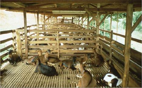
Figure 10.12 In the tropics, goats are often housed in raised pens on slatted floors as a means of controlling gastrointestinal parasitism. If the goats are fed fresh-cut herbage, however, they may still be exposed to infective larvae. Source: Courtesy of Dr. David M. Sherman.
In intensive management systems in temperate regions, goats are often continuously housed in barns on solid floors, fed stored feeds such as hay and silage, and allowed exercise in well-drained dry lots. Under these conditions, goats, in most cases, can be maintained relatively free of gastrointestinal nematode parasites, although certain species, e.g., Skrjabinema, Trichuris, and Capillaria, are adapted to successful transmission away from moist herbage. To be totally effective, confinement should be absolute for all age groups in the herd. However, a drawback of total confinement systems is that, if not carefully managed, the advantages gained by controlling nematodiasis may be lost to an increase in coccidiosis and respiratory problems.
Many small dairy herd owners practice only semiconfinement rearing, turning goats out on pasture after winter confinement. These pastures may contain some level of overwintering parasite larvae if stocked with goats the year before. Harsh winters with extended periods of cold or freezing weather sharply reduce larvae on pasture, but even under such conditions, a small proportion of larvae may still survive. After mild winters, the population of overwintering larvae is greater and goats turned out in spring can become infected. By late summer, when weaned kids begin grazing in earnest, pastures can be heavily infected.
Under conditions of seasonal pasturing, two strategic worming interventions may be justified and beneficial, based on the prevailing herd situation. Pregnant does should be dewormed within one month of kidding to minimize the exposure of kids to infective larvae associated with the periparturient egg rise, and all goats, including kids, should be treated with anthelmintics before turnout in the spring. Drugs employed should be effective against both adult and arrested larval stages. When possible, in temperate climates, spring turnout should be onto pastures not grazed by goats or sheep for a full year. Monitoring of infection levels by fecal egg counts from composite fecal samples should be done periodically during the grazing period to determine if there is a need for tactical anthelmintic therapy.
Some strategies for pasture management can reduce parasite burdens and risk to goats in spring. Pasture rotation is important and should be practiced when there is sufficient pasture area available. Avoid using the same pastures for goats two years in a row. Grazing cattle or horses the preceding year, growing and harvesting a hay or grain crop, and leaving the land fallow are some options to reduce the risk of dangerous overwintering larvae on a given pasture. Other tools include maintaining forage height greater than two inches, providing areas of browse within pastures or along fence lines for goats, maintaining a low stocking rate, grazing goats along with cattle, avoiding creation of wet patches associated with leaky waterers, and fencing off naturally occurring wet patches in pasture (Hale et al. 2007).
In some situations goats are grazed year-round, but confined at night in corrals for protection. In such a system in Tanzania, with a single rainy season, significant, superior weight gains were achieved in grazing goats when a strategic anthelmintic treatment was administered at the end of the rainy season (Connor et al. 1990).
In extensive grazing systems, continuous or prolonged grazing is the fundamental management scheme, as is often the case in fiber or meat goat production. The extent and severity of parasite infection depend on multiple factors, including stocking rates, herbage quantity and quality, season, and weather conditions. For example, Angora goats on the Edwards Plateau in west Texas exhibit a low prevalence of H. contortus infection, probably because range is extensive, browse is abundant, and rainfall is limited. When these goats are moved to east Texas and forced to graze intensively on improved pasture with little browse, and much more rainfall, clinical haemonchosis is widespread (Craig 1982).
Careful observation and monitoring of body condition, fecal consistency, and fecal ova counts are advisable in grazing animals to identify the effects of management and weather changes on the incidence of parasitic infection. In the case of haemonchosis, if 5-10% of the herd has fecal egg counts of 500 epg of feces or more, then tactical treatment is justified before the onset of clinical signs. Where H. contortus is the principal concern, FAMACHA scores can be used for monitoring and/or planning intervention.
When the predominant parasites and their behavior are well known, problems may be anticipated without monitoring. In Texas, tactical anthelmintic therapy has been automatically given to grazing Angora goats after heavy rains, on the reasonable presumption that acute haemonchosis is likely to ensue because of rapid, synchronous larval development. The use of tactical treatments must be weighed against concerns of promoting anthelmintic resistance. For best effect, tactical treatments should be accompanied by movement of stock to clean pastures to avoid immediate reinfection on contaminated herbage, though some animals should be left untreated, with parasites in refugia, to slow the development of anthelmintic resistance. If strip grazing is practiced, care must be taken that restricted grazing areas are of sufficient size for the animal population. Otherwise, the rate and intensity of larval buildup may be severe enough that the presumed beneficial effect of strip grazing is negated. The smaller the strip, the more frequently animals should be moved.
Drought affects the use of pastures. Management changes made in response to drought, such as congregation of animals for supplemental feeding and watering, may precipitate an increased incidence of parasitism. Supplemental feed should be fed in feeders, not from the ground, and should be provided at multiple sites, spread out to avoid the intensification of parasite ova in places of high animal density. This is even more important with point-source water supplies. If runoff or seepage from tanks or troughs is not properly avoided or drained away, heavy contamination of the soil with infective larvae can occur at points where goats congregate to drink.
In tropical regions, extensive grazing is often practiced and the lack of seasonal changes in temperature and humidity allows infective larvae to be continuously present in the environment. In this situation, the frequent, regular use of anthelmintics is often practiced to limit resident worm populations and minimize the effects of parasitism in goats. This approach is labor intensive and costly, and can accelerate the development of drug resistance. If such suppressive treatment regimens are necessary, it may be helpful to use limited-spectrum anthelmintics when the local parasite population permits it. For example, salicylanilides could be useful where the primary nematode problem is H. contortus and broad-spectrum therapy is not required. Treatment intervals of three weeks are suggested, so that larvae ingested since the preceding treatment will not have yet matured to adulthood and begun producing ova. Rotation of anthelmintics among the different classes of available drugs should be kept to a minimum, because too frequent rotation of drugs within an active season or cycle of parasite development can itself promote resistance. Also, when haemonchosis is the main concern, as it often is in tropical environments, selective deworming of anemic animals only based on FAMACHA scores can help keep parasites under control, while reducing the use of anthelmintics and increasing the proportion of nonresistant larvae on pasture from the goats that were not dewormed (in refugia). Whenever local conditions allow, producers should be encouraged to raise goats in confinement in raised pens on slotted floors, and not feed them fresh-cut herbage. Effective grazing systems have been developed for the tropics wherein small ruminants are moved to a new area of pasture after three to four days of grazing and not returned to the previous area for about one month (Barger 1999; Waller 2006). A longer rest period is required in cooler climates, because the larvae survive longer on pasture.
Where grazing of goats is practiced, mixed grazing of different livestock species is being recommended as a means of parasite control, on the presumption that even though cross-species transmission can occur, the parasites infecting one species are less suited to an alternative host and will therefore result in a less severe infection. The net effect is one of dilution of the parasites most infective for each host species. Another effect is less competition for available herbage and browse among grazing animals with different feeding preferences, leading to a better plane of nutrition for all. In Texas, goats make up 20-40% of the grazing population and may be mixed with sheep, cattle, deer, or any combination of the three. Mixed grazing, however, does open the door to potential cross-transmission of other diseases such as paratuberculosis.
In the face of growing anthelmintic resistance, other approaches to parasite control are also being adopted or explored. Selective deworming is one such approach, aimed at increasing the number of non-resistant larvae in refugia, as discussed above. Other approaches include breeding and selecting stock for parasite resistance, using forages with anthelmintic properties, administering copper oxide wires, and using anti-nematodal fungi. They are discussed briefly as follows.
Resistance can be defined as the ability of an animal to suppress establishment and/or subsequent development of worm infection. As discussed earlier, there is good evidence of a genetic basis for helminth resistance in small ruminants, and some heritability estimates have been reported for different breeds of goats relative to different parasites under a variety of environmental conditions. At present, there is little documentation in the literature that farmers are applying selective breeding to their herds and flocks for improved parasite control, but the experimental evidence indicates that it has potential as a useful parasite management tool and no doubt some are practicing it. Owners can select a breed with known resistance characteristics, or they can attempt selective breeding for resistance within their own herds. Veterinarians can work with farmers on the development of these breeding programs. In the published studies, various criteria or markers were used as a basis for selection, including fecal egg counts, PCV, total protein, serum pepsinogen, body condition scoring, or FAMACHA score based on the type of goat, production system, predominant parasites, and other factors. One important caveat in such an undertaking is that correlations of parasite resistance to other desirable production traits have not been well studied in goats, and the risk exists that selection for parasite resistance might also select for loss of desirable production traits. Breeding schemes for small ruminants relative to the development of resistance or resilience to endoparasites in the tropics have been reviewed (Baker and Gray 2004).
Host nutrition can play a significant role in mitigating the effects of parasites. Adequate and proper nutrition, particularly the provision of supplemental protein, can boost host resilience in the face of parasite infestation, and help minimize the adverse effects of the parasite burden (Coop and Kyriazakis 2001). Furthermore, some feeds may have a direct anthelmintic effect, for example forages high in condensed tannins such as sulla (Hedysarum coronarium), birdsfoot trefoil (Lotus corniculatus), big trefoil (Lotus pedunculatus), sainfoin (Onobrychis viciafolia), sericea lespedeza (Lespedeza cuneata), and chicory (Cichorium inty- bus). Several controlled studies have demonstrated the capacity of condensed tannin forages to produce an anthelmintic effect on trichostrongyles in goats compared to other forages. These studies include the grazing of sericea lespedeza at pasture in Oklahoma (Min et al. 2004), as well as the feeding of sericea lespedeza as pellets or hay in Georgia (Shaik et al. 2006; Terrill et al. 2007). The latter work indicated that the anthelmintic effect is retained through drying, pelleting, and storage of the forage. The mitigating effects produced included a direct anthelmintic effect on the adult worms in the gastrointestinal tract, as well as a reduction in parasite egg viability and/or larval development in feces (Shaik et al. 2006). Sericea lespedeza is well suited to the growing conditions of the southern United States and offers a possible adjunct to anthelmintic therapy for parasite control in that region, where the number of goats raised for meat has increased dramatically in recent years, along with a growing problem of gastrointestinal nematodiasis.
Other studies demonstrating anthelmintic effects against trichostrongyles in goats involved the feeding of tannin- rich sainfoin as hay in France (Paolini et al. 2003, 2005) and the feeding of dried leaves of Acacia karoo in Zimbabwe (Kahiya et al. 2003).
Another aspect of nutrition with the potential to be exploited for parasite control relates to observations that macrominerals and trace elements may modify the hostparasite relationship. In particular copper, in the form of copper oxide wire particles (COWP), was demonstrated to have anthelmintic activity against some nematodes in experimentally infected sheep, notably H. contortus (Bang et al. 1990). Subsequently a number of studies with COWP have been carried out in goats (Chartier et al. 2000a; Martinez Ortiz de Montellano et al. 2007; Burke et al. 2007b).
The source of COWP in these studies is generally a commercially available 2 g gelatin capsule containing COWP with a 1.7 g copper metal content that is marketed for control of copper deficiency and congenital swayback in lambs. Others have used 12.5 or 25 g COWP boluses marketed for cows, but the boluses are repackaged into small gelatin capsules to produce a dosage suitable for small ruminants (Hale et al. 2007). The results of these studies have been varied. The main anthelmintic effect is directed against H. contortus, as measured by reductions in fecal egg counts or parasite counts at necropsy. Little or no effect has been reported against other abomasal or intestinal trichos- trongyles such as T. circumcincta or T. colubriformis in goats. Duration of effect in pastured goats appears to be about three to four weeks, possibly up to six weeks, and the effects are more evident in young goats than adults.
Potential toxicity is a concern, though goats are less susceptible to copper toxicity than are sheep. Copper oxide is preferred over other copper salts because of its limited absorption from the intestine, thus reducing the risk of toxicity. Dosage is empirical, but is generally reported as 0.5 g for kids and 2 g for adults. Dosing can be repeated during the grazing season in kids up to a maximum of 2 g total dose. It has been suggested that COWP be administered selectively to heavily parasitized goats based on FAMACHA scores or other parameters, rather than dosing the whole flock because of concerns for development of resistance to COWP. However, the mechanisms of action for COWP are unknown, so risk of resistance is unclear. What is clear is that COWP can be a useful adjunct to parasite control where H. contortus is the main concern, but it must be coupled with other interventions in an integrated parasite management program to be most beneficial.
Biologic control of parasitic nematodes using nematopha- gous fungi is another tool currently under evaluation. A number of such fungi, which naturally occur in soil and feces, are known to kill infective third-stage and second- stage larvae, thus reducing the larval burdens in pastures. Some of these fungi, most notably Duddingtonia flagrans, are also capable of surviving passage through the alimentary tract of livestock. Therefore, research has been conducted in a variety of livestock species to determine if feeding spores of D. flagrans can reduce the number of free-living stages of gastrointestinal nematodes in feces and therefore pastures.
Results in species other than goats have been variable but promising. Several experimental studies have now been carried out in goats in the laboratory or in pilot trials (Paraud and Chartier 2003; Waghorn et al. 2003; Terrill et al. 2004; Paraud et al. 2005), which indicate, in general, that the fungus is capable of reducing the number of developing larvae of important parasitic nematodes in the goat. In a field trial (Paraud et al. 2007), young grazing goats in France were given a daily oral dose rate of 106 spores/kg bw for just over three months via inclusion in a mineral mix. Compared to controls, kids receiving the spores showed lower fecal egg counts and serum pepsinogen levels at the end of the grazing season, as well as a higher growth rate. D. flagrans spores are now marketed commercially as Bioworma® (International Animal Health Products, Huntingwood, NSW, Australia) in Australia, New Zealand, and the United States for feeding to grazing livestock to substantially reduce the numbers of infective worm larvae emerging from manure onto pasture.
The quest for effective vaccines against gastrointestinal nematode parasites has been long and arduous. One promising focus was the use of parasite gut membrane proteins as antigens for control of H. contortus (Knox et al. 2003), and indeed the first vaccine derived from such antigens became available against H. contortus in 2014. The vaccine was initially approved in Australia under the name Barbervax® (Moredun Foundation, Midlothian, UK) and now is also available in South Africa (Wirevax®) and the United Kingdom. It is approved for use only in sheep. The vaccine does not prevent or eliminate infection, so it is an addition to, not a replacement for, a comprehensive parasite control program.
Proper usage of the vaccine requires multiple applications. In advance of the high-risk season for Haemonchus infection at pasture, lambs should receive three “priming” vaccinations three to four weeks apart to confer immunity, and then a series of vaccinations six weeks apart over the season of risk to maintain protection. This vaccination protocol, though labor intensive, can reduce Haemonchus egg counts in lambs at pasture by 85%, and in some cases may actually be less costly than the repeated drenching with anthelmintic that might otherwise be required.
If the vaccine were to become available for goats, its use might have to be managed differently. Goats do not develop age-related immunity to gastrointestinal parasites the way sheep do, and therefore the vaccine might have to be given to grazing goats of all ages and not just kids (Kearney et al. 2016).
Paramphistomiasis or Rumen Flukes
Paramphistomes are trematode parasites commonly referred to as rumen flukes, stomach flukes, or conical flukes. Adult flukes may be present in the rumen of goats in large numbers, but are essentially non-pathogenic. Clinical paramphistomiasis is associated primarily with the immature forms that feed voraciously in the small intestine before moving to the rumen to mature.
Etiology
Numerous genera in the family Paramphistomatidae infect domestic ruminants. In goats, Paramphistomum cervi, Paramphistomum explanatum, Fischoederius elongatus, Gastrothylax crumenifer, and Cotylophoron cotylophorum are found. Cotylophoron travassosi, Cotylophoron bareil- liensis, and Cotylophoron fullerborni also have been reported from goats in Brazil (Cavalcante et al. 2000) and Paramphistomum daubneyi from goats in France (Silvestre et al. 2000). The taxonomic classification of paramphistomes is currently in flux. For example, P. daubneyi reported from France in 2000 is reported from Ireland in 2014 as Calicophoron daubneyi (Zintl et al. 2014).
These trematodes all have a similar, indirect life cycle involving water snails as intermediate hosts (Soulsby 1982). The size of mature flukes ranges from 5 to 20 mm, making them visible to the unaided eye. They are pink to red. One paramphistome of Asia, Gigantocotyle explanatum, migrates to and matures in the bile ducts. It is essentially non-pathogenic, and should not be confused with the pathogenic liver fluke Dicrocoelium dendriticum.
Adult paramphistomes inhabit the rumen and lay clear, operculated eggs that are passed in the feces. When feces are passed into water, miracidia develop from these eggs over a period of 12-21 days, depending on temperature. Swimming miracidia enter water snails of various genera, including Planorbis, Bulinus, Lymnaea, and others. Mature sporocysts containing rediae develop in snails over a period of 11 days. Rediae are released within the snail and after an additional maturation of 10 days, contain multiple cercar- iae. In turn, cercariae are released from rediae and mature within the snail for approximately 13 more days. Mature cercariae are released from snails into water under stimulation of strong sunlight. Liberated cercariae attach to herbage and encyst as metacercariae, remaining viable for as long as three months.
When ruminants ingest contaminated herbage, metacer- cariae excyst in the small intestine, where they feed aggressively and mature for a period of six to eight weeks. Young flukes then migrate anteriorly to the rumen, where they undergo additional maturation after attachment to the rumen mucosa, begin laying eggs, and complete the life cycle. Migration to the rumen and subsequent development in that organ can be delayed or prolonged up to several additional months when fluke infestations are heavy.
Development of flukes varies in goats, cattle, and sheep. After experimental infection of the three ruminants with metacercariae of Paramphistomum microbothrium, migration of maturing flukes from the intestine to the rumen was virtually complete in sheep and cattle by 34 days after challenge, but was just beginning in goats at that point, and subsequent egg laying began two weeks later in goats than in cattle or sheep (Horak 1967). This prolonged residence of maturing paramphistomes in the small intestine may contribute to increased pathogenicity of the fluke in the goat.
Epidemiology
Though paramphistome infection of domestic ruminants occurs throughout the world, it has been mainly from tropical and subtropical areas. Until recently, reports of clinical disease have been limited to Africa, Asia, Australasia, eastern Europe, Russia, and the Mediterranean countries (Horak 1971). However, the distribution of paramphistomes is spreading into temperate zones, with more recent reports of clinical disease in cattle and sheep from the UK and Ireland (Zintl et al. 2014). While most reports involve cattle and sheep, reports of caprine paramphisto- miasis from India (Katiyar and Varshney 1963; Chhabra et al. 1978; Rao and Sikdar 1981), Sardinia (Deiana et al. 1962), Bulgaria (Denev et al. 1985), and Pakistan (Mohiuddin et al. 1982) suggest that goats are at risk wherever sheep and cattle are affected. However, one abattoir survey from Iran (Moghaddar and Khanitapeh 2003) identified paramphistome infection in sheep up to 4% but no infection in goats, while a survey in the former Zaire (Chartier et al. 1990) showed an infection rate of 54% in sheep but only 14.8% in goats, so species differences in exposure or response to exposure may occur.
In general, outbreaks of paramphistomiasis are increased during dry seasons, when snail and livestock populations become concentrated around shrinking natural water sources with restricted grazing and browsing (Horak 1971). Animals grazing in low-lying, poorly drained, swampy areas or with access to irrigation ditches and other standing water supplies are at high risk of infection caused by increased contact with metacercariae produced by water snails.
In north central India, most outbreaks are seen in small ruminants between late September and January after the rains that promote increases in snail populations. During subsequent dry periods of decreased forage availability, goats may be fed rice paddy straw and other grasses grown underwater that contain large numbers of metacercariae. The disease can be costly. Morbidity rates in goats in India during such outbreaks have been reported in the range of 35-79% and fatality rates in the range of 45-88% (Katiyar and Varshney 1963). Slaughterhouse surveys in the region show that mature rumen fluke burdens are highest from March to October and lowest from November to February, while immature fluke burdens are highest from September to April, the time period correlating to clinical outbreaks (Gupta et al. 1985).
Pathogenesis
The immature fluke developing in the upper small intestine is most responsible for the pathologic effects of paramphistome infection. These immature flukes embed deeply in the intestinal mucosa via suckers and feed by drawing a plug of mucosa into the sucker. This mucosal plug becomes necrotic and sloughs, leaving an erosion and petechiation. The pathogenicity of the paramphistomes is correlated directly with the immature fluke burden. It is estimated that attachment of 50 000 immature flukes would completely denude the upper 3 m of small intestine, where most paramphistome infections are confined during the developmental stage. Worm burdens of this intensity occur under natural conditions. Large numbers of simultaneously feeding, immature flukes cause marked intestinal irritation and mucosal disruption, leading to hypoproteinemia, diarrhea, and general debilitation.
Clinical Signs
The clinical signs of paramphistomiasis are similar to those of nematode gastroenteritis and the two diseases commonly occur together. Young goats are more frequently and seriously affected than older goats. Affected goats are listless and have diminished appetite. They may be polydipsic, and stand for long periods with their muzzles in the water. They will show a fluid diarrhea that may be projectile at first, but later may just drip from the rectum, staining the hindquarters. The diarrhea may contain mucus, epithelial shreds, and immature flukes. It has a characteristic and pronounced fetid odor. Intermandibular edema is pronounced and may extend over the face and brisket. Anemia, if present, is usually mild or moderate. The course of the disease is approximately 5-10 days in goats. During that time the animals lose weight, get progressively weaker, and eventually die. They may pass nothing but copious mucus from the rectum in the terminal stages. A prolonged form may occur where animals survive but persist in the cachexic state.
Clinical Pathology and Necropsy
Marked hypoproteinemia and hypoalbuminemia occur. Anemia may also be present. Because clinical disease is caused by immature non-egg-laying flukes, fecal sedimentation techniques for egg identification have little value, because false-negative results would be common. As an alternative to checking for ova, diarrheic feces can be passed through a sieve with 53 μ apertures and the residue examined either microscopically or macroscopically against a black background for immature flukes that are commonly passed in the feces. The immature flukes are pinkish-white with a large, prominent sucker (Horak 1971). Development of new techniques for detection of liver fluke infestation through examination of feces such as coproantigen ELISA (Kajugu et al. 2015) and loop-mediated isothermal amplification (LAMP) assay (Martinez-Valladares and Rojo-Vasquez) for detection of DNA suggest that these techniques may become available for identification of rumen fluke infestation as well.
At necropsy, the carcass is thin and the hindquarters are soiled with diarrheic feces. Subcutaneous edema, ascites, hydrothorax, hydropericardium, and lung edema may be present. Significant gross lesions of the alimentary tract are limited to the pyloric portion of the abomasum and the first 2-3 m of the small intestine. The abomasal folds are enlarged and edematous, and immature flukes may be seen attached to the mucosa, which also contains numerous erosions and petechiae. The affected intestine is thickened and edematous. The pitted mucosal surface is covered with catarrhal exudate. Careful inspection may reveal flukes deeply embedded in the mucosa, with only the external ends present in the lumen. The mucosa in this affected region appears corrugated, with many elevated ridges and multiple foci of hemorrhage. Immature flukes may also be found free in the gut lumen. Mature flukes may be present in the rumen and omasum. Multiple intestinal diverticula measuring 2 cm in diameter have been reported as an unusual finding associated with paramphistomiasis (Prajapati et al. 1982).
Histologically, affected areas of intestine show hypertrophy, edema, inflammatory cellular infiltrates, and fibrosis in the mucosa and submucosa (Sharma Deorani and Katiyar 1967). Histopathologic effects of adult flukes in the rumen are limited to epithelial desquamation of the papillae (Singh et al. 1984).
Diagnosis
Presumptive diagnosis is difficult, because of the clinical similarity of paramphistomiasis to nematode gastroenteritis and because both may occur simultaneously. Haemonchosis is more likely to produce anemia and less likely to produce diarrhea. However, other trichostrongyle infections may mimic paramphistomiasis exactly. Definitive diagnosis depends on ruling out nematode gastroenteritis by fecal flotation or necropsy, and ruling in par- amphistomiasis by identification of immature flukes in feces or at necropsy. Chronic fascioliasis must also be considered in the differential diagnosis. It occurs under similar environmental circumstances and may appear similar to the chronic form of paramphistomiasis.
Treatment
Elimination of the immature flukes with appropriate anthelmintic therapy is the major therapeutic objective and is lifesaving if treatment is started early in the course of disease. Animals must be removed from the source of infection before treatment, or they will be immediately reinfected. Morantel citrate at a dose of 6 mg of morantel base/kg bw has been shown to be 99.5% effective against immature paramphistomes (Srivastava et al. 1989). Other drugs with a reported efficacy of more than 95% against immature flukes in sheep and goats include bithionol (25-100 mg/kg), niclo- folan (6 mg/kg), niclosamide (50-100 mg/kg), and resoran- tel (65 mg/kg) (Rolfe and Boray 1988). Resorantel is also 100% effective against adult paramphistomes in goats (Sahai and Prasad 1975). Oxyclozanide (15 mg/kg) has a slightly less consistent efficacy range of 85-100% against immature flukes, but is also 100% effective against adult flukes. In general, the benzimidazoles are minimally effective against paramphistomes despite their relative effectiveness against liver flukes. Bithional may be toxic to goats at the higher end of the effective dose range (Boray 1985).
Directing treatment toward adult flukes is controversial. It has no direct benefit in the face of clinical outbreaks. Elimination of adult flukes reduces fecal egg counts and subsequent infection of snails, thereby reducing the general environmental burden of paramphistomes. Elimination of adult flukes, however, may also reduce the level of immunity that they induce in infected hosts, thereby increasing the risk of clinical disease in subsequent exposures (Horak 1971).
Control
The major thrust in the prevention of paramphistomiasis is to keep grazing animals away from areas with heavy concentrations of infected snails and from herbage contaminated with metacercariae. This means avoiding low-lying, poorly drained, flooded, or swampy areas, ponds, ditches, and paddy fields. Small ruminants should not be grazed with cattle under such circumstances, because the latter are known to shed large numbers of eggs and promote increased infection of snails.
Forages from contaminated areas can be used safely if ensiled or made into hay before feeding. When local conditions do not permit restricted grazing or harvesting, molluscicides may be used to reduce snail populations, but the relative cost-effectiveness of this remains to be proven. Water for drinking can be pumped from contaminated areas, treated with molluscicides, and provided in raised troughs, but this too is capital intensive.
Strategic use of anthelmintics can be helpful in controlling paramphistomiasis. In India it is recommended that livestock be treated in June and August to reduce mature fluke burdens and thereby reduce egg laying, and again in November, January, and March to control immature flukes (Gupta et al. 1985).
Though not yet available for widespread practical application, immunization may be a major future tool in param- phistomiasis control. Oral vaccination with irradiated metacercariae significantly reduced paramphistome burdens in goats on subsequent challenge compared with unvaccinated control goats (Horak 1967; Hafeez and Rao 1981).
Intestinal Cestodiasis or Tapeworms
Intestinal tapeworms occur in goats throughout the world. Relative to gastrointestinal nematodiasis, cestodiasis in goats usually has little clinical or economic significance. However, goat owners are often keenly aware of tapeworms because the proglottids, or egg packets, shed by the adult worm in the goat's feces are visible to the naked eye.
Etiology and Pathogenesis
The major intestinal tapeworm of goats worldwide is Moniezia expansa, though other Moniezia spp. may also infect goats. Avitellina spp. occur in goats in Europe, Africa, and Asia, often in conjunction with Moniezia spp., and are sometimes the predominant cestode (Raina 1973). Thysaniezia giardi infects goats in Europe, the former USSR, Africa, India, and North America. Stilesia globi- puncta occurs in goats and other ruminants, mainly in tropical regions of Africa and Asia. It is potentially the most pathogenic of the ruminant tapeworms, producing inflammatory nodules at the sites of attachment in the duodenal and jejunal mucosa that can lead to enteritis and diarrhea. Nevertheless, clinical illness in goats attributable to Stilesia infection is poorly documented.
Another Stilesia tapeworm, Stilesia hepatica, occurs in the bile ducts of ruminants in Africa and Asia, and several new or additional Stilesia spp. have been identified from goats in India as well. Thysanosoma actinioides, the fringed tapeworm, can be found in the small intestine, though it is more commonly located in the bile and pancreatic ducts. It occurs only in western North America and South America. Principally a tapeworm of sheep and deer, its prevalence in goats is poorly documented. There is one slaughter survey from Mexico in which the fringed tapeworm was found on average in about 1.5% of the livers of goats examined, compared to about 15% for sheep livers (Cuellar Ordaz 1980). It has been cited as a caprine parasite in the United States as well (Guss 1977). The liver tapeworms are discussed in detail in Chapter 11.
These anoplocephalid tapeworms all use ruminants as definitive hosts and various species of oribatid or psocid mites as intermediate hosts. Adult tapeworms developing in the goat intestine can be up to several meters long (Figure 10.13). They consist of a head or scolex and short neck followed by a long, segmented body composed of proglottids. The most posterior proglottids are packed with eggs. These gravid segments separate from the worm and are passed in the feces. These egg packets are white and 1-1.5 cm in length. They are visible in the fecal pellets and have the appearance of rice (Figure 10.14). The proglottid casing gradually disintegrates and oribatid or psocid mites, common in soils and on herbages, ingest the eggs. Infective cysticercoids develop within these arthropod intermediate hosts over a period of four months. Infective mites are consumed by ruminants while they feed. Cysticercoids are released from the ingested mites in the intestine and mature tapeworms begin to develop. The prepatent period in the goat is around 40 days for Moniezia spp.
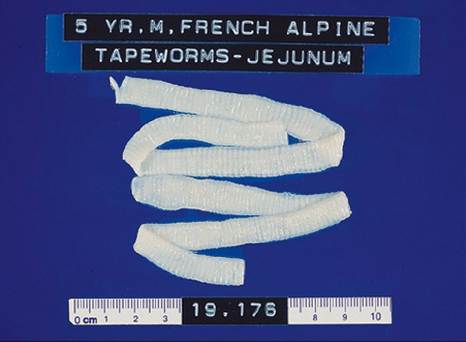
Figure 10.13 Adult Moniezia tapeworm from the intestine of a goat. Source: Courtesy of Dr. T.P. O'Leary.
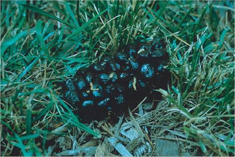
Figure 10.14 Tapeworm proglottids in goat feces. Source: Reproduced by permission of Dr. C.S.F. Williams.
Table 10.9 Overview of cestode infections of goats.
| Cestode | Definitive host | Intermediate hosts | Form in goat | Locations in goat | Impact |
| Moniezia expansa, other Moniezia spp. | Goats, other ruminants | Mites | Adult tapeworm | Intestine | Usually non-pathogenic |
| Avitellina spp. | Goats, other ruminants | Mites | Adult tapeworm | Intestine | Usually non-pathogenic |
| Thysaniezia spp. | Goats, other ruminants | Mites | Adult tapeworm | Intestine | Usually non-pathogenic |
| Stilesia globipunctata | Goats, other ruminants | Mites | Adult tapeworm | Intestine | Occasionally diarrhea |
| Stilesia hepatica | Goats, other ruminants | Mites | Adult tapeworm | Bile ducts | Liver condemnations |
| Thysanosoma actinoides | Goats, other ruminants | Mites | Adult tapeworm | Bile ducts | Liver condemnations |
| Taenia multiceps | Canids | Goats, other ungulates, humans | Metacestode cysts | Brain, spinal cord, muscle | Coenurosis, lameness, trim at slaughter, zoonosisa |
| Taenia hydatigena | Canids | Goats, other ruminants, pigs | Metacestode cysts | Liver, mesentery, omentum | Liver condemnation, trim at slaughter |
| Taenia ovis | Canids | Goats and more commonly sheep | Metacestode cysts | Heart, diaphragm, other muscle | Trim or condemnation at slaughter |
| Echinococcus granulosus | Canids | Goats, other ungulates, humans | Metacestode (hydatid) cysts | Liver, lungs, central nervous system, bone, muscle | Liver disease, slaughter condemnation, zoonosisa |
a The zoonotic potential of these cestodes does not derive from contact with goats or other intermediate host ruminants, but from contact with feces of definitive host canids.
The goat also serves as an intermediate host for a number of cestode parasites that use canids as definitive hosts. In many cases, these infections are more economically and clinically significant than the adult tapeworm infections just listed. All of these intermediate metacestode infections are addressed elsewhere in this text, in conjunction with the organ system most often affected. A summary overview of the various cestode infections is given in Table 10.9.
Light infestations of goats by the common tapeworms are generally non-pathogenic (Soulsby 1982; Williams and Schillhorn van Veen 1985). This is largely because tapeworms do not feed destructively with active mouthparts, but rather absorb nutrients from the intestinal lumen through their integument. In goats, at least 50 worms are required to produce deleterious effects. Several hundred may be present in an individual goat (Guss 1977). They may compete significantly for nutrients with the host, leading to ill-thrift. They may produce luminal distension in the intestine, resulting in a distended, pot-bellied appearance. Their presence may prolong transit time of ingesta in the gut. In feedlot lambs on grain rations, this is believed to promote the development of clostridial enterotoxemia. A similar interaction has not been described in goats. Massive tapeworm infections may sometimes even occlude the intestinal lumen. This results in signs of colic and may lead to spontaneous rupture of the intestine in young lambs and kids, with dire consequences.
Epidemiology
Infections are most often observed in young kids at pasture during their first summer grazing season. Pasturing is not a prerequisite for infection, however, because the infective mites may be present in the barnyard or on carried forage. The pattern of infection may vary with climate and geography. In Nigeria, no seasonal variation was seen in the incidence of sheep and goat tapeworms in the dry savannah and desert regions, while the incidence was increased during the rainy season in the rain forest zone (Enyenihi et al. 1975). A natural resistance to cestode infection develops with age, and in populations of goats with constant exposure to tapeworms, worm burdens are always less severe in older goats.
Because tapeworms are often derived at pasture, heavy tapeworm burdens may also be a sentinel for heavy nematode infections. Therefore, when owners notice tapeworm proglottids in goat feces, deworming for tapeworms may be used as an opportunity to treat more serious nematode infections when broad-spectrum anthelmintics are used. Multiple parasitic infections are the rule, not the exception, in goats.
Clinical Signs
When clinical disease is associated with tapeworms, it usually involves young goats under 6 months of age. Affected animals may show poor growth rates and a pot-bellied appearance. Proglottids are present in voided feces that are usually normal in appearance, but may be soft or unpelleted. Constipation may also occur. In complete luminal obstruction caused by tapeworms, kids have symptoms of colic and decreased fecal output. Animals that have ruptured the intestine may be profoundly depressed, or found moribund or dead.
Clinical Pathology and Necropsy
Proglottids observed in the feces are diagnostic, but proglottids sometimes degenerate before being shed. Examination of feces using fecal flotation methods will reveal characteristic tapeworm eggs. M. expansa ova are distinctly triangular. There are no other specific clinical chemistry or hematologic abnormalities associated with tapeworms.
At necropsy, the long, white-segmented tapeworms are strikingly obvious in the lumen of the small intestine. In obstructions, they pack the lumen and may be free in the abdominal cavity when rupture of the intestine has occurred.
Diagnosis
Tapeworm infections are definitively diagnosed by the presence of proglottids in the feces or worms in the intestine. Attributing disease to tapeworm infection is more problematic. It is essential that nematode parasitism, par- amphistomiasis, and liver fluke disease all be thoroughly ruled out before clinical disease is ascribed to tapeworms.
Treatment
A wide range of anthelmintics are currently available for eliminating adult tapeworms in goats. Oral niclosamide (50 mg/kg bw) is highly effective and has a wide margin of safety; it is non-toxic at five times the recommended dose. Praziquantel (5 mg/kg bw) is also effective, but the injectable form can be highly irritating to goats at the injection site. Oral febantel (5 mg/kg) is effective against M. expansa and a range of nematodes. The newer benzimidazoles are also effective against tapeworms and nematodes. These include mebendazole (15 mg/kg), fenbendazole (15 mg/ kg), cambendazole (20 mg/kg), and oxfendazole (10 mg/ kg). Albendazole (10 mg/kg) is effective against tapeworms, nematodes, and the adult liver fluke Fasciola hepatica. Cautions associated with the use of benzimidazoles, particularly with regard to the development of anthelmintic resistance, are discussed in the section on treatment of nematode gastroenteritis.
Older treatments for tapeworms in goats include copper sulfate, nicotine sulfate, and phenothiazine with lead arsenate. Though reasonably effective, these compounds all are potentially toxic if misused or improperly dosed.
Control
It is difficult to justify a preventive program aimed at adult tapeworm control on economic grounds, because the clinical impact on goats is usually minimal. When the appropriate broad-spectrum anthelmintics are used for nematode parasite control, however, tapeworm control can be included in the program as a bonus. The group of major concern in both cases is the young goat on pasture for the first time. Given current concerns about the development of anthelmintic resistance in nematode parasites, it would not be prudent to administer benzimidazoles expressly for the control of tapeworms, unless there was also some strategic or tactical justification for their use against gastrointestinal nematodes at the same time.
Controlling the intermediate host mites is difficult because these arthropods are ubiquitous and often present in enormous concentrations. Plowing and reseeding are considered to reduce mite populations at pasture, but the overall effect on tapeworm populations in goats is questionable. Pasture rotation with at least a one-year layoff may be helpful, because mite populations decrease over winter.