Hiatal hernia
A hiatal hernia results from an abnormal or stretched phreni- coesophageal ligament, which allows herniation of the abdominal esophagus, the gastroesophageal junction, parts of the stomach, and/or other abdominal organs into the thoracic cavity via the hiatus.
A sliding, axial hiatal hernia occurs when the abdominal esophagus and part of the stomach slide up into the chest as a unit. A paraesophageal hiatal hernia occurs when part of the stomach and /or other abdominal organs enter the chest next to the esophagus.74 A congenital form of a sliding hiatal hernia has been described in the Chinese Shar Pei and appears to be the most common form.74,75 Sliding hiatal her-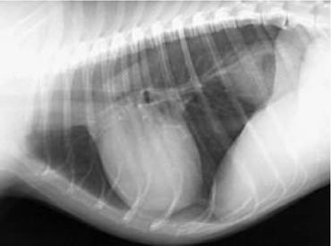
Figure 3.12:
Radiograph of a dog with intermittent vomiting and regurgitation. Lateral thoracic radiograph of a 4-year-old castrated male Beagle with intermittent bouts of vomiting and regurgitation.
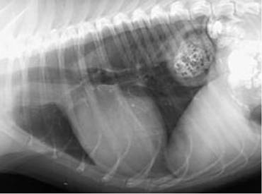
Figure 3.13:
Hiatal hernia. Barium contrast study of the same dog as shown in Figure 3.12 delineating the hiatal hernia.
nias can occur in cats, but are uncommon overall.76,77 Paraesophageal hiatal hernias are rare. Hiatal hernias can be acquired due to trauma.
The most common clinical signs of hiatal hernias are a result of GER and include occasional or persistent regurgitation, vomiting, dysphagia, dyspnea, and/or ptyalism.75-77 However, some animals with mild disease may be asymptomatic.74 The diagnosis may be evident from survey thoracic radiographs. A soft-tissue opacity may be noted in the caudodorsal thorax, in the area of the esophagus (Figure 3.12). Distal esophageal dilation may also be seen. A hiatal hernia is often a dynamic condition so that multiple radiographic exposures may be necessary to yield a diagnosis.In some cases,contrast videofluoroscopy may be needed to confirm the diagnosis (Figure 3.13). The condition is more difficult to diagnose via endoscopy.
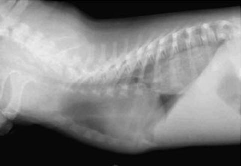
Figure 3.14:
Gastroesophageal intussusception. Lateral thoracic radiograph of a 4-month-old male Labrador Retriever showing a soft tissue mass effect in the area of the distal esophagus. The puppy was diagnosed at endoscopy and surgery with a gastroesophageal intussusception.
Surgical correction is usually required for animals with congenital disease.74-77 Those with acquired disease may respond to medical management consisting of small frequent meals, H2 receptor antagonists, prokinetic agents, and/or sucralfate suspension.74,77
3.3.10