Infections Due to Histoplasma capsulatum var. farciminosum
Following traumatic inoculation, the development of the subspecies farciminosum is usually responsible for nodular cutaneous lesions. In the skin, the fungus induces an inflammatory lesion containing granulocytes, macrophages and multinucleated giant cells.
A fibrous and oedematous peripheral reaction further surrounds the nodule. An abscess progressively develops and starts to discharge yellow pus containing yeasts, macrophages and granulocytes. Finally, a granulation tissue appears and an ulcer with inverted borders is formed. Infected leucocytes or yeasts are able to spread the infection via lymphatic vessels to adjacent tissues. Bacterial superinfection may occur.In equids, the subspecies farciminosum is responsible for a debilitating disease called epizootic lymphangitis (Guerin 2010; Scantlebury and Reed 2009). The incubation period ranges from 1 and 7 months. The classical presentation is a superficial lymphangitis. Sometimes genital organs or bones are involved. Hyperthermia is rarely reported. The lesions are present on the skin and more rarely on the mucous membranes (lips, conjunctiva, nasal or respiratory epithelium). Five types of lesions have been described:
1. The initial lesion is a cutaneous ulcer with inverted borders and painful outline; thick, yellowish and sometimes bloody pus is produced, mostly observed on the limbs, thorax, chest, neck and head.
2. The cord is a congested or knotted rope lymph vessel (Fig. 5.3).
3. The spots are hemispheric nodules, 0.5-3 cm diameter, tough and painless, isolated or lined on a cord.
4. The tumours are located on lymph nodes, up to 5-30 cm diameter, and may turn into fistula.
5. The engorgements are due to a diffuse reaction within the conjunctive tissue and are observed around the lower limbs.
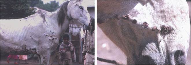
Fig.
5.3 Lesions of equine epizootic lymphangitis in Ethiopia. (Left) Ulcers and nodules; (right) cordlike lesions (courtesy from Christine Guerin)
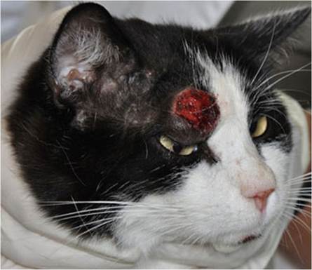
Fig. 5.4 A lesion of feline histoplasmosis due to Histoplasma capsulatum var. farciminosum (Fischer et al. 2013)
Infected horses become restless because of the disturbance due to numerous flies attracted by cutaneous lesions. There is a progressive loss of appetite and condition as severity of the disease increases. Because of its debilitating nature and its high case fatality, some horse-owners in Ethiopia have started to refer to equine histoplasmosis as “horse AIDS” (Ameni 2006). In a study investigating the economics of the carthorse industry in Ethiopia, Aklilu and Zerfu (2010) reported that losses to the owner due to morbidity of a horse with histoplasmosis resulted in more than a 50% reduction in daily earnings.
Infection due to H. capsulatum var. farciminosum has been reported in animals other than equids. Histoplasmosis in four dogs diagnosed in Japan lacked pulmonary or gastrointestinal lesions and was characterised by multiple granulomatous or ulcerated lesions on the skin and in the mouth. The subspecies farciminosum was reported as the causative agent in these cases (Murata et al. 2007). In Europe, similar observations have been made in badgers in Germany (Eisenberg et al. 2013) but also in cats in Switzerland (Fischer et al. 2013) and eastern France (Fantini et al. 2014, Guillot et al. 2015) (Fig. 5.4).
5.6