Introduction
The variety of diagnostic imaging modalities that have become available to veterinarians has led to the need for expanded expertise in ultrasound, computed tomography (CT), nuclear scintigraphy, and magnetic resonance imaging (MRI).
However, conventional radiography and ultrasonography remain essential, cost-effective, and readily available methods for diagnosing many gastrointestinal disorders in dogs and cats. In the past 10 years, ultrasonography has become an invaluable diagnostic tool, which has virtually replaced the need for barium contrast studies in animals presenting for vomiting and/ or diarrhea. Historically, the combination of plain and contrast radiography was considered the gold standard for the examination of the gastrointestinal tract. Currently, however, the combination of sonography and endoscopy has become a more frequently used approach.Radiography and ultrasonography of the abdomen should be performed together since they can each provide complementary information. Survey radiographs provide a global view of the abdomen that cannot be obtained with sectional imaging such as ultrasonography. Survey radiographs may also allow an immediate diagnosis to be made, as is the case in patients with an intestinal obstruction. Indications for plain radiography may include dysphagia, regurgitation, vomiting, acute abdomen, constipation, abdominal pain, abdominal distension, or a palpable mass. In animals with chronic diarrhea or marked abdominal effusion, abdominal radiographs are less beneficial. The spectrum of gastrointestinal diseases that can be detected by ultrasonography include intussusceptions, pancreatitis, peritoneal infiltrative disease, gastrointestinal wall infiltrations, abdominal neoplasia, and hepatobiliary disease. Ultrasonography may also provide functional information in intestinal motility and hemodynamic disorders.
Furthermore, ultrasound- guided, percutaneous tissue biopsy can be performed to collect samples for cytological or histological examination. However, the clinical usefulness of ultrasonography is highly operatordependent and the detection and interpretation of changes relies heavily on the ultrasonographer's expertise.Barium contrast studies of the gastrointestinal tract are generally only indicated when the combination of clinical information, survey radiography, and ultrasonography do not lead to a diagnosis.1 Contraindications to performing barium contrast studies include survey radiographic evidence of obstructive disease or free peritoneal gas or liquid. Although iodinated contrast agents may be used, they should be avoided in debilitated or dehydrated patients since they can worsen the condition of the patient or lead to a hypovolemic state. 2
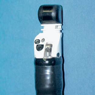
Endoscopic ultrasound is commonly used in human gastroenterology to examine the gastrointestinal tract and for performing guided transluminal biopsies. Although this technique has been underutilized in veterinary medicine, it can be applied in both canine and feline patients.3 A high frequency transducer mounted on the tip of a conventional video endoscope (Figure 1.8) allows the acquisition of high resolution images of the esophageal wall, stomach, liver, pancreas, lymph nodes, adrenal glands, intra-abdominal vasculature, kidneys, spleen, duodenum, jejunum, and proximal colon.
Figure 1.8:
Echoendoscope. This figure shows the tip of an Olympus GF-UC140-AL5 video gastroendoscope (Olympus Optical, Hamburg, Germany). The ultrasound transducer is mounted at the tip, while the optics and working channel are mounted at an angle on the side. The transducer is a multi-frequency (5-10 MHz) curved (180°) linear array transducer designed for guided tissue aspirations.
Contrast-enhanced ultrasonography is being used with increasing frequency in veterinary medicine.
Modern ultrasound contrast agents are gas-containing stabilized microbubbles that remain intact in the vascular space for several minutes after intravenous injection and increase the intensity of the backscattered ultrasound.4 In human medicine, contrast-enhanced ultrasound has been most widely used for the differentiation of malignant versus benign focal liver lesions.5 It has recently been shown that contrast harmonic ultrasound of the liver can be used to detect portosystemic shunting in dogs.6CT, MRI, and nuclear scintigraphy are alternative methods for the investigation of the abdomen in cats and dogs.7,8,9 However, there are only a few reports about the use of these modalities in small animal gastroenterology and these methods have not yet attained common usage for investigating gastrointestinal disorders. CT may be performed for the detection of abdominal neoplasia, pancreatic imaging, or for the detection of portosystemic shunts.10,11 Only a few reports concerning the use of MRI for the examination of the abdomen in dogs and cats are available and more work is clearly needed in this field.12 Nuclear scintigraphy provides functional information about the gastrointestinal tract and has been well established in veterinary medicine. In the hands of an experienced user, rectal scintigraphy has significantly improved the rapid diagnosis of portosystemic shunts and allows for the quantification of the shunting fraction both pre- and post- operatively.13,14 Nuclear scintigraphy has also been used for the diagnosis of hepatobiliary disease and the quantification of gastric emptying time.7,15
This chapter provides guidelines for a diagnostic imaging approach in dogs and cats with gastrointestinal disorders. Each section discusses the imaging of different anatomic regions according to specific clinical signs. Imaging for the evaluation of hepatobiliary and pancreatic disorders is described in a separate section. Although radiography and ultrasonography are emphasized, indications for alternative imaging modalities are also described.
1.3.2