Laboratory Diagnosis
The ideal clinical specimen to be sent to the laboratory is a biopsy in both humans and dolphins (Vilela and Mendoza 2015). However, skin smears of the granulomas collected with a scalp blade (Talhari et al.
2009) or the Scotch tape technique (Miranda and Silva 2005) are also valuable tools for the microscopic visualization of the pathogens. Both L. loboi and P. brasiliensis var. ceti resist culture; thus the use of traditional laboratory media has to be performed with the purpose only of ruling out other pathogens.To perform the wet mount technique, the biopsy is divided into 2-3 mm cubes and placed onto a slide containing two drops of 10 or 20% KOH (Vilela and Mendoza 2015). The addition of calcofluor enhances sensitivity when observed under fluorescence. The samples are heated for 5 min and then left on the counter. In wet mount preparations, L. loboi from humans and P. brasiliensis var. ceti from dolphins appear as single spherical or oval yeast-cells developing branching chains of cells (Fig. 9.6). The yeast-like cells in dolphins measure ~2-10 μm, whereas in humans ~4-14 μm, according to Haubold et al. (2000). In 10% KOH, the cells appear hyaline with a thick cell wall (Fig. 9.6).
Experimental inoculation of L. loboi has been successfully obtained in several mammalian species including humans (Belone et al. 2001; Borelli 1962; Madeira et al. 2000). The mouse model proposed by Madeira et al. (2000) showed better results using propagules from human lacaziosis. Inoculated mice on the injected pads developed small granulomas that slowly enlarged. In histopathology, diffuse infiltrate-containing macrophages, lymphocytes, plasma cells, fibrosis, and numerous yeast-like cells can be observed. There are no official reports of P. brasiliensis var. ceti experimental infection in mice. Incidentally, few years ago, one of the authors (LM) was informed of a successful experimental inoculation in a mouse injected with yeast-like cells from an infected dolphin (Dr. Libero Ajello, personal communication).
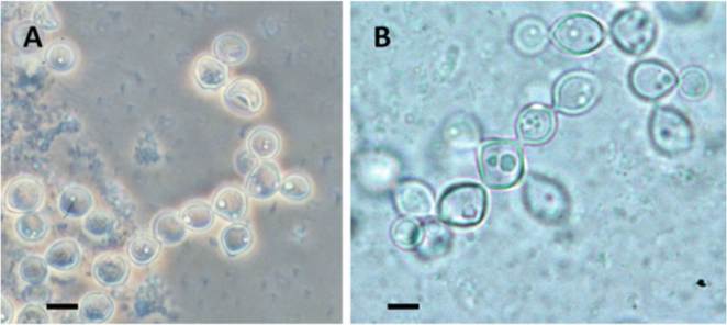
Fig. 9.6 (a) Paracoccidioides brasiliensis var. ceti yeast-like cells in 10% KOH wet mount preparation from a case of dolphin with cutaneous granulomas (bar = 10 μm). Presence of uniform single cells and branching chains of yeast-like cells (Nomarski microscopy). (b) A wet mount (10% KOH) preparation from a human lacaziosis case (bar = 10 μm). Presence of yeast-like cells in chain
9.7