Lid Abnormalities
Lacerations of the eyelids should be repaired using standard techniques. Skin diseases involving the lids (e.g., contagious ecthyma, capripox, mange, dermatomycosis, dermatophilosis, staphylococcal dermatitis, zinc deficiency, ticks) are described in Chapter 2.
When skin lesions are restricted to the eyelids, the possibility that they are caused by ocular discharge or rubbing in response to ocular discomfort should be investigated. Injuries to the face may occur as a consequence of blindness. Small palpebral fissures, thickened non-pliable lids, and partial prolapse of the nictitating membrane occur in Nubian kids with inherited beta mannosidosis (Render et al. 1989), which is discussed in Chapter 5.Entropion
Entropion is the turning inward of the lower lid or both eyelids so that the eyelashes rub on the cornea. The condition is painful and results in tearing and retraction of the eyeball. The lids are partially closed; the lashes may be stuck together with exudate.
Congenital or Primary Entropion
When a kid's eyelids are malformed from birth, the condition is usually recognized within the first few days of life. The longer the entropion remains uncorrected, the more likely it is that a serious infection or corneal ulceration will result. It is probable, though unproven, that this condition has a hereditary component. Thus, if surgical treatment is required on humane grounds, the identity of the goat should be recorded so that it can be slaughtered for meat or sterilized rather than being permitted to reproduce.
Spastic Entropion
Older animals may develop entropion as the result of prolonged squinting when another painful eye condition is present (Figure 6.5). Typical examples are severe keratoconjunctivitis or the presence of a foreign body in the eye. If one goat continues to suffer from “pinkeye” after the rest of the herd has recovered, a point should be made of examining its eyelids.
When the entropion is of spastic origin, surgically altering the eyelids is not always necessary. Instead, the lid position can be corrected by sutures or wound clips long enough for the initial and secondary problems and pain to be resolved.Other Causes of Secondary Entropion
It is possible for entropion to follow a previous injury to and deformation of the eyelid. A small or collapsed globe also permits the lids to roll inward, as does retraction of a painful eyeball deeper into the orbit. Severe dehydration and emaciation (as from starvation or parasitism) are perhaps the most common causes of secondary entropion.
Corrective Measures
The literature is replete with techniques for correction of entropion in goats and in other species (Rook and
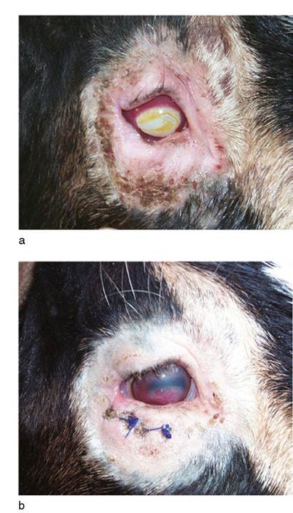
Figure 6.5 (a) Chronic spastic entropion with severe keratitis and hair loss. (b) Same goat, 12 days after surgical correction of the entropion by removal of an ellipse of skin below the eye. Source: Courtesy of Dr. M.C. Smith.
Cortese 1981; Wyman 1983; Baxendell 1984; Moore and Whitley 1984; Whittaker et al. 1999; Pearce and Moore 2013). In general, the veterinarian should not rush into a complicated and therefore expensive technique such as the Hotz-Celsus procedure (Donnelly et al. 2014) without giving consideration to one of the more temporary but often effective methods developed by ingenious shepherds and private practitioners.
One such simple but versatile technique is the use of Michel wound clips (Miltex Instrument Co., New York, USA; Eales et al. 1984) or metal surgical staples, three or four per affected lid, to pinch up a fold of skin adjacent and parallel to the lid margin. No anesthesia is required; it is simple to remove and replace a clip if the effect of its placement is not immediately satisfying, and the clips fall out on their own over time. The owner of a newborn kid may be able to achieve correction simply by drying the area and manually rolling the lid margin outward as often as possible over the course of one or two days.
Another version of this approach is to roll superglue onto the skin below the eyelid with a toothpick (which serves only as the applicator), causing the skin to evert and adhere to itself (Mongini 2007). Tensing of the lid by injection of penicillin (0.5 mL) in the same site where clips would be placed is also effective. Sometimes a small cut is needed at the lateral canthus. A slightly more traumatic approach is to crush a fold of skin with hemostats; the ensuing swelling everts the lid. Any additional causes of ocular pain should be identified and treated. An antibiotic ointment is administered for several days, until the eye appears comfortable.Surgical entropion repair can be conducted if conservative measures fail. Sedation is applied as necessary. The skin and subcutaneous tissues are anesthetized with a depot of 1 or 2% lidocaine aligned parallel with the margin of the affected lid(s). A thin ellipse of skin, oriented parallel with the lid margin and located 1-2 mm away from the margin, is sharply excised. Inclusion of the deeper orbicularis oculi muscle is rarely necessary. The wound is sutured with simple interrupted or horizontal mattress sutures, taking care to (i) not penetrate the underlying conjunctiva, and (ii) limit the length of suture remaining in the knot (Irby 2017). This method may create an ectropion when the scar contracts, although this complication appears rare.
Tumors
A hemangioma of the third eyelid has been reported in a goat in England (Matthews 1992). Tumors of the third eyelid (type not specified) have been observed in Saanen goats in Australia (Baxendell 1984) and other regions where animals are exposed to high-intensity sunlight (Ahmed and Hassanein 2012). Conjunctivitis, lacrimation, and even a purulent secretion may result. The tumor is surgically removed with the aid of tranquilization, local anesthesia, and (if available) electrocautery. Eye ointments are used postoperatively. The Angora goat (because of lack of protective pigmentation) is also considered to be predisposed to ocular squamous cell carcinoma.
Warts may involve the eyelids. They are discussed in detail in Chapter 2. Most warts on the face regress spontaneously, but excision may be desirable in select cases. They need to be distinguished from contagious ecthyma lesions, which are also proliferative and self-limiting, but have an important zoonotic potential. Almost any lesion on the eyelid, including warts and other tumors, can become secondarily infected. Corneal squamous cell carcinomas with Papillomavirus-like DNA sequences have been reported in two goats with pigmented eyelids from Italy (Mara et al. 2005).