Lymph Node Enlargement
Although enlargement of one or more lymph nodes in a goat typifies caseous lymphadenitis, other etiologies may be involved. In particular, other bacterial infections may cause hyperplasia or abscessation of the regional node (Gezon et al.
1991). Lymph node enlargement is common in animals with joint infection, including caprine arthritis encephalitis virus infection. Skin diseases such as sarcoptic mange and contagious ecthyma are often accompanied by enlarged nodes. Finally, lymphosarcoma occasionally causes enlargement of external lymph nodes.Caseous Lymphadenitis
Caseous lymphadenitis is a chronic contagious disease that mainly affects sheep and goats (Brown and Olander 1987; Williamson 2001) and is increasingly recognized in camel- ids (Anderson et al. 2004). The condition occurs worldwide and is well known in many regions of North and South America, Australia, New Zealand, Europe, and South Africa. The disease in sheep has been reviewed (Fontaine and Baird 2008).
Etiology
Corynebacterium pseudotuberculosis (previously known as Corynebacterium ovis) is the causative agent of caseous lymphadenitis. Its cultural characteristics are described under diagnosis. Horses are occasionally infected with the organism and may develop ulcerative lymphangitis or chronic abscesses, but a different biotype from the one infecting sheep and goats is apparently involved (Aleman et al. 1996). Experimental intradermal inoculation of goats with strains of equine origin has caused abscesses at the injection site and in draining nodes, but not in visceral locations (Brown et al. 1985). Most equine strains reduce nitrate to nitrite, whereas small ruminant strains do not (Costa et al. 1998).
Pathogenesis
The organism enters the goat's body through wounds or small breaks in the skin or mucous membranes and eventually becomes localized in a regional lymph node.
Experimentally, tiny abscesses are detectable in lymph nodes by eight days after intradermal inoculation (Kuria et al. 2001). Cell wall lipid permits the organism to resisl. digestion by cellular enzymes, and C. pseudotuberculosis can survive as a facultative intracellular parasite, even in activated macrophages (Holstad et al. 1989). Sphingomyelin-specific phospholipase D exotoxin produced by the organism is largely responsible for its spread.The incubation period until abscesses are noted in superficial lymph nodes is typically two to six months or longer (Ashfaq and Campbell 1980). These abscesses may rupture and drain spontaneously. The environment and curious herd mates thereby become contaminated, but the initially infected goat's abscess heals. It its common for one or more additional lymph nodes, following the lymphatic drainage pattern, to develop abscesses several months later. Experimental intradermic inoculation with small numbers of bacteria has demonstrated that spontaneous cures, without abscess rupture but accompanied by development of antitoxin titers, can occur (Langenegger and Langenegger 1988).
Internal (visceral) abscesses, especially in the lungs, may develop if the organism reaches the blood, including via the thoracic lymph duct, or if it is inhaled. The association of internal abscesses with respiratory disease and wasting disease is discussed in other chapters. Other adverse economic effects of the infection in a herd include decreased market value of stock for sale and of hides from slaughtered goats. Milk production may be decreased.
Clinical Signs
The goat without internal abscesses typically shows no clinical signs other than enlargement and abscessation of one or more peripheral lymph nodes (Figure 3.2). Many of the external lymph nodes that may be involved in caseous lymphadenitis are pictured in Figure 3.3. Exactly which nodes are infected depends on the location of the wound that allowed entry of the organism into the body.
Thus, most dairy goats (75-87%) with external abscesses have lesions on the head and neck (Ashfaq and Campbell 1979a, b; Holstad 1986b; Schreuder et al. 1986; parotid, mandibular, and superficial cervical “prescapular” nodes), presumably because injuries from thorns, splinters from wooden feeders, combat wounds, and scratching at lice most commonly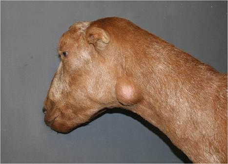
Figure 3.2 Abscessation of a parotid lymph node due to caseous lymphadenitis. Source: Courtesy of Dr. M.C. Smith.
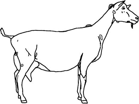
Figure 3.3 Location of common swellings caused by caseous lymphadenitis and caprine arthritis encephalitis. An abscess in the location of external lymph nodes (stippled) suggests caseous lymphadenitis. Enlargement of atlantal or supraspinous bursae (cross-hatched) may occur with caprine arthritis encephalitis.
occur here. Contact of these skin lesions with milking stands, feeders, or scratching posts contaminated by draining abscesses of other goats leads to infected nodes about the head and neck. By contrast, a popliteal lymph node (Figure 3.4) would only become enlarged if infection occurred in the distal hindlimb. Mammary lymph node involvement can result from infection of skin lesions on the udder or from mastitis. Slaughterhouse studies typically show the highest prevalence in the prescapular node, among the nodes routinely inspected (Ghanbarpour and Khaleghiyan 2005).
Diagnosis
There is no perfect test for the diagnosis of caseous lymphadenitis (Oreiby 2015). Diagnosis of the condition is based on the presence of a firm to slightly fluctuant subcutaneous swelling in the anatomic location of a lymph node (Burrell 1981). In a herd with a history of caseous lymphadenitis, the clinical findings alone are considered presumptive evidence. When a herd history is lacking (no previous veterinary evaluation, assembled herd, individually purchased animal), then laboratory assistance is indicated for confirming the diagnosis.
A sample for culture is obtained by inserting a sterile needle through shaved, disinfected skin into the mass. If aspiration yields nothing, the needle is withdrawn and saline is flushed through the needle to obtain material for a stained smear or culture. Even when the point of skin penetration is intentionally offset from the point where the needle passes through the wall of the abscess, some pus can leak back and serve as a focus of infection to others in the herd. Thus, if pus is present, the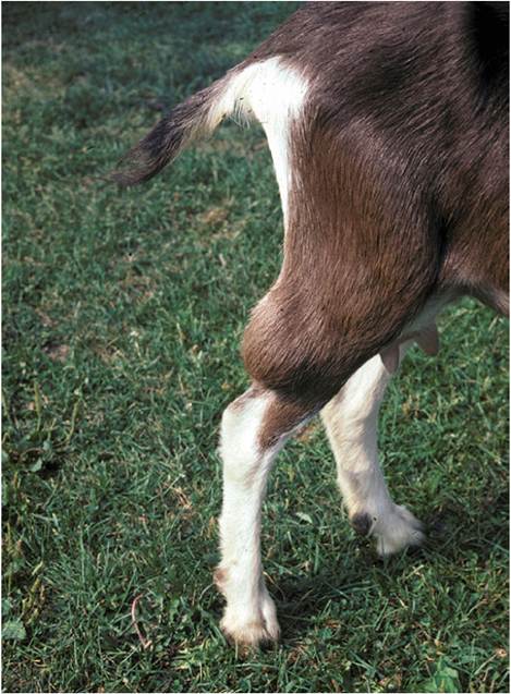
Figure 3.4 Abscessation of a popliteal lymph node. Source: Courtesy of Dr. M.C. Smith.
lump and the goat should be handled as if caseous lymphadenitis were present (see below) until culture results become available. Cultures of pus from draining caseous lymphadenitis lesions are frequently overgrown by non- pathogenic organisms or secondary invaders such as Proteus species. However, other bacterial infections such as anaerobic Staphylococcus aureus, which is sometimes called Morel’s disease (Alhendi et al. 1993; Szalus-Jordanow et al. 2010), and Trueperella pyogenes and neoplasia, including lymphosarcoma, must be ruled out if C. pseudotuberculosis is not isolated. Oil granulomas from previous vaccination against paratuberculosis should also be considered (Windsor 2011). The other conditions causing subcutaneous swellings, as discussed in this chapter and reviewed elsewhere (Williams 1980; Fubini and Campbell 1983), also should be considered in the differential diagnosis.
The pus in an abscessed node in a goat may be creamy white, yellowish, or greenish; it is typically odorless and is more pasty (less inspissated) than in sheep. A lamellar “onion ring” configuration, as seen in sheep with this disease, is rarely present in goats (Batey et al. 1986), but has been described in computed tomography images of internal abscesses in a goat (Grosso et al. 2018).
The organism (a facultative anaerobe) grows readily but slowly on blood agar (Brown and Olander 1987).
The colonies are very tiny or invisible after 24 hours (Lindsay and Lloyd 1991), and a longer incubation period is recommended before declaring that no growth has occurred. The colonies are still tiny and button-shaped after 48 hours, but are surrounded by a narrow zone of hemolysis. The colonies can be easily moved around on the surface of the agar plate and splatter when placed in a flame, because of high content of lipid. The organism is catalase positive, whereas Trueperella pyogenes, which also produces tiny colonies, is catalase negative (Quinn et al. 2002). Gram-stained smears show Gram-positive or Gram-variable small coccoid rods. A longer rod form is sometimes seen in smears of pus.Serologic tests such as the bacterial agglutination test and synergistic hemolysis inhibition (SHI) test (Zaki 1968; Brown et al. 1985, 1986b; Holstad 1986a) have been used for identifying goats with early or internal forms of caseous lymphadenitis. The SHI test is commercially available in the United States, through the University of California, Davis, and several other laboratories, and has a sensitivity of 98% in goats (Brown and Olander 1987). However, in the same study population, 28% of goats with no demonstrable abscess were also SHI positive, indicating a lack of specificity. In another study using an SHI result ≥1 : 8 as positive, a positive serologic test was a poor predictor of the future development of clinical disease (Washburn et al. 2013). These animals might have been previously exposed, recovered, or positive because of internal abscesses. Laboratory personnel report that a titer of 1 : 256 or greater correlates well with internal abscesses, but that occasionally an animal with a walled-off abscess tests negative. Experimental infections have not been detectable serologically until at least 15 days after infection (Kuria et al. 2001), so purchased animals should be retested at the end of their quarantine period. Colostrally derived antibodies may cause false-positive test results in kids under 6 months of age (Williamson 2001).
Elsewhere in the world various enzyme-linked immunosorbent assay (ELISA) tests are available and considered to be economical as well as easier to perform than other serologic tests for infection (reviewed by Oreiby 2015). The antigens used in these ELISA tests include whole cell extracts, total secreted antigens, exotoxin, and recombinant phospholipase D exotoxin (Hoelzle et al. 2013). Most tests have not been specific enough for current infection to justify their use in culling programs. A decreasing titer on a retest taken two to four months after the first sample suggests that an active infection is not present. An ELISA test to detect antibodies against exotoxin and used for disease eradication in the Netherlands (Dercksen et al. 1996) has been improved to increase sensitivity to 94%, with specificity of 98% (Dercksen et al. 2000). Assays to detect phospholipase D may be less sensitive in sheep than in goats (Hoelzle et al. 2013).
Because the organism is a facultative intracellular pathogen, cell-mediated immunity is involved in the immune response. Preliminary work with a commercially available bovine interferon gamma ELISA (Bovigam, Pfizer Animal Health, New York, USA) for cell-mediated immunity to C. pseudotuberculosis has suggested that this ELISA test is sensitive to prior infection and is unaffected by vaccination (Menzies et al. 2004). Caprine interferon gamma crossreacts in the assay. This test may be useful to detect carriers in vaccinated herds, although it may not reflect the extent of the infection.
The increases in total serum proteins and gamma globulin that are reported for goats with caseous lymphadenitis (Desiderio et al. 1979) are also non-specific. An intradermal allergic test using a water-soluble protein “lymphad- enin” has been used to differentiate goats with abscesses from uninfected goats (Langenegger et al. 1986). Injections were made in the shoulder region and the maximum increase in skin-fold thickness occurred after 48 hours.
Surgical Treatment
Treatment of individual animals involves either draining or surgically removing the abscessed nodes. Such animals should be closely examined for additional abscesses, including ultrasound examination for enlarged internal lymph nodes if possible, before undertaking surgery. Ripened abscesses can be incised generously at a ventral point and the cavity flushed with dilute disinfectant. Because the infection is potentially zoonotic, people performing this procedure should wear gloves. The pus should be collected and burned and the goat should be kept strictly isolated until the lesion is completely covered by healthy skin, typically 20-30 days later (Ashfaq and Campbell 1980). Allowing an abscess to rupture in the main goat pen or returning the animal to the herd where herd mates will lick the wound will only serve to contaminate the environment and spread the infection. Stanchions, milking stands, and keyhole feeders in particular will become contaminated. The advantages of surgical removal of the encapsulated abscess are that the treated animal does not need to be quarantined afterward and spread to other nodes is less likely to occur, but veterinary expertise is required and there are dangers associated with anesthesia and dissection near large blood vessels and cranial nerves. Extirpation of abscessed parotid nodes is particularly dangerous, and treatment of a retropharyngeal abscess requires marsupialization.
An alternative but controversial treatment for carefully chosen abscesses is to inject them with 10% formalin at the point where the ripening abscess has become fixed to the overlying skin. A 16-gauge needle is used and approximately 20 mL of formalin is repeatedly injected into and withdrawn from the abscess until the cloudiness of the for- malin/pus mixture in the syringe is no longer increasing; larger abscesses require a larger initial volume of formalin. This causes sloughing of the node within a few weeks, but raises the specter of meat or milk contamination or carcinogenesis in the minds of many veterinarians. Formalin is rapidly converted to formic acid, and formate is an intermediate in normal metabolism in the body. If the abscess is not fixed to the skin, formalin leaking out of the injected abscess will damage surrounding tissues as well as cause pain to the animal. Injection into caseous lymphadenitis abscesses of either 10% tincture of iodine or 2.5% sodium hypochlorite has been shown to be ineffective (Santiago et al. 2013).
Antibiotic Therapy
In the past, long-term treatment with antibiotics or isonia- zide of abscesses inaccessible to surgical management has not been rewarding. Antibiotic sensitivity testing is of little benefit, because most antibiotics do not penetrate into the abscessed lymph node and the organism itself may be intracellular. Administration of penicillin or tetracycline for a few days after spontaneous rupture or lancing of an abscess is suggested to prevent dissemination of the organism to other lymph nodes, but the value of this treatment has not yet been determined in controlled clinical trials. Tulathromycin has been administered either intralesion- ally or subcutaneously after closed lavage of abscesses, to avoid the environmental contamination that results from lancing, with short-term success (Washburn et al. 2009), but the goats were only followed for 30 days.
Success in treating foal pneumonia caused by Rhodococcus equi with a combination of erythromycin and rifampin led to renewed interest in medical treatment of valuable sheep and goats. Pharmacokinetic data derived from other species suggest that an oral dose of 10-20 mg/kg rifampin s.i.d. (once daily) might be appropriate (Sweeney et al. 1988; Frank 1990; Jernigan et al. 1991). A possible erythromycin dosage is 4 mg/kg administered intramuscularly (IM) or subcutaneously. Because erythromycin is highly irritating, a rifampin/penicillin combination might be preferable. Treatment should be continued for four to six weeks. Reports of the efficacy of rifampin in goats with caseous lymphadenitis are not yet available. One report of rifamyin at 10 mg/kg IM b.i.d. (twice a day) for 10 days, combined with long-acting oxytetracycline at 20 mg/kg IM every third day, gave a promising reduction in the size of affected lymph nodes, though the animals were only followed for one month (Senturk and Temizel 2006). There is no information available for estimation of meat or milk withdrawals to avoid antibiotic residues after the use of rifampin.
More recently, azithromycin has become popular for treatment of rhodococcal pneumonia in foals because of good intracellular penetration and long half-life (Chaffin et al. 2008), and the pharmacokinetics of this antibiotic in goats have been studied (Carceles et al. 2005). To date, there is no information on the efficacy of azithromycin in goats with caseous lymphadenitis.
Herd Eradication and Control Programs
Eradication of caseous lymphadenitis from a herd is difficult. The owner must be willing to cull goats, sheep, and camelids with multiple abscesses and forgo purchase of animals from infected herds. Introduction of the disease into the United Kingdom by imported goats has underscored the risks associated with an open herd status (Gilmour 1990; Lindsay and Lloyd 1991). Ideally, a negative serologic test should be required before purchase, even from a herd believed to be free of the disease. Newly acquired animals, including camelids, should be examined for lymph node enlargement at least monthly for one year or more after arrival. An eradication program based on culling of serologically positive animals was successful in 53 herds (approximately 13 000 adult goats) in the Netherlands (Dercksen et al. 1996). Monitoring herds in Norway for reinfection after an eradication program has been done using bulk tank milk ELISA tests (Nagel-Alne et al. 2014).
Valuable infected animals may be kept isolated; their kids should be removed at birth and fed heat-treated or bovine colostrum to avoid transmission of the disease as the newborn searches for the udder. Commercial pasteurization is known to kill C. pseudotuberculosis in milk (Baird et al. 2005). Division of an enzootically infected herd into infected and apparently uninfected groups has also been proposed as a means of limiting spread (Mullowney and Baldwin 1984). The housing facilities should be free of nails, wire, and other objects that might induce breaks in the skin. Control of external parasites is very important, because pruritic goats will rub themselves on nails and posts. Needles, tattooers, and surgical instruments should be sterilized between animals. Shearing equipment that has been used on another farm should likewise be disinfected.
All wounds should be treated promptly with a disinfectant, and the umbilical cords of kids should be dipped in iodine at birth. In infected herds of Angora or Cashmere goats, animals should not be dipped for control of external parasites during the two weeks immediately after shearing; topical pour-on insecticides can be substituted. Animals with chronic respiratory disease or wasting should be culled, or at least isolated from the herd. Natural transmission from lung lesions via discharge of pus into airways has been demonstrated in sheep (Ellis et al. 1987).
An environment contaminated with pus may be the source of new infections for weeks or months. Recovery of the organism from wood surfaces, straw, hay, and soil has been demonstrated by various researchers (Augustine and Renshaw 1986; Brown and Olander 1987). Thus, the isolation facilities used to contain a goat after rupture or lancing of an abscess should have a concrete floor. The bedding should be burned and the pen thoroughly cleaned (pressure washed) and disinfected between animals. Chlorhexidine is bacteriocidal to the organism at 0.625%; this concentration of chlorine and of quaternary ammonia is effective against most isolates (Sa et al. 2013). In a successful herd eradication program in Norway, housing areas used by infected animals were left vacant for three months after disinfection, and the upper 10 cm of soil in the paddocks was removed to decrease the risk of soil-borne infection (Nord et al. 1998).
Vaccination
Numerous studies using mice have evaluated immune responses to the organism. Cell-mediated immunity has been demonstrated to restrict bacterial proliferation. Neutralization of exotoxin (phospholipase D) produced by the organism is believed to limit spread from the primary site of infection.
The value of vaccination as an aid in controlling the disease in ruminants has been frequently questioned. A decrease in the prevalence of abscesses in the herd has often been noted where autogenous bacterins have been used, but concerned owners have simultaneously culled known infected goats and generally improved hygiene measures. As a note of caution, if not properly prepared and tested, an autogenous vaccine against this organism may contain enough free toxin to kill the vaccinated goat. The SHI test does not distinguish between naturally infected and vaccinated goats, so test and cull is difficult to use once a vaccination program has been implemented. Any vaccinated goat may also be infected. Likewise, vaccinated goats test falsely positive in an ELISA for antibodies against exotoxin and cell wall antigens (Sting et al. 1998). Additionally, vaccination can cause false positives in some ELISA tests for paratuberculosis (Manning et al. 2007).
A commercial vaccine (Glanvac®, Commonwealth Serum Labs, Melbourne, Australia) developed in Australia has been evaluated in goats (Anderson and Nairn 1984a, b; Brown et al. 1986a). This product is a formalinized exotoxin with incomplete Freund's adjuvant. In one study, challenge by swabbing a live culture onto abraded skin revealed that either one or two doses of vaccine gave good protection; 3 of 20 vaccinated goats developed abscesses, as compared to 10 of 10 control goats (Anderson and Nairn 1984a). Colostrally derived immunity protected young kids against challenge (Anderson and Nairn 1984b). In enzootically infected herds, serologic titers in kids disappear by 3-4 months of age, only to reappear after exposure to the organism. Vaccination should probably be performed before 4 months of age (Holstad 1986c), but colostrally derived antibody may interfere when vaccination is begun before 3 months (Paton et al. 1991). The successful use of Glanvac in sheep in Australia when two initial doses followed by annual boosters are given has been summarized by Windsor (2011).
A different commercial vaccine (Case-Bac® and Caseous-DT®, Colorado Serum Co., Denver, CO, USA) is available in the United States. This bacterin-toxoid preparation is only labeled for sheep, where its efficacy has been documented (Piontkowski and Shivvers 1998), but it has been used on goats. There are numerous anecdotal reports of adverse reactions, including severe milk drop, lameness, anorexia, fever, and depression for one to two days after vaccination in adult dairy goats in infected herds, but many owners and practitioners report that vaccination of young stock beginning at 2 or 3 months of age has been helpful in reducing disease prevalence. Vaccination should probably be continued for many years, including annual boosters, because of the possibility that one or more members of the original herd remain carriers and shedders of the organism.
More recently, a provisionally licensed bacterin has become available for kid goats in the United States (Texas Vet Lab, San Angelo, TX, USA). It is not licensed for lactating or pregnant goats. After two initial doses, does and bucks should be revaccinated each year before breeding. Field evaluation of this vaccine relative to efficacy or adverse reactions is lacking, and the vaccine appears to have disappeared from the market as of 2021.
Field evaluation of crude filtrated C. pseudotuberculosis toxoid combined with whole killed cells has been disappointing in goats (Holstad 1989), whereas another experimental whole cell vaccine gave a non-statistically significant trend for fewer cases in the vaccinated goats in a field trial in Canada (Menzies et al. 1991). A formalin- inactivated whole cell vaccine with aluminum gel phosphate adjuvant gave partial protection (estimated at 77%) in a field trial under extensive conditions in Brazil (Ribeiro et al. 1988). Whole cell preparations of the organism adju- vanted with mycobacterial components have been evaluated (Brogden et al. 1990). A modified live intradermal vaccine has been developed by EMBRAPA in the State of Bahia, Brazil.
Zoonotic Potential
Human lymphadenitis caused by C. pseudotuberculosis has been reported, especially from Australia (Mills et al. 1997; Peel et al. 1997), and the literature has been reviewed by Bastos et al. (2012). The course was often protracted and diagnosis delayed until a culture was performed. Recovery usually required surgical removal of the affected lymph node, with supplemental antibiotics. Owners, slaughterhouse workers, and veterinarians should handle infected animals and abscesses with caution.
Melioidosis
In tropical areas such as Southeast Asia, India, China, and northern Australia, a zoonotic disease caused by Burkholderia (Pseudomonas) pseudomallei is seen in goats and must be differentiated from caseous lymphadenitis. The disease seen in humans has been reviewed (Cheng and Currie 2005) and the global distribution of the organism, including in the Americas, has been updated (Currie et al. 2008).
Etiology
Burkholderia (Pseudomonas) pseudomallei is a Gramnegative bacillus with polar flagella that may show bipolar staining. It closely resembles Pseudomonas aeruginosa. The organism may be cultured on sheep blood agar or MacConkey agar (up to four days at 37 °C). Some strains are hemolytic.
Epidemiology and Pathogenesis
The organism resides in soil and contaminated water. Survival in soil for up to 30 months has been reported (Thomas and Forbes-Faulkner 1981). Human cases often occur shortly after heavy rains or flooding, and soils in the southeastern United States are considered to be suitable for survival of the organism (Portacci et al. 2017).
Percutaneous infection of goats has been reproduced experimentally (Soffler et al. 2014). Infected animals, including rodents, pass the organism in feces. Spread from animal to animal by biting insects also occurs. Vertical transmission is possible, as natural and experimental infection of pregnant goats has led to infection of aborted and live-born kids (Retnasabapathy 1966; Thomas et al. 1988a) and the organism can be transmitted through the milk (Choy et al. 2000).
Clinical Signs
Initial bacteremia is followed by formation of abscesses and granulomas in superficial lymph nodes, lung, and other internal organs. Prescapular lymph nodes are commonly involved and contain grayish yellow, creamy pus (Sutmoller et al. 1957). Chronic mastitis (van der Lugt and Henton 1995), weight loss, polyarthritis, and meningoencephalitis are also reported, as is sudden death from rupture of an aortic aneurysm (Choy et al. 2000).
Diagnosis and Control
Lung lesions can be demonstrated radiographically (Soffler et al. 2014). The indirect hemagglutination test (positive at 1/40 or higher) is considered most suitable for screening, because it has a sensitivity of 98%. The 100% specific complement fixation test (positive at 1/8 or higher) is used for confirmation (Thomas et al. 1988b). Sensitivity of the complement fixation test is lowered (82%) in chronic infections. The final diagnosis is made by culture. Older lesions in goats are sometimes sterile (Thomas et al. 1988b).
Antibiotic treatment is ineffective. Infected goats are to be eliminated and the herd monitored by serologic testing (Baxendell 1984). Sodium hypochlorite (0.5%) and Virkon® (Antec International, Sudbury, UK) disinfectant (1%) are bacteriocidal (Portacci et al. 2017).
Other InfectiousAgents
Goats with arthritis because of a variety of causes may have enlargement of the associated lymph node. Regional lymph node enlargement is a constant finding with caprine arthritis encephalitis virus infection (Robinson and Ellis 1986), which is a common cause of lameness and enlarged joints in dairy goats in North America, Europe, and Australia. The reader should refer to Chapter 4 for an in-depth discussion.
Dermatophilus congolensis, a Gram-positive organism that forms branching filaments in pus, has been isolated from abscessed superficial lymph nodes of Beetal goats. Other breeds housed with infected goats were not involved (Singh and Murty 1978). This organism more typically causes a superficial dermatitis. A single Nubian goat with clinical signs of weight loss and a cough had Actinomyces hyovaginalis cultured from a necrotic tracheobronchial lymph node (Schumacher et al. 2009). Goats with contagious ecthyma (soremouth) virus infection may have noticeable enlargement of lymph nodes draining the head. Mange can also cause marked lymph node enlargement. See Chapter 2 for discussion of skin diseases.
In regions where trypanosomiasis is endemic, Trypanosoma brucei, and to a lesser extent Trypanosoma congolense and Trypanosoma vivax, cause a marked lymphadenopathy in goats. Theileria is another blood-borne parasite that causes enlargement of lymph nodes. These diseases are discussed in Chapter 7.
Infectious agents causing mastitis (including tuberculosis) may cause enlargement of the supramammary lymph nodes and are discussed in detail in Chapter 14.
Lymphosarcoma
Lymphosarcoma in goats is a neoplastic disease of unknoλwι etiology, and in some studies is the most commonly identtfed tumor in caprine submissions (Lohr 2013). It has many siιm- Iarities to sporadic or juvenile bovine iyιmP3°sarcoma. Neither the bovine leukemia virus nor antibodies against that virus associated with adult bovine lymphosarcoma have been found in goats with naturally occurring lymphosarcoma, although two experimentally produced goat cases have been reported (Olson et al. 1981; Kettman et al. 1984). T-cell lymphomas, often involving the mediastinum, predominated over B-cell lymphomas in one study (Kiser andLohr 2017).
Lymphadenopathy of superficial nodes is not a consistent finding; only 3 of 10 goats in one report were thus affected (Craig et al. 1986). In these animals, a lymph node aspirate yielding many lymphoblasts might confirm the diagnosis (Duncan and Prasse 1986). However, as least in cattle, fine needle aspirates have relatively low sensitivity for diagnosis of lymphosarcoma (Washburn et al. 2007). In one yearling Nubian goat in Texas with lymphosarcoma diagnosed from lymph node aspirates, the yeast Histoplasma capsulatum was identified in the same samples (Schlemmer et al. 2019).
Goats affected with lymphosarcoma are generally more than 2 years of age. They may have a variety of clinical signs, including high fever, emaciation, diarrhea, or dyspnea. The organs most frequently involved are liver, spleen, lungs, and lymph nodes, although the reproductive tract has also been involved (DiGrassie et al. 1997). Rapid deterioration occurs when clinical signs are noted, with death or euthanasia supervening in one week to two months in most cases. This is in contrast to the long and usually benign course of external caseous lymphadenitis.
Published reports of successful treatment of lymphosarcoma in goats are lacking. For those special cases where the owner will not permit euthanasia, an oncologist should be consulted for a current canine protocol. Meat and milk withdrawals will have to be established.
Prominent Lymph Nodes of Normal Size
A distinction must be made between increased size and increased ease of palpation of lymph nodes. Animals that are emaciated because of malnutrition, parasitism, or chronic infectious diseases have very prominent nodes because subcutaneous fat is absent.