Swellings with Unrestricted Distribution
Certain conditions can cause local swelling over almost any part of the goat's body. They will be considered before conditions with a more regional distribution. Ultrasound examination may provide clinically useful information.
Masses limited to the dermis or epidermis, joint distension, and changes involving the mammary gland and scrotum are discussed in other chapters.Hematoma and Seroma
A fresh hematoma is easily diagnosed by aspiration of non- odorous, sterile red fluid. A yellow sterile fluid containing few erythrocytes is present in a seroma or organized hematoma. Diagnostic aspiration, however, is not without risk. Even with very careful attention to skin preparation, the needle is likely to carry a few bacteria into the accumulated fluid. Unless the hole in the skin is offset from the point where the needle enters, a small tract will permit leakage of fluid and retrograde entry of more bacteria. It is hard to imagine better growing conditions for a bacterial culture than in whole blood or serum at body temperature.
Goat Medicine, Third Edition. Mary C. Smith and David M. Sherman. © 2023 John Wiley & Sons, Inc. Published 2023 by John Wiley & Sons, Inc.
The practitioner might choose to drain a fluctuating lump, whether it be hematoma, seroma, or abscess. This procedure should be delayed a week or more if a hematoma is suspected to avoid renewed bleeding when the pressure of containment is relieved. If the swelling is not over a large blood vessel and antibiotic residues in milk or meat are of no concern, then small fluid pockets may be drained through a needle and subsequently injected with penicillin or other antibiotic. A pressure bandage may prevent recurrence. Larger pockets must be opened widely and packed with gauze soaked in an antiseptic or provided with indwelling drains if proper healing is to occur.
Seromas on the poll of breeding bucks, resulting from regular head bashing with other males, are best left totally alone.Cellulitis and Abscess
Cellulitis is an acute, diffuse, edematous, suppurative inflammation of deep subcutaneous tissues or muscle. The swelling may be painful and accompanied by fever. The cause is often foreign-body penetration or injection of irritant drugs. Aspiration for cytology and culture and sensitivity testing is warranted if the goat is systemically ill.
If encapsulation and abscess formation occur, signs of local inflammation (pain, heat) decrease (Figure 3.1). Given time, many abscesses “ripen” or develop a soft spot in the capsule where rupture is likely to occur. This helps to distinguish an abscess from a hematoma. The hematoma is initially fluctuant, but later develops a firm capsule as the blood organizes and resorption begins. Abscesses usually heal rapidly when drainage is supplied. Lancing an abscess at its softest spot reduces both pain and hemorrhage associated with the surgical procedure. Another, more ventral location may be more appropriate for incision, however, to allow better drainage. Animals should be removed to an isolation or hospital pen before draining or rupture occurs. Caseous lymphadenitis should be ruled out (by culture or with less certainty by absence of lymph node involvement in any animal in the herd) before the animal with a draining abscess is returned to the herd.
Emphysema
When crepitation is palpable in subcutaneous or deeper tissues, it is important to determine the origin of the air or
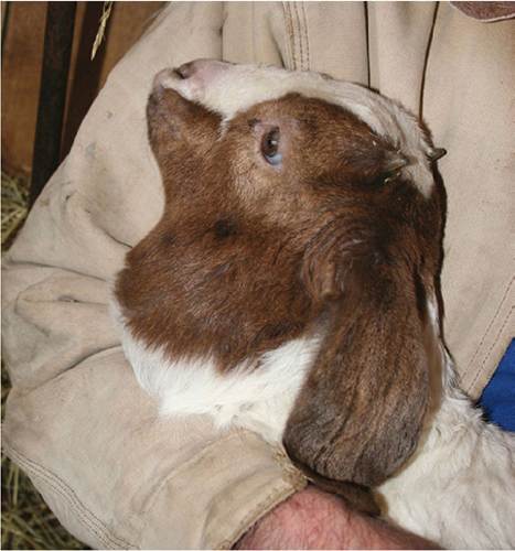
Figure 3.1 Large, thin-walled abscess in the upper neck of a 1-month-old kid from which a Streptococcus sp. was cultured. Source: Courtesy of Dr. M.C. Smith.
other gas that is present. Air may be escaping from the respiratory tract (tracheal laceration, severe pulmonary emphysema) or it may be entering through sucking skin wounds (Tanwar et al.
1983; Barman et al. 2017). Attention to the primary problem should stop the inflow of air, and resorption will occur gradually over days or weeks, but may be aided by aspiration.A crepitant, painful swelling caused by infection of wounds with gas-forming clostridia such as Clostridium chauvoei may lead to death of the animal (van Tonder 1975). The condition can be diagnosed by cytologic examination of aspirates, anaerobic culture, or immunofluorescence testing. Aggressive treatment with systemic and intrale- sional penicillin is successful in the early stages. Gangrenous tissues slough later if the animal survives. In regions where clostridial infections are common, multivalent clostridial vaccines (C. chauvoei, Clostridium septicum, Clostridium novyi) provide inexpensive protection.
Edema
Pitting edema without crepitation may occur in wounds infected with C. septicum (malignant edema) or C. novyi (swelled head in bucks). Other causes include trauma without tissue infection, frostbite, hypoproteinemia (parasitism, kidney disease, paratuberculosis), and congestive heart failure. Udder edema is discussed in Chapter 14.