Microsporum canis
Microsporum canis is the most common dermatophyte in cats and dogs (Fig. 3.6). Cats are considered to be the most important reservoir, but the species is also found regularly in rabbits and horses (Sharma et al.
2007; Cafarchia et al. 2013a; Pasquetti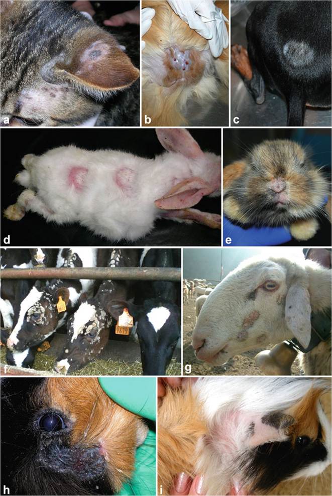
Fig. 3.6 Clinical presentation of infections caused by zoophilic dermatophytes. (a-c) Microsporum canis: irregular areas of alopecia covered by scales and scabs in a cat (a), pseudomycetoma in a cat (b), annular area of alopecia in a dog (c). (d-e) Trichophyton mentagrophytes: annular erythematous areas of alopecia on the back, extremities, and ears of a
et al. 2013). It has been occasionally reported in a number of other domestic and wild animals, e.g., cattle, sheep, goats, ferrets, camelids, marmots, eastern cottontails, foxes, etc. (Gallo et al. 2005a, b; Chermette et al. 2008; Pignon and Mayer 2011). Isolates coming from horses were previously classified as M. equinum, a species later synonymized with M. canis based on phylogenetic and population genetic analyses (Graser et al. 2000a; Kaszubiak et al. 2004; Sharma et al. 2007). Microsporum canis is known to reproduce mainly asexually, although it is also capable to produce sexual state under laboratory conditions (Hironaga et al. 1980).
Microsporum canis is diffused worldwide and plays an important zoonotic role. In some countries it tends to overpass antropophilic dermatophytes as cause of human ringworm episodes (Chermette et al. 2008; Ameen 2010). It is the most frequent agent of tinea capitis in many European countries, the Eastern Mediterranean, South America, and China; it also causes highly inflamed lesions on glabrous skin and infrequently onychomycosis (Ginter-Hanselmayer et al. 2007; Seebacher et al. 2008; Skerlev and Miklic 2010; Uhrlaβ et al.
2015; Zhan et al. 2015).Dermatophytosis by M. canis is a pleomorphic and usually not a localized disease in cats despite appearances to the contrary (DeBoer and Moriello 2006). In addition, many infected cats have no or only few lesions. In particular, long-haired breeds can be subclinical carriers or have only minimal clinical symptoms; sometimes lesions become evident after shaving of hair. Isolation of M. canis from the haircoat in the absence of obvious lesions indicates either infection or fomite carriage from exposure to a contaminated environment. Distinguishing is often impossible, and Wood's lamp can help to detect minimal lesions invisible at naked eye. A mechanical carriage will only be revealed through fungal cultures (a plate with one or few CFU is usually indicative of fomite carriage). Dogs more often exhibit the classic annular areas of peripherally expanding alopecia, scale, crust, and follicular papules and pustules, with sometimes a central area of hyperpigmentation. It is quite clear that the response to infection in cats, more often than in dogs, tends to resemble that described in human patients with chronic infection by antropophilic dermatophytes. This is illustrated by the high number of cats which develops minimal and persisting lesions, just due to a “tolerant” immune response. This is an evidence of the strong adaptation of the fungus to the feline host. It is known that Persian cats are predisposed to M. canis infection (DeBoer and Moriello 2006; Miller et al. 2013) and to the development of more aggressive forms such as pseudomycetoma (Zimmerman et al. 2003; Bianchi et al. 2017). Apart from a genetic predisposition, this may also reflect a less efficient grooming of the haircoat because coat length has been reported as an important factor in the carriage of M. canis spores (Sparkes et al. 1993). A genetic predisposition to develop a generalized form of M. canis infection seems to exist also in Yorkshire Terrier dogs (Sparkes et al.
1993).
Fig. 3.6 (continued) rabbit (d), scaling lesion on the snout and upper lip of a rabbit (e). (f-g) Trichophyton verrucosum: discrete, scaling patches of hair loss located on the head and neck of cattle (f) and sheep (g). (h-i) Trichophyton benhamiae: weeping lesion under the eye of a guinea pig (h), itchy area of alopecia behind the ear of a guinea pig (i)
Available data show that M. canis cause >90% of dermatophytoses in cats worldwide. It is generally also the most prevalent dermatophyte isolated from dogs but with greater variation. For instance, in the USA, dermatophytosis was diagnosed in 14.9% of cats and 3.8% of dogs with cutaneous lesions, and M. canis is accounted for 92 and 43% of feline and canine cases, respectively (Lewis et al. 1991). In Brazil, dermatophytosis was confirmed in 27.8% of cats presented with dermatological problems, with M. canis responsible for 100% of cases; the prevalence in dogs was 9.8%, i.e., 68.5% of all cases of dermatophytosis (Copetti et al. 2006). Similar long-term studies from Europe showed comparable or even higher prevalence of M. canis in animals with skin disorders. Dermatophytes were isolated from 40.7% of cats in Croatia, and M. canis represented 98.7% of the isolates (Pinter et al. 1999). In Italy, dermatophytosis was diagnosed in 24.7% cats and 18.7% of dogs with M. canis representing 97 and 83% of the isolates, respectively (Mancianti et al. 2002). Climatic conditions appear to play a significant role in the diffusion of the pathogen. The prevalence is basically higher in hot and humid climates as shown in studies from climatically different regions of the USA and Italy (Moriello et al. 1994; Romano et al. 1997; Cafarchia et al. 2004; Proverbio et al. 2014). Seasonal differences in the incidence of infection have been found in many countries in animals as well as in humans (Simpanya and Baxter 1996; Cafarchia et al. 2006; Lee et al. 2012; Uhrlaβ et al. 2015). With similar climates, prevalence varies in relation to other factors, firstly the lifestyle of animals taken in consideration.
In the UK, show cats were reported to have a carrier rate of 12.5%, while in household cats, the isolation rate was 2.2% (Quaife and Womar 1982; Sparkes et al. 1994). In a report from Belgium, 2.1% of pet cats were found to be asymptomatic carriers, while the prevalence in cats in shelters was 16% (Mignon and Losson 1997).Small outbreaks (usually 1000) was reported over an 8-year period (Strachan and Blank 1963). Circumstances that led to this outbreak were not completely clarified, but it was concluded that cats and dogs played a major role in the spread of infection. Other interesting episodes reported in literature are nosocomial epidemics, infections in schools, outbreak in a nursing home for elderly people, and veterinary clinic (Shah et al. 1988; Snider et al. 1993; Drusin et al. 2000; Yu et al. 2004; Gurtler et al. 2005; Grills et al. 2007; Kopel et al. 2012; Pasquetti et al. 2012; Hillary and Suys 2014; Subelj et al. 2014). In all these contexts, the infection spreads without an animal intervention, which shows that human-to-human transfer of M. canis, although considered rare and self-limiting, can be occasionally very efficient. Rarely, M. canis causes outbreaks in rabbit farms, and their origin usually came from animals imported from abroad and integrated into the local farms (Gonzalez et al.
1988; Cabanes et al. 1997); outbreaks in laboratory mice (Difonzo et al. 1986) and a porcine farm have also been described (Cabo et al. 1995).
In general, studies of the fungal flora of asymptomatic cats and dogs highlight a very important point. Although many animals have been found to act as healthy carrier of M. canis, this fungus should not be considered part of the normal fungal flora. If it was, it would have been isolated routinely from healthy animals regardless of geographical region, lifestyle (indoor or outdoor), or status (pet or stray) (DeBoer and Moriello 2006).
3.7.2