Myotatic Reflexes
Myotatic or tendon reflexes are tested by sharply striking the tendon of a specific muscle (or sometimes the muscle itself) and evaluating the strength of the reflex contraction.
The afferent component of the reflex arc involves the muscle spindles, which are stretch detectors; sensory fibers in the peripheral nerve; the dorsal nerve root and its ganglion; and the connection from the sensory nerve fiber to the lower motor neuron in the ventral horn of the same spinal cord segment (Fig. 8.6). The efferent component of the reflex arc involves the lower motor neuron, ventral nerve root, motor fibers in the peripheral nerve, the neuromuscular junction, and the muscle being tested. Lesions in the spinal cord cranial to the level of the reflex arc and lesions of the brain result in normal to increased myotatic reflexes after a period of several days. Lesions in either the afferent or the efferent components of the reflex arc result in decreased to absent myotatic reflexes.In adult horses and many adult cattle, myotatic reflexes can be tested only in recumbent animals. Some cattle can be cast with ropes for this purpose, and young animals and small ruminants can be placed in lateral recumbency for testing. These reflexes should be tested only in the limbs that are uppermost when the animal is lying on one side. The animal must be turned over to test the limbs on the opposite side; however, reflexes are often diminished in limbs that have been laid upon for prolonged periods of time (hours). In adult horses and cattle, only patellar reflexes can be reliably tested. The reflex responses are assigned a qualitative clinical score as follows: 0—No reflex activity
1— Hypoactive
2— Normal
3— Hyperactive
4— Hyperactive and clonic
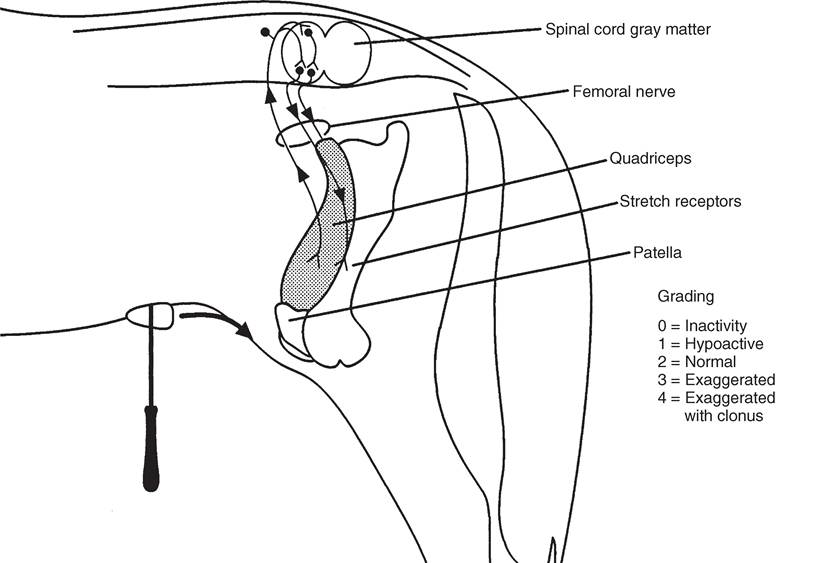
FIG. 8.6 Pathways governing patellar tendon reflex.
■ TABLE 8.4
Innervation of the Thoracic Limbs of Large Animals
| Spinal Cord Segment | Peripheral Nerve | Muscle(s) |
| C7 | Suprascapular | Supraspinatus, infraspinatus |
| C6, C7 | Subscapular | Subscapularis |
| C7, C8, T1 | Pectoral | Subscapularis, pectoral muscles |
| C6a, C7, C8 | Musculocutaneous | Biceps brachii, coracobrachialis, brachialis |
| C8, T1, T2 | Median | Flexor carpi radialis, deep digital flexor, superficial digital flexor |
| C8a, T1, T2 | Ulnar | Flexor carpi ulnaris, deep digital flexor, superficial digital flexor |
| C7, C8, T1 | Radial | Triceps, extensor carpi radialis, ulnaris lateralis, lateral and common digital extensors |
| C6b, C7, C8 | Axillary | Deltoideus, teres minor, subscapularis, cleidobrachialis |
| C7, C8 | Long thoracic | Serratus ventralis |
| C8, T1, T2b | Thoracodorsal | Latissimus dorsi |
| C8, T1 | Lateral thoracic | Cutaneous trunci |
aContributes innervation in the ruminant only.
bContributes innervation in the horse only.Clonus is a phenomenon observed with severe upper motor neuron lesions: the response of the muscle being tested is a series of rapid, repeated contractions rather than a single contraction. The innervation of the limbs is listed in Tables 8.4 and 8.5.
THORACIC LIMB MYOTATIC REFLEXES
Triceps Reflex. Hold the limb moderately flexed at the elbow, and percuss the triceps tendon just above the olecranon using a heavy instrument. The normal response is a contraction of the triceps muscle, leading to retraction of the upper limb and extension of the elbow. The triceps reflex measures the integrity of the radial nerve and the C7 to T1 spinal segments.
Biceps Reflex. Hold the limb moderately extended at the elbow and place the supporting hand over the attachment of the biceps muscle on the dorsomedial aspect of the limb at the level of the elbow joint. Percuss the biceps tendon or the taut biceps muscle with a heavy instrument. Contraction of the muscle may be perceived visually or by palpation. A slight flexion of the elbow and extension of the carpus is normal. The test measures the function of the musculocutaneous nerve and spinal cord segments C6 to C8 in ruminants and C7 and C8 in horses.
Lesions cranial to C6 result, within several days from the time of injury, in general hyperreflexia of both thoracic limbs and pelvic limbs. Lesions located in spinal segments C5 to T2 result in immediate hyporeflexia to areflexia of the thoracic limbs and, within several days, hyperreflexia of the pelvic limbs.
PELVIC LIMB MYOTATIC REFLEXES
Patellar Reflex. Flex the stifle moderately, and sharply percuss the middle patellar ligament with a heavy instrument. The normal reflex is a sharp contraction of the quadriceps
■ TABLE 8.5
Innervation of the Pelvic Limbs of Large Animals
| Spinal Cord Segment | Peripheral Nerve | Muscle(s) |
| L3a, L4, L5, L6b | Femoral | Quadriceps |
| L5a, L6, S1 | Cranial gluteal | Gluteals, tensor fascia lata |
| S1-S5 | Caudal gluteal, pudendal | Biceps femoris, middle and superficial gluteals |
| L5a, L6, S1, S2 | Sciatic, peroneal | Lateral digital extensor, long digital extensor, short digital extensor, cranial tibial |
| L5a, L6, S1, S2 | Tibial | Gastrocnemius, popliteus, superficial and deep digital flexor, interosseus |
| L5a, L6, S1, S2 | Pudendal | Retractor penis |
| S3-Cd5 | Caudal rectal | Rectum, anal sphincter, bladder |
aContributes innervation in the horse only.
bContributes innervation in the ruminant only.femoris muscle resulting in extension of the stifle and a forward jerk of the lower part of the limb. The patellar reflex measures the function of the femoral nerve, the quadriceps femoris muscle, and L3 to L5 and L4 to L6 spinal cord segments in horses and cattle, respectively.
Cranial Tibial Reflex. The cranial tibial reflex is elicited by flexing the hock and sharply striking the belly of the cranial tibial muscle. The reflex consists of a slight extension of the digit. The cranial tibial reflex is mediated through the peroneal and sciatic nerves and spinal cord segments L5 to S2 and L6 to S2 in the horse and the ruminant, respectively. Lesions of the spinal cord anterior to L3 segments result in hyperreflexia, whereas lesions of L3 to L6 spinal segments result in hypo- reflexia or areflexia.
FLEXOR (WITHDRAWAL) REFLEXES. The flexor reflexes are elicited on the upside front and hind limb in the recumbent large animal. For safety, the examiner should be positioned on the dorsal side of the animal or well cranial to the limb being tested. A painful stimulus is applied to the uppermost foot or distal limb. The normal response consists of two phases: (1) rapid reflex limb flexion, and (2) slower conscious perception of and response to the stimulus, characterized by attempts to assume sternal recumbency, vocalization, ear and eye movements, and/or kicking. The thoracic limb flexor reflex tests the integrity of the axillary, median, and musculocutaneous nerves and spinal cord segments C5 through T2, as well as the flexor muscles of the limb. The pelvic limb flexor reflex is mediated by the sciatic, peroneal, and tibial nerves; the pelvic limb flexor muscles; and spinal cord segments L6 to S2.
Spinal cord and peripheral nerve lesions may be localized further by testing the integrity of the sensory innervation of the skin of the limbs. Areas of decreased or absent cutaneous sensation reflect lesions of the peripheral nerves innervating the regions of the skin or of the spinal cord segments in which those sensory nerves terminate. The skin over the trunk and much of the limbs is innervated by more than one peripheral nerve. Some areas of the limbs derive sensory innervation from a single peripheral nerve. These areas are termed the autonomous zones for those peripheral nerves. Damage to a peripheral nerve innervating the skin of a limb therefore will result in decreased to absent cutaneous sensation in the autonomous zone for that nerve. This information can be used to localize lesions. Decrease in or loss of sensation to an entire limb or to limbs on both sides of the body suggests a lesion affecting several local spinal cord segments or a transverse spinal cord lesion rostral to the affected limbs.