Neoplastic diseases of the large intestines
Carolyn J. Henry
Large intestinal cancer is primarily a disease of middle-aged to older dogs, occurring at a median age of 8 years for epithelial tumors and 11 years for mesenchymal masses.1-4 Adenomatous polyps and carcinoma in situ account for the majority of canine large intestinal neoplastic lesions, but only occur infrequently in the small intestine.1,5-8 Other canine non-lymphoid large intestinal masses include leiomyomas, leiomyosarcomas, GI stromal tumors, mast cell tumors (MCT), plasmacytomas, rectal ganglioneuromas, neurilemomas, and carcinoids (see 9.4).2-4,9-16 Of the canine large intestinal malignancies, leiomyosarcomas and GI stromal tumors usually occur in the cecum (Figure 6.12), whereas carcinomas occur more commonly in the rectum and colon.7,8,15,16 In the dog, adenocarcinomas affect the rectum more commonly than the colon.8,17,18 In most reports, there has been a slightly higher prevalence of large intestinal cancer in males than in females.8,17,18 Also, most colorectal tumors occur in purebred dogs, with German Shepherds, West Highland White Terriers, and Collies reportedly being over-represented.1,2,8,19 However, a true breed predisposition is difficult to discern, given the paucity of reports that are accompanied with population demographics.
In two series of visceral MCT, miniature breeds, particularly Maltese, were most frequently affected, but large bowel lesions represented the minority of cases.9,10Previous reports have indicated that feline non-lymphoid cancer is far more common in the small bowel than in the large intestine20. However, more recent data suggest that a site predilection for the feline small intestine may no longer exist (personal unpublished data). The mean age of cats with colonic neoplasia is 12.5 years, with adenocarcinoma being the most common tumor.7,21 Other reported non-lymphoid masses include MCTs, neuroendocrine carcinomas (see 9.4), leiomyosarcomas, fibrosarcomas, and hemangiosarcomas.7,20-24 This section will focus on the more common non-lymphoid large intestinal masses.
The reader is referred to Chapter 9.3 for a discussion of GI lymphoma.Clinical signs
Dogs with large intestinal tumors are commonly presented because of hematochezia, tenesmus, and dyschezia, or intermittent rectal bleeding that is not associated with defeca- tion.1,6,25 Colorectal polyps may cause a secondary rectal prolapse.1,19 Because feline large intestinal masses are more likely to occur in the proximal segments (i.e., in the cecum or colon rather than in the rectum), clinical signs in cats may be less obvious for large bowel disease than they are in dogs.20 The most commonly reported presenting complaints in cats with non-lymphoid large intestinal neoplasia are weight loss, anorexia, vomiting, and diarrhea.21,24 Hematochezia and tenesmus are reported less frequently.6,21 Canine large intestinal leiomyosarcoma, leiomyoma, and GI stromal tumors are most often located in the cecum and generally lack mucosal involve- ment.3,16 As such, they are less likely to cause hematochezia, but may be detected due to clinical signs from intestinal obstruction or septic peritonitis secondary to tumor rupture. Paraneoplastic syndromes, including hypoglycemia and eryth- rocytosis, have been reported with large intestinal smooth muscle tumors.3,11,26 When paraneoplastic hypoglycemia occurs, dogs can present with central nervous signs, including seizures and ataxia.3 Paraneoplastic neutrophilic leukocytosis has been reported secondary to canine rectal adenomatous polyps.25
Diagnosis
In over half of all affected cats and dogs, rectal examination is useful for identifying colorectal masses.1,17,21 Most dogs present with solitary masses that tend to be polypoid or annular stenotic lesions, although diffusely infiltrative large bowel adenocarcinoma has been reported in some patients.1 For animals with non-palpable masses, useful imaging techniques include abdominal radiographs, ultrasonography, CT, rigid proctoscopy, and flexible colonoscopy.1,21,27 Survey radiographs should be obtained first and may be adequate for the identification of a mass.
A case series of dogs with colorectal leiomyoma indicated that contrast studies were required for mass identification in only one of six dogs.28 The increased availability of ultrasonography in private practice has made this modality an attractive alternative to contrast studies. In one report of cats with intestinal cancer, abdominal ultrasound permitted mass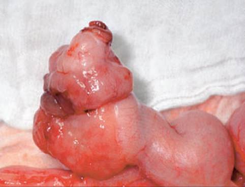
Figure 6.12:
Cecal leiomyosarcoma. Although cecal malignancies are uncommon in dogs, this is the most common site for leiomyosarcomas and gastrointestinal stromal tumors in the canine large intestine.
localization in 84% of imaged cases.21 Ultimately, diagnosis relies upon biopsy, obtained either surgically or by endoscopy. Caution is advised when relying on endoscopic biopsy for definitive diagnosis, as one report indicated discordant results between surgical and endoscopic biopsy specimens in three of ten dogs. In all three cases, the diagnosis determined from the surgical biopsy specimen was more severe than that noted on the original endoscopic biopsy.1 The appearance of lesions on endoscopy or at surgery may help to predict the prognosis. In one report of dogs with colorectal adenocarcinoma, those patients with single, pedunculated polypoid masses had the longest mean survival time (32 months), compared to dogs with masses with a nodular or cobble-stone appearance (12 months), or those with annular masses causing strictures (1.6 months).17
As with small intestinal tumors, immunohistochemistry may aid in differentiating intestinal tumor types. Recent data suggests that most tumors previously reported as canine GI leiomyosarcomas were, in fact, GI stromal tumors.16 In one report of 50 canine GI tumors originally diagnosed as leiomyomas, leiomyosarcomas, or spindle cell sarcoma, 21 (42%) were reclassified as GI stromal tumors on the basis of histological review and immunohistochemistry.
Ten of these occurred in the large intestine, with seven in the cecum.16 The use of immunohistochemistry to detect desmin, smooth muscle actin, and KIT (CD117) helps to differentiate true smooth muscle tumors from GI stromal tumors.Staging
Colonoscopy and proctoscopy are essential for complete tumor staging in dogs with colorectal cancer.17 In one report, four dogs that were treated by local excision without presurgi- cal endoscopy were later found to have lesions proximal to the surgical site.17 In addition to assessment of local disease extent, full staging to rule out metastatic disease is warranted. Metastases occurred in 16 of 19 cats with non-lymphoid large intestinal masses in one study, with metastatic sites including colonic and mesenteric lymph nodes,liver,omentum, peritoneum, spleen, bladder, urethra, mesocolon, lungs, and duodenum.21 Reported metastatic rates for canine rectal carcinoma vary widely. Two large series of cases dating back to 1975 or earlier reported metastatic rates of 64% or greater, whereas no metastases were noted in a series of 78 dogs diagnosed with colorectal adenocarcinoma between 1973 and 1984.14,17,18 When metastasis occurs, the common sites include the regional lymph nodes, lungs, liver, and spleen.14,18 Metastasis via presumed tumor cell implantation has also been reported with colorectal carcinoma, resulting in lesions in the periproctal area.18,27 As in humans, canine GI stromal tumors tend to metastasize within the abdominal cavity, with sites including the liver, spleen, mesentery, serosa, and mesenteric lymph nodes. The metastatic rate in dogs in one report was 29%.16
Treatment
Surgery is the treatment of choice for most large intestinal masses and may provide excellent long-term outcome for patients with solitary masses.29 The surgical approach chosen depends on tumor location, but may entail local resection after eversion of the mass through the anus, anal pull-out, or ab- dominoanal pull-through procedures.
Alternatively, laparotomy or sagittal pubic osteotomy approaches may be necessary. The latter has been described for treatment of various inaccessible intrapelvic masses in 24 dogs.29 This approach facilitated surgical exploration of the affected region in a report with seven dogs with masses involving the colon or rectum. Excision was attempted only in cases where regional metastatic disease was not identified. Alternatively, treatment of benign or inaccessible rectal masses using transanal endoscopic excision and cautery has been reported for six dogs and resulted in a cure in three dogs and a significant improvement in two.30 Rectal perforation is, however, a potential complication of this and other colonic surgical procedures.Dogs undergoing excision or cryosurgery for colorectal adenocarcinoma live significantly longer (by an average of 7 to 9 months) than those undergoing biopsy alone.17 Mass excision, along with margins of 4 to 8 cm of normal surrounding intestinal tissue is recommended to ensure complete tumor removal.25 The mean post-surgical survival in 57 dogs with colorectal tumors was 20.6 months and was influenced by tumor type and surgical margins. Dogs with an adenoma or carcinoma in situ survived significantly longer than those with an invasive carcinoma. Also, dogs with clean surgical margins lived significantly longer than those with tumor cells within the surgical margins.8
In cats, aggressive surgery provides for longer survival times than do more limited procedures. Subtotal colectomy resulted in a median survival time of 138 days, compared to 68 days for cats that received a lesser resection and only 10 days for cats that only had a biopsy taken.21 A thorough surgical exploration of the abdomen is advised, as metastatic disease warrants a poor prognosis. In one study, cats with metastasis to the lymph nodes or abdominal organs at the time of surgery had a median survival time of 49 days, compared to 259 days for those without metastasis.21
The use of colostomy has been reported infrequently for dogs with colorectal tumors.27,31 In one report, an incontinent endon colostomy was used after resection of an annular rectal adenocarcinoma.
The device required twice daily management by the owner, but the management was reported to be relatively simple and not associated with any complications.27Reports of chemotherapy for the treatment of canine and feline large intestinal cancer are sparse. One dog treated with cyclophosphamide and 5-fluorouracil after surgery for diffuse colonic adenocarcinoma had persistence of clinical signs and died within three months.32 More promising results have been reported for a limited number of cats treated with chemotherapy. The reported median survival time for four cats receiving adjuvant doxorubicin for colonic carcinoma was 280 days, compared to 56 days for 12 cats that did not receive chemotherapy.21 Adjuvant therapy that has been investigated for treatment of large intestinal cancer in dogs and humans includes NSAIDs in an attempt to capitalize on their antiinflammatory and putative anti-neoplastic properties and to target COX-2 receptors, which are up-regulated in the majority of canine patients with colorectal neoplasms.33,34 In a report of eight dogs with tubulopapillary rectal polyps, all but one dog treated with piroxicam (dose equivalent: 0.24-0.46 mg/ kg/day administered per rectum q 3 days or 0.34 mg/kg PO on alternate days) demonstrated symptomatic improvement including reduction in hematochezia and tenesmus.33 This therapy has not yet been evaluated in cats with large intestinal neoplasia. Targeted therapy of GI stromal tumors using receptor tyrosine kinase inhibitors has shown promise in human oncology, but awaits further evaluation in companion animals.
Follow-up and prognosis
Recommended follow-up procedures for dogs undergoing removal of colorectal masses include a rectal examination every three months and colonoscopy every six months in order to allow for early detection of recurrent or new lesions.1,35 Recurrence of clinical signs is not uncommon, having been reported in 75% of dogs with multiple masses at surgery and all dogs with diffuse lesions in one case series.1 For dogs with carcinoma in situ or solitary masses, recurrence of clinical signs is less common. When recurrence occurs, it generally does so within the first year after surgery (270 to 365 days post sur- gery).1
Malignant transformation of benign lesions (adenomatous polyp) to more aggressive ones (carcinoma in situ or invasive carcinoma) is a concern in human patients and is the basis for regular follow-up examinations. A similar progression has been reported in a small percentage (18%) of dogs in one report, although the mechanisms underlying this progression are un- clear.1
The overall survival time for dogs with colorectal tumors varies by tumor type and location. The best prognoses are offered for dogs with rectal polyps or smooth muscle tumors and the worst (11 days or less) are reported for patients with MCT.10 Although the reported median survival times for cats with large intestinal cancer are poor (< 3.5 months), individual survival times of up to 28 months have been reported.6,25,32 Attempts to identify appropriate biological markers to predict tumor behavior have not yielded clinically useful options to date. The detection of p53 overexpression in tumor tissue is not of prognostic significance for canine epithelial colorectal tumors or gastrointestinal MCT.8,9 Pending the discovery of more accurate markers for tumor behavior, histological type, tumor staging, and surgical margin assessment are the most reliable tools for prognostication and should be used in conjunction with clinical patient reassessment to develop a sound plan for case management.
O Key Facts
■ Adenomatous polyps and carcinoma in situ are the most common large intestinal tumors in dogs, whereas adenocarcinomas predominate in cats.
■ Many canine gastrointestinal mesenchymal tumors that were once classified as leiomyomas or leiomyosarcomas have more recently been correctly classified as gastrointestinal stromal tumors.
■ Hematochezia, tenesmus, and dyschezia are clinical signs typically noted in dogs with large intestinal tumors.
■ Because large intestinal tumors in cats tend to occur in the more proximal segments, clinical signs may be less obvious than in dogs, where large intestinal masses occur more commonly in the rectum.
■ Colonoscopy and proctoscopy are advised as part of tumor staging to rule out additional lesions prior to surgery.
References
1. Valerius KD, Powers BE, McPherron MA et al. Adenomatous polyps and carcinoma in situ of the canine colon and rectum: 34 cases (1982-1994) J Am Anim Hosp Assoc 1997; 33: 156-160.
2. Holt PE, Lucke VM. Rectal neoplasia in the dog: a clinicopathologic review of 31 cases. Vet Rec 1985; 116: 400-405.
3. Kapatkin AS, Mullen HS, Matthiesen DT et al. Leiomysarcoma in dogs: 44 cases (1983-1988). J Am Anim Hosp Assoc 1991; 201 (7): 1077-1079.
4. ter Haar G, van der Gaag I, Kirpensteijn J. Canine intestinal leiomyosarcoma. Vet Quart 1998; 20 Suppl 1: S111-S112.
5. White RAS, Gorman NT. The clinical diagnosis and management of rectal and pararectal tumors in the dogs. J Small Anim Pract 1987; 28: 87-107.
16. Birchard SJ, Couto CG, Johnson S. Nonlymphoid intestinal neoplasia in 32 dogs and 14 cats. J Am Anim Hosp Assoc 1986; 22: 533-537.
7. Head KW Else RW Dubielzig RR. Tumors of the alimentary tract. In: Meuten DJ (ed), Tumors in Domestic Animals, 4th ed. Ames, Iowa State Press 2002; 401-481
18. Wolf JC, Ginn PE, Homer B et al. Immunohistochemical detection of p53 tumor suppressor gene protein in canine epithelial colorectal tumors. Vet Pathol 1997; 34: 393-404.
6. Ozaki K,Yamagami T, Nomura K et al. Mast cell tumors of the gastrointestinal tract in 39 dogs. Vet Pathol 2002; 39: 557-564.
7. Takahashi T, Kadosawa T, Nagase M et al. Visceral mast cell tumors in dogs: 10 cases (1982-1997) J Am Vet Med Assoc 2000; 216: 222226.
8. Cohen M, Post GS, Wright JC. Gastrointestinal leiomyosarcoma in 14 dogs.J Vet Intern Med 2003; 17: 107-110.
9. Reimer ME, Reimer MS, Saunders GK et al. Rectal ganglioneuroma in a dog. J Am Anim Hosp Assoc 1999; 35: 107-110.
10. Singleton WB. An unusual neoplasm in a dog (a probable neurilemoma of the caecum). Vet Rec 1956; 68: 1046.
11. Patnaik AK, Hurvitz AI, Johnson GF. Canine intestinal adenocarcinoma and carcinoid. Vet Pathol 1980; 17: 149-163.
12. Gibbons GC, Murtaugh RJ. Cecal smooth muscle neoplasia in the dog: report of 11 cases and literature review. J Am Anim Hosp Assoc 1989; 25: 191-197.
13. Frost D, Lasota J, Miettinen M. Gastrointestinal stromal tumors and leiomyomas in the dog: A histopathological, immunohistochemical, and molecular genetic study of 50 cases. Vet Pathol 2003; 40: 42—54.
14. Church EM, Mehlhaff CJ, Patnaik AK. Colorectal adenocarcinoma in dogs: 78 cases (1973-1984).JAm Vet MedAssoc 1987; 191: 727-730.
15. Schaffer E, Schiefer B. Incidence of canine rectal carcinomas.J Small Anim Pract 1968; 9: 491-496.
16. Sieler RJ. Colorectal polyps of the dog: a clinicopathologic study of 17 cases. JAm Vet Med Assoc 1979; 174: 72-75.
17. Turk MAM, Gallina AM, Russell TS. Nonhematopoietic gastrointestinal neoplasia in cats: a retrospective study of 44 cases. Vet Pathol 1981; 18: 614-620.
18. Slawienski MJ, Mauldin GE, Mauldin GN et al. Malignant colonic neoplasia in cats: 46 cases (1990-1996).JAm Vet MedAssoc 1997; 211: 878-881.
19. Barrand KR, Scudamore CL. Intestinal leiomyosarcoma in a cat. J Small Anim Pract 1999; 40: 216-219.
20. Patnaik AK, Liu SK, Johnson GF. Feline intestinal adenocarcinoma: a clinicopathologic study of 22 cases. Vet Pathol 1976; 13: 1-10.
21. Cribb AE. Feline gastrointestinal adenocarcinoma: a review and retrospective study. CanVet J 1988; 29: 709-712.
22. Phillips BS. Tumors of the intestinal tract. In: Withrow SJ, MacEwen EG (eds.), Small Animal Clinical Oncology, 3rd ed. Philadelphia, WB Saunders, 2001; 335-346.
23. Sato K, Hikasa Y, Takehito M et al. Secondary erythrocytosis associated with high plasma erythropoeitin concentrations in a dog with cecal leiomyosarcoma. J Am Vet Med Assoc 2002; 220: 486-490.
24. Kumagai D, Shimada T,Yamate J et al. Use of an incontinent end-on colostomy in a dog with annular rectal adenocarcinoma. J Small Anim Pract 2003; 44: 363-366.
25. McPherron MA, Withrow SJ, Seim HB et al. Colorectal leiomyomas in seven dogs. J Am Anim Hosp Assoc 1992; 28: 43-46.
26. Davies JV, Read HM. Sagittal pubic osteotomy in the investigation and treatment of intrapelvic neoplasia in the dog. J Small Anim Pract 1990; 31: 123-130.
27. Holt PE, Durdey P. Transanal endoscopic treatment of benign canine rectal tumors: preliminary results in six cases (1992 to 1996). J Small Anim Pract 1999; 40: 423-427.
28. Hardie EM, Gilson SD. Use of colostomy to manage rectal disease in dogs. Vet Surg 1997; 26: 270-274.
29. Feeney DA, Klausner JS, Johnston GR. Chronic bowel obstruction caused by primary intestinal neoplasia: a report of five cases. J Am Anim Hosp Assoc 1982; 18: 67-77.
30. Knottenbelt CM, Simpson JW, Tasker S et al Preliminary clinical observations on the use of piroxicam in the management of rectal tubulopapillary polyps. J Small Anim Pract 2000; 41: 393-397.
31. McEntee MF, Cates JM, Neilsen N. Cyclooxygenase-2 expression in spontaneous intestinal neoplasia of domestic dogs. Vet Pathol 2002; 39: 428-436.
32. Leib MS, Campbell S, Martin RA. Endoscopy case of the month: rectal bleeding in a dog. Vet Med 1992; 526-532.
7