Nutritional and Metabolic Diseases
Nutritional Muscular Dystrophy
NMD, or white muscle disease, is the most common manifestation of selenium and/or vitamin E deficiency in goats. Suboptimal levels of these nutrients impair the animals' ability to control oxidative reactions in cells, which can result in extensive muscle necrosis.
Cardiac muscle, the diaphragm, and other skeletal muscles may be affected, resulting in a wide range of clinical presentations that can make accurate diagnosis a clinical challenge.Etiology and Pathogenesis
Naturally occurring pro-oxidants, including hydrogen peroxide, superoxide radical, and hydroxyl ion, are generated through normal metabolic activities. These so-called reactive oxygen species can be toxic if allowed to accumulate. In particular, they can damage polyunsaturated fatty acids that are an integral part of cell membranes and the limiting membrane of cellular organelles through the process of oxidation. Muscle cells may be particularly prone to damage by oxidation because of their high metabolic activity. Both selenium and vitamin E participate in enzymatic reactions that control oxidative processes in cells of mammalian tissues via oxidation-reduction reactions.
Selenium is a cofactor of the enzyme glutathione peroxidase that reduces H2O2 to H2O. The selenoprotein glutathione peroxidase is found in cells of numerous tissues, including muscle and red blood cells. While the enzyme activity in red blood cells is not directly related to the pathogenesis of nutritional myopathy, whole blood can be readily sampled and assayed for such activity as an indirect measure of selenium content in the body.
Vitamin E, or alpha tocopherol, also functions independently as an antioxidant, primarily protecting membrane polyunsaturated fatty acids against oxidation. Rather than reducing existing peroxides, it prevents their formation.
The antioxidative role of vitamin E increases in importance with increased levels of polyunsaturated fats in the diet. Further details of the pathophysiology of selenium and vitamin E deficiency as they relate to cellular oxidation are available elsewhere (Van Metre and Callan 2001).Dietary deficiencies of vitamin E and/or selenium can lead to reduced enzymatic activity and reduced protection of tissues from the adverse effects of peroxides. Muscle damage can be severe, with hyaline and granular degeneration of muscle fibers, necrosis, and replacement of muscle by proliferating connective tissue. Deficiency of only selenium or only vitamin E may not lead to clinical abnormalities if the other nutrient is available in abundant supply because their mechanisms of action, though distinct, may provide an adequate degree of physiologic cross-protection.
The location and severity of muscle damage dictate the various clinical manifestations observed in NMD. Sudden death may be observed when cardiac muscle is involved or, if less acute, there may be signs of congestive heart failure. When the diaphragm is involved, there is labored respiration that may mimic pneumonia. When the limb muscles are involved, affected animals become stiff-gaited initially or recumbent. Muscles of the thigh are most commonly affected. Involvement of the tongue or pharyngeal muscles may affect nursing and eating and suggest neurologic disease. Death may occur as a direct result of the effects of myopathy or due to secondary complications such as predation, starvation, exposure, or supervening infections, including inhalation pneumonia.
Epidemiology
Selenium deficiency is associated with selenium-deficient soils and the inadequate uptake of selenium by forages grown on those soils. Legumes are less able to extract soil selenium than are grasses, and the increased calcium in legumes interferes with selenium absorption from the gastrointestinal tract. Selenium-deficient areas in North America are discussed in Chapter 19 (see Figure 19.3).
In addition, the following countries have regions with selenium-deficient soils: most countries of Latin America, northern Europe, Australia, New Zealand, Indonesia, the Philippines, China, and in Africa Kenya, Sudan, Uganda, Swaziland, and South Africa (National Research Council 2007). In Australia, the disease in goats is associated with regions of high rainfall and acid soils and the application of superphosphate fertilizers (Baxendell 1988). Acidic soil pH and a high soil content of sulfates or phosphorus inhibit the absorption of Se by plants.Vitamin E deficiency is independent of soil type and more closely reflects forage quality. Prolonged storage of feedstuffs results in degradation of vitamin E content. Housed goats fed stored feeds with no access to green feeds are more likely to experience deficiency. The feeding of excessive polyunsaturated fats in the diet may tax the ability of marginal or even adequate vitamin E levels to successfully perform their antioxidant function.
Numerous clinical syndromes of vitamin E/selenium deficiency are recognized in ruminants, horses, and pigs (Constable et al. 2017), as well as poultry. In goats, NMD is the most common disease manifestation and is recognized as a consequential caprine disease in the United States (Guss 1977), Australia (King 1980b), New Zealand (Thompson 1986), and some European countries (Tontis 1984; Roncero et al. 1989), as well as on the Mexican plateau, where it was identified as the main cause of death in extensively managed kids between 8 and 90 days of age (Ramirez-Bribiesca et al. 2001). All breeds appear susceptible and the condition may develop under extensive or intensive management systems. In the former case, animals are grazing selenium-deficient pastures; in the latter, they are consuming feeds grown on selenium-deficient soils. The condition has been recorded in wild mountain goats (Oreamnos americanus) in Canada (Hebert and Cowan 1971) and in zoo goats (Biolatti and Vigliani 1980).
Goats may be more susceptible to NMD than other ruminants, since cattle and sheep in New Zealand grazing the same areas as goats did not show the condition (Anonymous 1987b; Rammell et al. 1989).Clinical disease is most common in young kids up to 6 months of age and less likely in adults. The selenium and vitamin E status of the dam is critical to the occurrence of NMD in her offspring. Stillborn and weak newborn kids have been attributed to NMD in Australia (King 1980b). In parts of New Zealand, NMD is the most common recognized cause of mortality in kids between 1 and 9 weeks of age (Buddle et al. 1988). Mortality in New Zealand outbreaks is as high as 20% on some premises (Anonymous 1987b). Sudden, unaccustomed exercise is a common prelude to clinical muscular dystrophy in kids. This may occur when kids are turned out for cleaning of pens or put out to pasture for the first time.
In feeding trials with adult goats, selenium deficiency has been shown to impair reproductive performance and milk production. Conception rates were lower in selenium- deficient goats compared with control goats. Though no abortions were noted, kid survival rates were also reduced. During the first two months of lactation, selenium- deficient goats gave 23% less milk than control goats, with relative reductions of 11% in milk fat content and 12% in milk protein content (Anke et al. 1989). A seleniumresponsive dermatosis has been recognized in goats and is discussed in Chapter 2. Other clinical manifestations of vitamin E/selenium deficiency in adult goats have been suggested. These include fetal resorption, dystocia caused by decreased uterine tone, retained placenta, postpartum recumbency, generalized ill thrift, and reduced semen quality.
Selenium deficiency may also increase susceptibility to infection in goats by reducing neutrophil function (Aziz et al. 1984; Aziz and Klesius 1986) and alter immune responses by inhibiting production of leukocyte migration inhibitory factor in caprine lymphocytes (Aziz and Klesius 1985).
More is known about the effects of selenium and vitamin E on the immune responses of other domestic animal species and the subject has been reviewed (Finch and Turner 1996).Clinical Findings
NMD is most often brought to the attention of veterinarians when previously healthy kids from 3 days to 6 months of age are found dead, recumbent, having difficulty rising, or exhibiting a stiff-legged gait. Faster-growing kids are more often affected. Recumbent kids are usually mentally alert, but may be depressed. They may struggle to rise, and these efforts may be interpreted as convulsions or neurologic signs by the owner. If assisted to rise, they may be unable to stand or they may exhibit muscle tremors from the effort made, particularly in the hindlimbs. Fever is a variable finding, usually associated with secondary infections. A history of unaccustomed exercise in the preceding few days can often be elicited. If untreated, kids with NMD often will die within several days of the onset of signs.
Other clinical presentations may not immediately suggest NMD but are consistent with localized muscle damage. The inability to nurse or swallow, with milk exiting through the nostrils or quidding of solid feed, may be seen when the tongue or pharyngeal muscles are involved. Dyspnea, increased respiratory rate, moist lung sounds, and coughing can occur when the diaphragm or left heart muscle is involved. Delivery of stillborn or weak, poorly suckling kids can be caused by severe in utero deficiencies of selenium or vitamin E. In this instance, a herd history may include complaints of dystocia and a high prevalence of retained placenta in adult does.
Clinical Pathology and Necropsy
Direct confirmation of selenium or vitamin E deficiency in the live animal can be obtained by measuring the concentration in blood. In practice, however, this is not commonly done because the assays are complex, expensive, and not widely available. Reference values for goats have been published (Van Metre and Callan 2001).
Adequate selenium concentrations in caprine serum are reported from different laboratories to be 0.05-0.16 part per million (ppm) or 80-100 ng/mL, with values less than 60 ng/mL representing deficiency. For caprine whole blood the normal range for selenium is 0.15-0.25 ppm, with less than 0.05 considered deficient. For vitamin E, 60-150 μg∕dL in caprine serum is considered an adequate concentration. Experimental studies in goats indicate that plasma vitamin E concentrations less than 1.5 μmol∕L in does and less than 1 μmol∕L in kids increase the risk of myopathy (Jones et al. 1988).An alternative but indirect method of determining selenium deficiency is to measure glutathione peroxidase activity in red blood cells from heparinized whole blood. Normal mean activity in goat kids has been reported as 113 IU activity/mL of red blood cells (Jones et al. 1988). When using this assay as an indicator of response to selenium therapy, it should be noted that there is a lag period of at least two weeks before parenterally administered selenium leads to measurable increases in glutathione peroxidase activity in whole blood. This is because selenium is incorporated into developing red cells in the bone marrow before their release into the blood and has no effect on cells already in circulation. Concerns have been raised about the glutathione peroxidase assay, including instability of the enzyme in storage even when samples are refrigerated, inconsistent expression of enzyme activity among laboratories, and wide variation in values between laboratories. As a result, use of the test has declined (Van Metre and Callan 2001).
Measurements of serum CK and serum AST are commonly used as indicators of muscle necrosis for making a presumptive diagnosis of NMD. CK is a muscle-specific enzyme. Mild to moderate elevations of serum CK may be seen secondary to trauma or prolonged recumbency from any cause, including birth, but marked elevations are seen in the acute phase of NMD. Normal serum CK for goats is reported to be 49.1 ± 2 IU/L (Stevens et al. 1994), while other sources report a normal range of 14-62 IU/L (Garnier et al. 1984). Serum CK levels of 1000 IU/L to more than 50 000 IU/L may be observed in NMD, depending on acuteness and severity. Subclinically affected animals may also have elevations exceeding 1000 IU/L. Serum AST will also be elevated, but AST is not muscle specific, so elevations of AST without concurrent elevations of CK may not indicate muscle damage. The serum half-life of CK is several hours, while the AST half-life is in days. Therefore, when both are increased muscle damage is probably ongoing, whereas a decrease in CK with a persistent elevation of AST is more favorable, suggesting resolution of active muscle necrosis.
At necropsy, muscles of the limbs, diaphragm, heart, tongue, and pharynx should be carefully examined. Affected muscle appears white to gray, like cooked poultry, and stands out in contrast to adjacent, normal, red- brown muscle. The surface of the lesion may appear chalky when calcium has accumulated in degenerated muscle. The lesions in limb muscles are bilaterally symmetrical. Large muscle bellies may be abnormal, such as the semimembranosus muscles in the thighs, and patchy hemorrhage may occur in addition to pale discoloration (Figure 4.10). Especially in the heart and diaphragm, whitish yellow streaks or patches may be present within normal muscle masses. Heart lesions may extend through the myocardium to the endocardium and involve the pap - illary muscles as well. When the heart is involved, there may be evidence of congestive heart failure such as
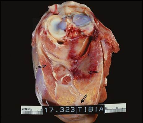
Figure 4.10 Gross postmortem lesions in the rear limb muscles of a goat with nutritional muscular dystrophy. Normal muscle is seen at the left (open arrow). The white chalky areas of muscle at the bottom of the picture are severely affected (black arrow). The lesion on the right is demarcated by an area of hemorrhage (open arrow). Source: Courtesy of Dr. T.P. O'Leary.
ascites, hydrothorax, pericardial effusion, pulmonary edema or congestion, and/or a swollen, congested, friable liver. Fibrin tags have been observed on the liver surface (Thompson 1986). Aspiration pneumonia may be observed secondary to swallowing difficulties. Tongue and pharyngeal muscles should be examined carefully in these cases.
Histologically, the muscle lesion is characterized by hyaline degeneration of striated muscle cells with coagulation necrosis, a pattern of degeneration referred to as Zenker's necrosis. Ultrastructural studies indicate that the lesion primarily involves the contractile components of the muscle cell cytoplasm.
Determination of selenium levels in liver at necropsy can give some indication of the severity of selenium deficiency. Normal liver selenium level in the goat has been reported at 1.00-4.80 ppm dry weight, with concentrations of less than 0.40 ppm considered deficient (Van Metre and Callan 2001). The same reference reports normal liver vitamin E concentration as greater than 250 pg/100 g wet weight in goats.
Diagnosis
Definitive diagnosis of NMD depends on the identification of typical muscle lesions in affected kids at necropsy or decreased levels of selenium and/or vitamin E in blood or tissues. A presumptive diagnosis in the field can be made on the basis of typical clinical signs, supportive laboratory findings, and a favorable response to treatment with parenteral administration of vitamin E and selenium.
When stillborn or weak kids are involved, other causes of infectious and non-infectious abortion must be considered, as discussed in Chapter 13. Sudden death in young kids may also be caused by gastrointestinal parasitism, especially hemonchosis and coccidiosis, colisepticemia, and enterotoxemia. Cases of NMD may appear very similar to enterotoxemia when the heart muscle is involved because of the presence of pericardial and peritoneal effusions. Histologic examination of the heart is necessary for diagnosis. When dyspnea, coughing, and other signs of respiratory distress are observed, bacterial, viral, and verminous pneumonias must be considered.
Difficulties in swallowing in young goats may be associated with cleft palate or neurologic diseases such as listeriosis or polioencephalomalacia. When a stiff-legged gait is observed, tetanus must be considered. The differential diagnosis for recumbency in young goats includes musculoskeletal trauma, enzootic ataxia, and the neurologic form of CAE. In enzootic ataxia and CAE virus infection, recumbency is usually preceded by a progressive ataxia that is characterized more by weakness than stiff-leggedness, and with a more prolonged course.
Treatment
Parenteral administration of combination sodium selenite/ alpha-tocopherol preparations is the treatment of choice in the acute phase of NMD. Administration of both compounds together results in a higher rate of recovery in kids than either compound administered alone (Baran 1966). Combination products are marketed widely for administration either SC or IM. Note, however, that the vitamin E in some of these products may not be present at a therapeutic level, as it is included in order to prevent oxidation of the selenium. When dosage information is not specifically given for goats, the recommended sheep dose can be given. A recommended dose for sheep is 1 mg selenium as sodium selenite and 50 mg (68 IU) of alpha tocopherol/18 kg (40 lb) bw, but label dosages can be followed for specific products (Van Vleet 2005). For products approved for use in sheep, the label may indicate that the dose for lambs 2 weeks of age or older is 1 mL per 40 lb bw, but with a minimum dose of 1 mL. This minimum dose can be a dangerous dose for a small kid, so 0.5 mL to small kids or 0.25 mL to dwarf breed kids when given at birth is recommended. Also, note that parenteral selenium/vitamin E injection has been associated with abortion and death in pregnant ewes and some commercial products are specifically labeled as not for use in pregnant ewes. Additional cautionary details on the use of injectable selenium/vitamin E products are given in Chapter 19.
Affected goats usually respond favorably to a single treatment within 24 hours, though recovery may not be complete, depending on the severity and extent of preexisting muscle damage. Animals that do not respond may be retreated once more at 24 hours. If no improvement is seen after the second treatment, then a poor prognosis must be offered, or an alternative diagnosis pursued. Excessive, repeated use of parenteral selenium can result in selenium toxicosis. In some herd situations, vitamin E deficiency may play a greater role in the development of disease than can selenium deficiency, and when combination products with limited vitamin E content are not producing a desired therapeutic effect, administration of additional vitamin E or nutritional supplementation of vitamin E should be considered (Byrne 1992).
As NMD indicates a nutritional deficiency, herd-wide administration of parenteral selenium/vitamin E preparations in the short term is justified at the time confirmed clinical cases are being treated in order to prevent additional cases. In the United States, recent reports of abortion in some sheep receiving parenteral injections have resulted in these products being disallowed for use in pregnant ewes. Abortions have not been reported in does, but caution should be exercised in using these products in pregnant does and clients be advised of the risk. Alternatively, for pregnant does oral administration of selenium can be initiated, up to 0.3 mg selenium/kg dry matter of the total ration.
Control
Selenium deficiency should be anticipated in goats in regions known to have selenium-deficient soils. Several approaches to correcting nutritional deficiency are possible, and the choice depends on economic, management, and regulatory factors. Feed supplementation with selenium is a desirable approach to control, though there may be regulatory constraints against the practice. In the United States, selenium supplementation of feed has been approved for use in sheep and beef for some time, but it was not until 2005 that allowance was made for goats. As of 2005, selenium can be supplemented in a complete ration for goats as selenium yeast at a level up to 0.3 ppm added selenium (FDA 2005). This is considered adequate to prevent the occurrence of NMD in selenium- deficient areas.
Where feed supplementation is not permitted by regulation or is impractical, strategically timed parenteral administration of selenium/vitamin E preparations is an effective means of control during periods of high risk. Does can be given therapeutic doses of a combination preparation at the onset of breeding season and again four to six weeks before kidding, observing the caveats mentioned above in the treatment section regarding pregnant does. Kids should be given a therapeutic dose at birth by SC injection or oral paste and again at 1 month of age. Over-the-counter oral preparations of selenium and vitamin E are available in the United States and labeled by the manufacturer for goats. Care should be taken not to overdose very small kids. In herds where clinical cases are observed in older kids, this can be repeated at 2 or 3 months of age. Bucks should receive the preparation twice a year, with the first administration timed to the onset of the breeding season. Drenching forms of sodium selenite are available in some areas. This allows selenium administration to be conveniently incorporated into other preventive medicine activities such as parasite control. Sheep doses can be used.
Other control methods are being increasingly applied to cattle and sheep, but have not been evaluated in goats. Injectable barium selenate, a repository form of selenium, maintains adequate selenium levels for as long as six months. Use of the preparation has been reported in goats in Spain. When given to dairy goats at a dose of 1 mg sele- nium/kg bw 15 days before breeding, seven- to eightfold increases in glutathione peroxidase activity were still measurable at the time of kidding, compared to no change in untreated controls (Sanchez et al. 2007). However, it is not clear if increased selenium levels were present in milk, which would be a significant regulatory concern in the United States.
Orally administered selenium pellets can provide adequate levels for up to one year in sheep. A sodium selenite, four-month, constant-release bolus is approved for oral use in cattle in the United States, but not for small ruminants.
Topdressing of pasture with sodium selenate at a rate of 10 g/ha may be an economic alternative to individual animal treatment and will prevent selenium deficiency for as long as 12 months (Kimberling 1988). The risk of inadequate vitamin E in the diet still exists when these techniques are used, and vitamin supplementation is indicated, as discussed in Chapter 19. In contrast to selenium, vitamin E is reported to be non-toxic to goats (Ahmed et al. 1990b).
Rickets
Rickets is a metabolic abnormality of growing bone. It is characterized by a failure of mineralization in newly formed bone matrix at the epiphyses of long bones. As such, it is a disease of young, growing animals. The condition is classically attributed to an absolute deficiency of vitamin D2 arising from the housing of young stock in buildings devoid of sunlight or the provision of green feeds not cured in the sun. However, chronic deficiencies of either calcium or phosphorus in the face of adequate vitamin D can also produce the condition. Inadequate mineralization at the epiphyses leads to structural weakness and abnormal growth, particularly in the long bones, which will show enlargements at the end plates and deformation due to the stresses of weight bearing.
Clinical and subclinical rickets have been described in young goats (Yousif et al. 1986). Clinically affected animals show stiffness of gait, a tendency to remain recumbent, a bowing deformity of the forelimbs, swollen carpi, and enlargements of the costochondral junctions, a lesion commonly described as the rachitic rosary. Loosening of the shoulder blade attachments and a dropped withers have also been identified with caprine rickets and may be the only signs seen (Guss 1977). Subclinically affected animals may show only signs of anorexia and a stunted growth rate.
The most consistent clinical pathologic finding in rickets is an elevation of serum AP. Abnormalities in serum calcium and phosphorus levels are less consistent, though in at least one report clinically affected goats had both hypophosphatemia (mean 2.76 ± 0.15 mg/dL) and hypocalcemia (mean 7.05 ± 0.18 mg/dL), with a ratio of serum calcium to serum phosphorus consistently more than 2 : 1. Affected goats were also hypoproteinemic and had decreased serum zinc, iron, copper, and magnesium levels compared with control goats (Yousif et al. 1986). Hypophosphatemia, hypocalcemia, hypovitaminosis D3, and increased serum AP were identified as the main biochemical abnormalities in 2- to 6-month-old goats with rickets in Egypt (El-Sayed and Siam 1992).
Radiographically, there is decreased density of affected bones, with widening and lipping of the non-mineralized growth plates. Antemortem biopsy of the costochondral junction or histologic examination of growth plates of long bones at necropsy confirms the failure of mineralization in the zones of provisional calcification in growth plates.
Successful treatment and control of rickets require accurate determination of the underlying cause through careful history taking, examination of housing and access to sunlight, and nutrient analysis of rations followed by correction of identified deficiencies. Nutrient requirements of vitamin D, calcium, and phosphorus are discussed in Chapter 19.
Osteoporosis
Osteoporosis is an osteodystrophy in which there is a progressive reduction of mineral and matrix content in bone. Like rickets, this condition is associated with dietary imbalances of calcium, phosphorus, and vitamin D, but is seen in mature animals rather than young growing ones.
Osteoporosis has been diagnosed in a herd of Oberhasli dairy goats in Switzerland associated with dietary deficiencies of calcium, phosphorus, and vitamin D, and aggravated by malnutrition attributable to gastrointestinal parasitism (Braun et al. 2009). The goats were provided with a salt lick, but it contained no calcium or phosphorus. The condition apparently had been present for some time before a definitive diagnosis was made, as 15 goats had been culled over a period of three years for stiff gait, lameness, chronic weight loss, and low milk production. Subsequently four adult goats 3-6 years of age were presented for clinical evaluation. The goats were thin, with depressed attitude and rough haircoat, showed arched backs and stiff gait, and shifted their weight from one forelimb to the other while standing. Laboratory findings included pronounced hypophosphatemia, marginal hypocalemia, and hypomagnesemia. Radiographs revealed decrease in mineralization of bone in the vertebrae, pelvic bones, and the head and shaft of the femur, where the bone cortex appeared to be very thin. At necropsy, the bones in one goat were palpably soft and the ribs could be cut with a knife. As enzootic calcinosis was known to occur in goats in the area, this was considered initially in the differential diagnosis after more obvious causes of lameness, such as foot problems, were ruled out. Following the confirmation of osteoporosis, it was recommended that a mineral supplement containing calcium, phosphorus, magnesium, and vitamin D3 be provided to the herd.
Epiphysitis
Epiphysitis is seen in young, rapidly growing goats that are fed excessive calcium. The lesion involves an unequal growth rate across the epiphyses of long bones, especially in the distal radial, distal metacarpal, and distal metatarsal epiphyses, and can result in premature closure of the growth plate either axially or abaxially. The result is a clinically obvious valgus or varus angular deformity of the limb around the affected epiphyses.
The exact pathogenesis of the lesion and its relation to excessive calcium feeding are not completely understood. The condition is similar to that seen in rapidly growing, overconditioned foals with a calcium/phosphorus imbalance in the ration. In one published report involving a 12-month-old pregnant Nubian doe, analysis of the total ration indicated a calcium-to-phosphorus ratio in the diet of 4.4 : 1 (Anderson and Adams 1983). The author (DMS) has seen severe bilateral bowing deformity of the carpi in a 12-month-old Saanen doe on a ration of alfalfa hay, a commercial goat chow supplemented with calcium, and free- choice dicalcium phosphate as a mineral lick (Figure 4.11). Elimination of the mineral and reduction of the goat chow in the diet resulted in some straightening of the forelimbs over the next two months.
Young, growing goats with painful epiphysitis may be reluctant to rise, have a stilted gait, or even walk on their knees. Overconditioned or pregnant goats may be especially painful due to the excessive burden of weight bearing. Joints associated with affected epiphyses may appear swollen. Angular limb deformities may be pronounced, depending on the duration of the condition.
Serum calcium, phosphorus, and AP levels are normal. Radiographs reveal an unequal growth rate across affected physes, with lipping, or overgrowth, of new bone on the margins of the physeal plate.
Management of epiphysitis in young goats requires ration analysis and correction of excessive feeding of calcium. The normal calcium-to-phosphorus ratio in the diet should be in the range of 1.5-2 : 1. In active cases, use of non-steroidal anti-inflammatory drugs such as flunixin meglumine, removal from hard surfaced flooring, and proper foot trimming may offer some clinical improvement.
Bentleg or Bowie
This is a condition most commonly seen in growing lambs grazed on pastures with phosphorus-deficient soils, notably in South Africa, New Zealand, and Australia. Affected animals develop genu valgum (bentleg) or genu varum (bowie). The condition has also been reported in 3-4-month-old Saanen kids in New South Wales, Australia
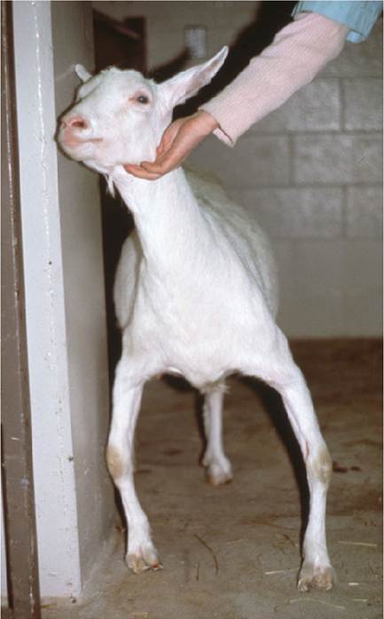
Figure 4.11 Bowing deformity of the forelimbs due to carpal epiphysitis associated with excessive calcium feeding in a yearling Saanen doe. Source: Courtesy of Dr. David M. Sherman.
(Murphy et al. 1959). Affected kids developed a severely knock-kneed (genu valgum) appearance at the carpi and were also bent in at the fetlock, with inward rolling on the medial claws. The condition was ascribed to an imbalance of calcium and phosphorus in the ration. In experimental trials, cases of bentleg were significantly reduced when a diet containing calcium and phosphorus in a ratio less than 1.4 : 1 was fed, compared to a diet with a Ca : P ratio greater than 1.8 : 1 (Murphy et al. 1959). In New Zealand, mature does in late pregnancy are reported to experience bentleg (Merrall 1985).
Bentleg, at least in goats, may be a manifestation of epiphysitis associated with excessive calcium feeding. However, the roles of other mineral imbalances, including iron excess, copper deficiency, and manganese deficiency, as well as plant toxicities, in the pathogenesis of bentleg have not been completely clarified. The etiology of bowie or bentleg in lambs is still considered to be unknown, though supplementing the diet with phosphorus or topdressing pastures with superphosphate usually results in disappearance of the disease (Constable et al. 2017).
Fibrous Osteodystrophy (Osteodystrophia Fibrosa)
Etiology and Pathogenesis
This bone disease is an example of nutritional secondary hyperparathyroidism that results from a chronic, sustained intake of excessive dietary phosphorus. Increased phosphorus intake leads to an analytically imperceptible hyperphosphatemia that suppresses serum calcium levels. The resulting hypocalcemia triggers an increased secretory response of parathyroid hormone. Parathyroid hormone mobilizes calcium from bone to restore normal serum calcium levels. In the face of chronic, excess phosphorus intake, this secondary hyperparathyroidism ultimately results in severe, generalized demineralization of bone and a fibrous reaction of the bony matrix.
Epidemiology
The condition is well known in goats (Carda Aparici et al. 1972; Naghshineh and Haghdoust 1973; Saha and Deb 1973; Andrews et al. 1983; Bandarra et al. 2011). It usually arises under circumstances of prolonged or monotonous feeding of rations with high phosphorus content. This often involves a diet high in grain and cereal hays, with little or no access to leguminous forages or calciumbased mineral supplements. Bran feeding is a common factor in many cases. The condition was described in the 1930s in goats experimentally fed flaked maize and bran with a minimum amount of hay (Glock and Murray 1939). All breeds and ages of goats are susceptible when weaned.
Clinical Signs
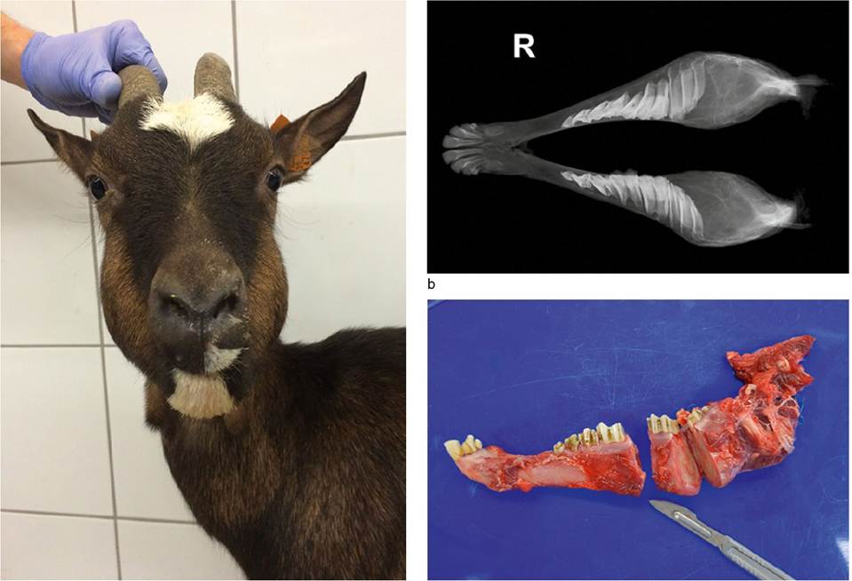
The initial signs may be non-specific and involve progressive lethargy, dyspnea, difficulty in eating and drinking, weight loss, and a preference for recumbency. Though all bones may be affected, there is a propensity for the mandibles first to show detectable abnormalities. There is a visible bilateral swelling of the mandibles, particularly over the rami, which may progress over several months and be quite pronounced (Figure 4.12a). These cases are frequently mistaken for lumpy jaw (actinomycosis), which is uncommon in goats. The jaw bone may be palpably soft. The goat's ability to open the mouth widely may be restricted. Salivation may be present and the tongue may hang from the mouth. Disruptions of the dental arcades with displacement of individual teeth is common. Teeth may actually point horizontally instead of vertically. Affected animals may have a stiff or painful gait, or be recumbent, due to pathologic fractures that may spontaneously occur in dystrophic bones.
Clinical Pathology and Necropsy
Enlargement and rarefaction are evident and displacements of the teeth can be identified in radiographs of the mandible (Figure 4.12b). Fractures of long bones, especially incomplete fractures, may also be confirmed radiographically. Serum chemistry results are variable. Elevation of serum AP is the most consistent abnormality. Hyperphosphatemia and hypocalcemia are also likely to occur, but their absence does not rule out fibrous osteodystrophy.
At necropsy, affected bones are soft, and in severe cases the mandibles can be cut easily with a knife (Figure 4.12c). The gross enlargement and distortion of the mandibles are evident on cross-section. Ribs are rubbery and flexible. Histologically, there is osteoporosis with marked demineralization of bony trabeculae, surrounded by a loose fibrous connective tissue matrix. Hyperplasia of the parathyroid glands is present, principally involving increased numbers of light chief cells, with extensive vacuolation and margin- ation of cell nuclei (Carda Aparici et al. 1972).
Treatment and Control
Reversal of clinical lesions is possible, if recognized early, by dietary management involving reduction of phosphorus levels and correction of the calcium-to-phosphorus ratio. Addition of leguminous hay and/or ground limestone to the diet is a practical way of increasing calcium in the diet without increasing phosphorus content. For grain-fed small ruminants in Brazil, addition of 1-1.5% calcium carbonate to the grains improved the Ca : P ratio and prevented fibrous osteodystrophy (Riet-Correa 2004).
Osteopetrosis
Osteopetrosis has been described by one authority as a bony abnormality of adult male goats fed diets excessive in calcium (Guss 1977). It is postulated that such diets promote hypercalcitoninism and the excessive deposition of calcium into bone. The condition is believed to be similar to the disease ankylosing spondylitis, which occurs in breeding bulls on high-calcium diets. However, this has not been confirmed experimentally.
The condition is said to occur in mature dairy bucks that are fed rations formulated for lactating does on the same farm. Such rations usually contain calcium in excess of that required for a non-lactating, non-growing male. Bucks show clinical signs only after they have ceased to grow and have been on the offending diet for many months. They develop proliferative calcification around the joints that leads to palpable enlargements, a stiff gait, and possible ankylosis, with reduced range of motion in affected joints. Radiographs confirm the proliferative lesion. When bony
a
c
Figure 4.12 Clinical appearance, radiographic and postmortem findings in fibrous osteodystrophy. (a) Typical swelling of the face caused by mandibular distortions associated with fibrous osteodystrophy. The ration of this goat consisted predominantly of oat grain, with little roughage provided. (b) Radiograph of advanced case showing enlargement and rarefaction of the mandibles, with the occlusal surfaces of the teeth directed medially. (c) Softened mandible easily sliced with a scalpel. Source: Reproduced by permission of Dr. Jaroslaw Kaba, Faculty of Veterinary Medicine, Warsaw University of Life Sciences, Warsaw, Poland.
proliferation is observed around joints in lame goats, CAE virus infection must also be considered in the differential diagnosis, particularly if mineralization of tendons and joint capsule is also present.
There is no treatment because the lesions are not reversible. Prevention is based on dietary management. Grass hay should serve as the basis of the diet, and calcium supplementation of the concentrate feed should not exceed 0.5%. If bucks are fed pure alfalfa hay, then no additional calcium supplementation should be offered, and addition of monosodium phosphate to the concentrate at a rate of 1% may be indicated.
Laminitis
Laminitis, or founder, is an aseptic inflammation of the sensitive laminae of the hooves. Acute and chronic forms occur in goats and result in lameness and possible deformities of the hoof.
Etiology and Pathogenesis
The causes and pathogenesis of laminitis in hoofed stock are not completely understood. The principal lesion appears to be vascular, associated with abnormal circulation within the corium, or dermis, of the hoof. Acute engorgement of vessels of the sensitive laminae leads to intense pain in the feet. Chronic circulatory dysfunction leads to both a breakdown of the connection between the corium and the hoof wall, allowing separation and rotation of the third phalanx within the hoof, and a distortion of new horny growth of the hoof.
Epidemiology
Laminitis in goats is seen more often in intensive management settings than in extensive ones. Its occurrence following sudden ration changes, excessive feeding of grain, or overt cases of engorgement toxemia suggests lactic acidemia as a predisposing factor. Experimental induction of lactic acidosis in goats by overfeeding of grain resulted in lameness (Tanwar and Mathur 1983). A clinical investigation of a dairy goat herd in the United Kingdom provided strong evidence for the association of prolonged concentrate feeding and chronic laminitis in the herd (Groenevelt et al. 2018). The occurrence of laminitis after kidding in association with retained placenta, metritis, pneumonia, mastitis, and enterotoxemia suggests the action of bacterial toxins in the pathogenesis of the condition. Laminitis has also been observed after normal kidding, and in association with unspecified allergic conditions (Guss 1977). Chronic laminitis is more often recognized clinically in goats than is acute laminitis.
Clinical Signs
Because acute laminitis often occurs in conjunction with other medical conditions such as grain overload that produce overt clinical signs, the presence of laminitis should not be overlooked. Affected goats may appear anxious and uncomfortable and grind their teeth from pain. Fever caused by underlying infectious diseases may be present. The goat may refuse to walk or even stand. The affected hooves are warm to the touch, particularly in the area just distal to the coronary band. Laminitis is almost always bilateral. The forelimbs are more often involved than the hindlimbs, but all four feet can be affected. Goats with acute laminitis of the forelimbs may walk on their knees.
In chronic laminitis, earlier episodes of acute laminitis may not have been seen, or recognized, so the onset may appear insidious. There is usually a vague lameness or an increasing tendency for animals to walk on their knees. The animal may stand with the hindlimbs placed well forward under the torso. Though heat is not palpable in the feet in chronic laminitis, the conformation of the hoof becomes distorted. The hoof wall becomes thickened, with a loss of distinction between the wall and the sole, and the feet become characteristically overgrown in a “slipper foot” or “sled runner” shape with the unworn toes turned upward. This is due to rotation of the third phalanx causing convexity of the sole, with the result that weight bearing is rocked back onto the heel (Pinsent 1989).
Clinical Pathology
There are no specific clinical laboratory abnormalities to support the diagnosis of laminitis, although the presence of lactic acidemia, endotoxemia, or a hemogram characteristic of inflammation may provide evidence of a likely underlying disease problem. Diagnostic radiography is rarely employed to confirm the diagnosis of laminitis in goats. Chronic lami- nitis is usually apparent from the gross appearance of the foot. In one report, longitudinal sections of the claws of goats with chronic laminitis at necropsy revealed downward rotation of the distal phalanx (Groenevelt et al. 2015).
Diagnosis
The presumptive diagnosis of acute laminitis is based on a history of predisposing management and disease factors, an acute reluctance to walk or stand, and heat in the feet, especially the front feet. Foot rot and puncture wounds must be ruled out. In chronic laminitis, arthritis must be ruled out when goats walk on their knees. Chronic selenosis must be considered in regions where it occurs when the hooves appear abnormal in association with lameness. Simple neglect of routine foot trimming must also be considered.
Treatment
In acute laminitis, it is important to identify and aggressively treat any predisposing disease process, such as engorgement toxemia or toxic metritis, correcting lactic acidosis, dehydration, and bacterial toxemias when present. Therapy directed at the laminitis consists primarily of analgesics to reduce pain in the feet and keeping the animal mobile. Non-steroidal anti-inflammatory drugs such as phenylbutazone and flunixin meglumine are particularly useful in this regard, because, as prostaglandin inhibitors, they probably also reduce the deleterious effects of endotoxins. Phenylbutazone can be given orally at a dose of 10 mg/kg once a day or flunixin parenterally at a dose of 1 mg/kg once a day, with the treatment tapering off over several days. Phenylbutazone should be avoided in animals for milk or meat production because of residue concerns, and in some countries its use in food animals is prohibited. In the United States it is prohibited only in female dairy cattle over 20 months of age, but its use is discouraged in younger cattle and other species intended for food. Enforced exercise may be helpful in the acute phase to promote normal circulation. Affected goats should be fed only grass hay while they recover and then brought back onto richer feeds cautiously. The value of antihistamines in the treatment of acute laminitis remains unproven. The use of corticosteroids is controversial, because they have been shown to induce laminitis in horses.
Management of chronic laminitis involves reduction of grain in the ration, avoidance of sudden ration changes, and frequent corrective foot trimming to approximate as normal a hoof conformation as possible. The foot is trimmed to reduce the height of the heel, to shorten the toes, and to remove the convexity of the sole to minimize the downward pressure of the tip of the third phalanx. When separation of the white line has occurred, the abaxial hoof wall should be trimmed back to the top of the pocket; otherwise dirt and debris will pack in and deepen the lesion. When necessary, administration of analgesics may help to control pain and promote mobility. For longterm analgesic therapy, aspirin is useful because of low
cost. Goats with a developed rumen can be sra.rled at a dose of 100 mg/kg orally twice a day and ltlra.led down to whatever lesser dose is needed to maintain comfort. Extended thera.py a.l. the starting dose could possibly lead to gasImal uIceration or anorexia. A good alternative is generic Ineloxicam, which is also low cost. It can be given ora∏y, Slturlinf twih a loading dose of 2mg∕kg followed by a daily dose of Img/kg until pain is controlled, and then i^i^:i^:i^t;a:i:i^t^d alt a dooe of 0.5-1mg∕kg every other day long lermt'MaU.hc∖vs2016J.
Control
To prevent laminitis in goats, abrupt feed changes should be avoided and grain feeding should be kept to a minimum. When high-energy rations are indicated for milk or hair production, addition of buffers such as sodium bicarbonate to the diet should be considered to reduce the risk of lactic acidosis. Regular hoof trimming should be carried out.
Zinc Deficiency
Zinc deficiency in ruminants is most often recognized as a proliferative dermatitis (parakeratosis), as discussed in Chapter 2. Zinc also plays a role in the normal development of bone and hoof. Zinc is most concentrated in bone in the Haversian system and participates in the calcification of preosseous tissue, either as an enzyme cofactor or as a metallic salt promoting crystal seeding during mineralization (Hidiroglou 1980). Dietary zinc requirements in goats are given in Chapter 19.
Zinc-deficient goats may display skeletal and hoof abnormalities in addition to dermatitis. Affected goats adopt an abnormal posture in which the back is arched and the feet are held closely together. Bowing of the hindlimbs and swollen hocks may also be seen. The dermatitis may involve the coronets, leading to inflammation of the hooves. Feet may be painful on palpation. Deep, transverse ridges may encircle the hoof wall. Zinc-deficient goats may also show poor growth rates, decreased feed intake, loss of condition, excessive salivation, and testicular dysfunction (Neathery et al. 1972). They may have impaired immune function, often dying of secondary pneumonic infections (Miller et al. 1964). Dwarfism has been reported in male goats in experimentally induced zinc deficiency. This was considered an indirect effect of zinc on pituitary function, rather than a direct effect of zinc on bone development (Groppel and Henning 1971).
In experimental feeding trials, goats on limited zinc diets had mean plasma zinc levels in the range of 0.49-0.79 ppm, while the range in normal control goats was 0.83-1.1 ppm (Neathery et al. 1972). In field cases of zinc deficiency in goats, plasma zinc levels in affected goats were in the range of 0.46-0.54 ppm (Nelson et al. 1984). Mean dry bone concentrations of zinc in goats on experimental zinc- deficient diets were 71 ppm, significantly less than the mean of 84 ppm in control goats (Groppel and Henning 1971). Hair samples can also be used to confirm zinc deficiency, with deficient goats having hair zinc concentrations less than 90 ppm (Neathery et al. 1972).
Successful treatment of hoof and postural problems was reported with oral administration of 250 mg of zinc sulfate daily for four weeks (Nelson et al. 1984). Commercial zinc methionine supplements are available in the United States.