Toxicological Diseases
Hypervitaminosis D
Vitamin D toxicity can result from excessive administration of parenteral or oral vitamin D. Such cases have been reported in cattle, horses, alpacas, llamas, and pigs (Constable et al.
2017). Excess vitamin D causes increased absorption of calcium from intestine, reabsorption from bone, and retention of phosphorus by the kidney. There is a tendency for thinning of bone and calcification of soft tissues to occur, especially muscle and vascular tissue, with multiple clinicopathologic consequences.The risk to goats of iatrogenic vitamin D toxicity has been demonstrated experimentally (Singh and Prasad 1987). Goats given cholicalciferol injections weekly for two to eight months developed clinical signs of dullness, depression, rough haircoat, inappetence, diarrhea, polyuria, polydipsia, and reduced growth rate. Muscle weakness and a stiff gait were observed in some animals. Hypercalcemia and hyperphosphatemia were consistent findings. Serum AP levels, however, were quite variable.
Enzootic calcinosis, caused by consumption of T. flaves- cens, is discussed later in this chapter. It is considered to be a naturally occurring form of vitamin D toxicity, because the plant contains high levels of 1,25 (OH)2D3. It is reported in goats in Europe.
Fluorosis
Fluorosis in goats is a chronic intoxication caused by prolonged exposure to fluoride compounds in feed, water, and soils. The incorporation of excessive fluorine into the skeletal or dental matrices causes abnormal development of teeth and bone, especially in growing animals.
Etiology and Epidemiology
Animals may come into contact with fluorine in many ways. The element is found in high concentration in phosphate-rich soils in some regions of the world, particularly in North Africa. Although plants do not take up fluoride readily, grazing animals may ingest soil along with plants while feeding, thus increasing their exposure to fluoride.
When vegetation does incorporate fluoride, a process enhanced by acid soils, the stems and leaves of plants have higher concentrations than grains. Deep well water is a source of excessive fluoride in Australia and North and South America. Volcanic ash is a source of pasture contamination in the Andes and Iceland (Constable et al. 2017).Rock phosphate, rich in fluoride, is mined commercially for manufacturing fertilizers and mineral supplements for animal feeds. Unless these mineral supplements are specifically defluorinated before incorporation into animal feeds, they may provide dangerous levels of fluoride. Water and tailings from mining operations can be sources of excess fluoride to animals living nearby due to runoff or careless disposal. This occurs in many parts of North Africa. In west central Morocco, for instance, where phosphate mining is a major industry, fluorosis is recognized as an important clinical problem in human and livestock populations and is known as darmous (Kessabi and Abdennebi 1985). Soils in endemic areas contain fluoride in the range of 1450-4085 ppm dry matter (DM). Pasturage in these regions has fluoride concentrations in the range of 95-260 ppm DM; hay, 85-180 ppm DM; and barley grain, 11-18 ppm DM. Clear water has an average of 1.4 ppm fluoride. However, drinking water in these areas is often turbid, containing much soil and rock particulate matter, and the average fluoride concentration of turbid water is 14 ppm. Fluorosis is likely to develop in livestock when the longterm ration concentration of fluorine exceeds 100 ppm DM. In the wet season grazing is identified as the major source of fluoride ingestion, while in the dry season it derives from increased water consumption. In Egypt, chronic fluorosis has been reported in goats kept in the vicinity of a superphosphate factory emitting airborne hydrofluoric acid as a byproduct (Karram 1984).
Fluoride is also found frequently in ores processed for aluminum and iron. Smelting and milling operations release fluoride into the air, thus contaminating fields, crops, and water sources through settling of particulates.
Animals grazing nearby can ingest sufficient deposited fluoride to develop toxicity, as reported in goats near an aluminum smelter plant in India (Sahoo and Ray 2004). In Inner Mongolia in China, industrial fluoride pollution has a serious impact on Cashmere goat production. The life span of goats may be reduced to two to three years due to impaired pasturing, mastication, and inanition resulting from teeth deformed by fluorosis (Wang et al. 2002).Fluorine exposure from the sources discussed above involve inorganic fluoride compounds and toxicity usually arises from chronic exposure. Acute fluorine toxicity may also occur in livestock accidentally exposed to large doses of organic fluorides such as sodium fluoroacetate, which is used as a rodenticide and in predation control. Acute fluoride toxicity from such accidental poisoning has not been reported in goats, though they are presumably susceptible.
An unusual form of acute monofluoroacetate poisoning of goats in South Africa occurs after ingestion of the plant Dichapetalum cymosum, also known as gifblaar, which accumulates monofluoroacetate. The clinical manifestation is usually rapid death due to heart failure, as discussed in Chapter 8. In northeastern Brazil, consumption of the plant Amorimia Septentrionalis, which contains sodium monofluoroacetate, causes considerable losses in grazing cattle due to cardiotoxic effects, including cardiofibrosis. Goats are susceptible and in experimental challenges, pregnant does fed the plant show embryonic death and abortion (da Silva et al. 2017). Kids fed colostrum from does consuming the plant may die acutely (Lopes et al. 2019).
Pathogenesis
Chronic fluorosis results from prolonged but not necessarily continuous ingestion of excessive fluoride compounds. Tolerance levels for goats have not been reported, but safe ration concentrations for breeding ewes up to 60 ppm and for feeder lambs up to 100 ppm have been established (Osweiler et al. 1985). In experimental studies involving daily administration of sodium fluoride, the response of goats appears to be similar to that of sheep (Milhaud et al.
1983). Fluorides are readily absorbable from the digestive tract, though the availability and digestibility of fluorides vary with the source. In goat studies, fluoride was 75% available from sodium fluoride, 65% from ground raw rock phosphate, 34% from defluorinated phosphate, and 38% from dicalcium phosphate (Clay and Suttie 1985).Though fluorine accumulates in all tissues, the manifestations of chronic fluorosis are related to fluorine accumulation in hard tissues, namely bone and teeth (Milhaud et al. 1980b).
Fluorine can be incorporated into bone throughout the life of the animal, but accumulates in teeth only during development. Therefore, the presence or absence of lesions in teeth erupting at different ages gives some indication of the duration and onset of exposure to fluoride. The effect of excessive fluoride on developing teeth is to impair normal mineralization of the preenamel, predentine, and prece- mentum matrices. There is evidence that high fluoride interferes with normal collagen synthesis to produce imperfect collagen or even non-collagenous protein, which alters the tooth matrix structure and results in abnormal tooth morphology (Wang et al. 2003). Affected teeth are softer, appear mottled, and wear more quickly. Typical staining of affected teeth results from oxidation of exposed organic material in the tooth. Affected animals may have difficulty drinking cold water, and due to excessive tooth wear may have difficulty in prehension and mastication. These animals grow poorly and are underconditioned as a result of impaired feed intake.
The adverse effects on bone include disruption of osteogenesis, acceleration of bone remodeling, development of exostoses and sclerosis, and osteoporosis. An important underlying mechanism for abnormal bone development in fluorosis is the impact of fluoride on collagen metabolism, leading to structural changes in collagen fibers that affect the extracellular matrix of bone. At the molecular level, collagen gene expression is altered in goats with fluorosis (Li et al.
2006, 2007). Animals with skeletal fluorosis may have visible swellings on bones and exhibit a stiff and painful gait or intermittent lameness. Chronic fluorosis can also produce anemia in affected livestock through accumulation in the bone marrow and suppression of erythropoiesis.Clinical Findings
Signs of chronic fluorosis may be non-specific and include anorexia, a stiff gait, a history of intermittent lameness, emaciation, a rough dry haircoat, and pale mucous membranes suggesting anemia. Morbidity rates can be high, especially in endemic areas. Careful observation of animals while they eat and drink may reveal difficult mastication or mouth pain. Specific examination of the teeth and palpation of the skeleton are necessary to correlate these general findings with a diagnosis of fluorosis. Incisors are most easily examined. Affected teeth show streaky or splotchy yellow to brown to black staining and have a mottled, chalky appearance to the enamel surface (Figure 4.13). Teeth may be worn beyond expectation for the age of the
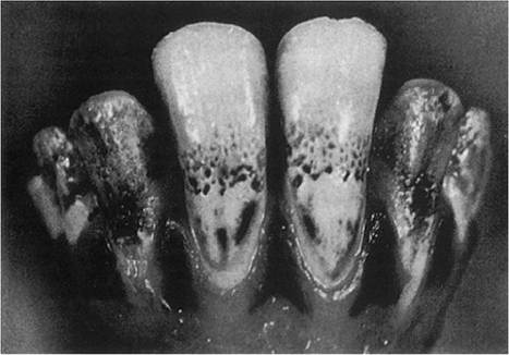
Figure 4.13 Typical dental lesions of chronic fluorosis. Note mottling and staining. Source: Milhaud et al. (1980a) / Recueil de Medecine Veterinaire.
animal, and some individual teeth may be prematurely lost. The number of teeth involved will vary depending on when exposure to fluorides began. However, pairs of teeth, for example, corner incisors, are bilaterally affected because they developed at the same time.
Palpation of the skeleton may reveal a general thickening of bones or focal swellings caused by exostoses, particularly on the mandibles, ribs, and metacarpal and metatarsal bones. Overt lameness or recumbency may be due to either bone pain or fractures occurring secondary to fluorotic changes in bone. Rib fractures are common in smaller does that have been mounted by large bucks at breeding.
Clinical Pathology and Necropsy
Normal plasma fluorine levels in goats have been reported in the range of 0.09-0.22 mg/L, while goats with signs of chronic fluoride intoxication have concentrations of 0.6-1.1 mg/L (Milhaud et al.
1980a). Urine fluoride levels of more than 15 ppm suggest chronic fluorosis in cattle, but diagnostic levels in goats have not been reported.Anemia may be documented in the hemogram of goats with chronic fluorosis (Karram et al. 1984). In other species, serum calcium and phosphorus levels are usually within normal limits, but serum AP is often elevated. In experimental chronic fluorosis in goats, serum AP levels were normal, as were serum calcium and phosphorus levels (Milhaud et al. 1980b). In a naturally occurring field outbreak, affected goats were hypocalcemic and hyper- phosphatemic, but AP was not measured (Karram 1984).
Radiographically, the severity and extent of detectable lesions depend on the duration, degree, and onset of exposure to fluoride. In the jaws, maxillary borders become hazy and irregular. Bone structure becomes more porous, with reduced density of cancellous bone. Compact or cortical bone is thinned. Molar and premolar tooth roots become less opaque and less substantial and abnormal tooth wear is apparent. In long bones, increased porosity and decreased density are also noted. Physeal cartilage of the metatarsal and metacarpal bones becomes hazy and double lines are noted at the growth plate. Osseous bridging across the growth plate may also be seen and joint spaces may be narrowed. Pseudoarthroses may be seen when rib fractures are present. Periosteal hyperostosis is a consistent finding in advanced, severe chronic fluorosis in cattle, but was not reported in experimental chronic fluorosis of goats (Milhaud et al. 1980b).
At necropsy, the carcass may be in poor flesh. There are no gross or histologic lesions in soft tissues. Bones may be brittle and have a chalky white appearance with a roughened, irregular surface. They may be thicker and heavier than normal. Mandible, metatarsal, and metacarpal bones are most often affected. Fluorotic molars and premolars may show staining, mottling, chalkiness, and excessive wear, similar to affected incisors. The staining, which involves the organic matrix of the tooth, cannot be scraped off, in contrast to superficial food staining. Microscopically, bones may show varying degrees of osteopetrosis or osteoporosis depending on the type, intensity, and duration of fluoride exposure. When fluorosis is suspected, bone should be analyzed for fluoride content. Mandible, rib, metatarsal, and metacarpal bones are suitable submissions. In normal goats, fluorine concentration in these bones is reported to be less than 17.5 ppm/fat-free dry matter (Milhaud et al. 1983).
Diagnosis
Presumptive diagnosis is based on a history of chronic exposure to fluorides, lameness or stiff gait, characteristic dental lesions, and consistent radiographic findings. Fibrous osteodystrophy, which occurs on high-phosphorus diets, may produce lameness, jaw swelling, and disruption of normal dental alignment, but mottling and chalkiness of teeth are absent. Diagnosis is most challenging in the early stages, when ill thrift or stiff gait may be the only presenting complaints. Definitive diagnosis requires confirmation of increased fluoride levels in bone samples taken by biopsy or at necropsy.
Treatment and Control
There is no specific treatment for chronic fluorosis beyond removal of animals from the fluoride source, which is difficult when the source is industrial or environmental. Attempts should be made to identify all sources of fluorine and their relative danger. When possible, management should allow young, growing stock access to the least contaminated pastures, feeds, and water supplies to reduce the incidence of dental fluorosis. Turbid water sources should be allowed to settle, and clear water only offered to goats. Water can be pretreated with slaked lime at a rate of 500-1000 ppm and allowed to settle for six days.
When mineral supplements are fed to goats, raw rock phosphate and superphosphate should be avoided or at least checked for fluorine levels before use. Defluorinated phosphate supplements are recommended. Suitable products should have a phosphorus-to-fluorine ratio of 100 : 1 or more (Osweiler et al. 1985). Extensively defluorinated products, however, are generally more expensive due to increased refining costs and may not be attractive to producers.
In parts of China, where industrial fluorosis is a major problem for grazing goats, a number of interventions have been attempted to reduce the problem, including removal of goats from high- to low-fluoride areas, use of stored green grass in the dry season as fodder, trimming of affected teeth, and mineral supplementation. However, the best results for controlling the wearing down of affected teeth was to supply supplemental protein-rich feed to goats in high-fluoride areas (Wang et al. 2002).
Chronic Selenium Poisoning
Chronic selenium poisoning is characterized by deformation and sloughing of the hooves, with marked lameness and secondary emaciation in grazing animals resulting from decreased locomotion.
Etiology and Epidemiology
Chronic selenium poisoning of livestock occurs mainly in regions of the world where soil selenium levels are high. Seleniferous soils are found in the Rocky Mountains and Great Plains of North America, parts of Australia and India, as well as Israel, Ireland, New Zealand, portions of the former Soviet Union, and elsewhere. Plants growing on seleniferous soils absorb selenium and prolonged consumption of these plants by livestock leads to toxicosis. Indicator or converter plants, such as Astragalus spp., require selenium for growth and contain particularly high concentrations. Though often unpalatable, livestock will eat these plants under conditions of deprivation or overgrazing. Facultative plants may also absorb selenium from the soil, but do not require it for growth. Grains such as corn, when grown on seleniferous soils, may contain toxic levels of selenium. More detailed information on selenifer- ous soil characteristics and soil-plant interactions is available elsewhere (Dhillon and Dhillon 2003).
Chronic selenium poisoning associated with prolonged ingestion of indicator plants assumes two clinical forms. The first, alkali disease, is characterized by weight loss and musculoskeletal abnormalities and is presented here. The second, blind staggers, is characterized by signs of neurologic dysfunction. In recent years the role of selenium in blind staggers has been called into question and the theory proposed that this condition is actually a manifestation of polioencephalomalacia resulting from excessive sulfur intake (O'Toole et al. 1996). Blind staggers is discussed further in Chapter 5.
Chronic selenium poisoning may also occur when selenium supplements are improperly mixed into feeds at excessive amounts. Experimental studies demonstrated that goats fed sodium selenite at a rate of 6 mg/kg bw daily became sick and died within 4-19 days after feeding began. Goats fed 3 mg/kg bw daily showed no adverse effects after 90 days (Pathak and Datta 1984). Acute selenium poisoning has been produced experimentally in goats. Single oral doses of sodium selenite in the range of 40-160 mg/kg bw killed goats in a matter of hours. Repeated daily doses in the range of 5-20 mg/kg bw produced signs of inappetence, diarrhea, hindlimb weakness, arching of the back, weight loss, recumbency, and death over a period of days to several weeks (Ahmed et al. 1990a).
Clinical Signs
Chronic selenosis seen in grazing goats in India appears clinically similar to the disease as seen in cattle and buffalo (Gupta et al. 1982). Animals introduced into seleniferous regions of the Punjab develop signs within six months after arrival. All ages and both sexes are affected. The earliest signs are visible cracks in the horns and hooves, with irregular new growth at the coronary bands. The cracks gradually deepen and separate the older distal segments of the hoof from the new growth above. Hooves become deformed and elongated. Infection often occurs in the deep cracks of the hoof wall. The foot emits a necrotic odor and becomes extremely painful as pus accumulates. Affected goats have increasing difficulty standing, move with a staggering gait, or exhibit overt lameness. Severely affected animals remain recumbent. Impairment of locomotion drastically reduces feed intake and goats become weak, depressed, and emaciated. Decreased conception and increased abortion rates are also seen. Death is by inanition, predation, or secondary infection.
Clinical Pathology and Necropsy
Necropsy lesions are not particularly helpful in the diagnosis of alkali disease, but tissue samples should be collected to determine selenium levels. Specific toxic tissue levels have only infrequently been reported in goats. In other herbivores, levels of selenium in liver and kidney in the range of 4-25 ppm are found in chronic poisoning. Affected hoof tissue has selenium levels in the range of 8-20 ppm (Osweiler et al. 1985). In goats with selenium toxicosis from eating maize grown on seleniferous soils in China, blood selenium concentrations greater than 0.2 μg∕g were an indicator of early selenium toxicity. Overt signs of selenium poisoning appeared when blood and hair values were over 0.5 and 0.3 μg∕g, respectively (Hou et al. 1994). Mean liver and kidney concentrations of selenium in these affected goats were 20.46 ± 0.89 μg∕g and 20.96 ± 1.21 μg∕g, respectively, while liver and kidney selenium concentrations in control goats were 0.50 ± 0.16 μg∕g and 8.92 ± 2.20 μg∕g, respectively.
Treatment and Control
There is no effective treatment for selenium poisoning. Some practical recommendations for rangeland management have been offered to aid in control (Davis et al. 2000). Soils should be tested to identify high selenium content and livestock fenced out of the worst-affected areas. Avoid overgrazing so as not to oblige livestock to consume sele- niferous plants. Practice rotational grazing to limit the length of time animals spend on soils with high selenium levels. Graze animals on pastures with higher selenium concentrations only in the fall and winter, when grasses are more mature and have lower selenium content. Practice weed control because broadleaf plants are greater selenium accumulators than grasses. Unless soil sulfate levels are already high, selenium uptake by plants can be reduced by applying sulfur or gypsum to soil, because sulfur is competitive with selenium in plant uptake. Avoid use of phosphate fertilizers, which themselves may have a high selenium content and can also enhance selenium uptake in plants by displacing selenium bound to soil. When economically feasible, supplementary feeding with feedstuffs derived from non-seleniferous regions is the best method to dilute the uptake of selenium in chronically exposed animals. Feeding of supplemental copper and sulfur in a mineral mix can be beneficial in counteracting a high-selenium diet, as can an increase in the overall protein content of the diet.
Plants Toxic to the Musculoskeletal System
Senna (Cassia) roemeriana
Ingestion of Senna (Cassia) roemeriana (twin leaf senna) has caused death due to skeletal myopathy in grazing cattle and sheep in the southwestern United States and northern Mexico. Experimental feeding of the dried plant to adult Spanish goats at a rate of 5 or 7 g∕kg bw for an average of 24 days resulted in toxic effects related to muscle damage. After about two weeks of intake, affected goats demonstrated decreased appetite, weight loss, increased heart and respiratory rates, and progressive weakness of the hindlimbs, leading to recumbency. Elevations in serum CK as high as 43 800 IU∕L were also detectable after two weeks. At necropsy, muscles were pale, especially in the hindquarters. There were also pericardial and pleural effusions and pulmonary edema in some goats. Histologically, affected muscle shows necrosis and fragmentation of myofibers, with infiltration of necrotic sarcoplasm by neutrophils and macrophages (Rowe et al. 1987). In naturally occurring cases, NMD would be the principal differential diagnosis.
Karwinskia humboldtiana
This shrub is indigenous to the southwestern United States and northern Mexico and is responsible for coyotillo poisoning, or limberleg, in livestock. The condition, which principally causes a neuropathy, is discussed in more detail in Chapter 5. However, it has been demonstrated that the fruits of K. humboldtiana can produce widespread skeletal and cardiac muscle degeneration in addition to neuropathy (Dewan et al. 1965). The lesion in skeletal muscle is one of hyaline degeneration, similar to that seen in NMD. At least some of the incoordination and weakness observed in limberleg may be due to muscle damage in addition to neurologic dysfunction.
Trisetum flavescens and Enzootic Calcinosis
In alpine regions of Europe, a condition of livestock known as enzootic calcinosis is ascribed to the ingestion of T. flavescens, or yellow oat grass, at pasture or in harvested forages. The plant contains high concentrations of 1,25-dihydroxycholicalciferol, the active metabolite of vitamin D. This metabolite markedly increases the intestinal absorption of calcium by herbivores. Chronic ingestion results in calcification of selected soft tissues, notably the great vessels, heart, lungs, tendons, and ligaments. A reluctance to walk and constant shifting of weight from leg to leg are common signs of tendon and ligament involvement. Further information on the cardiac effects of enzootic calcinosis are available in Chapter 8.
Enzootic calcinosis has been recorded in milk goats in Switzerland, on a farm where yellow oat grass comprised 8-34% of the herbage on various fields (Kessler 1982). Over a three-year period, 12 goats died. Affected goats showed progressive wasting, decreased milk production, increased heart and respiratory rates, and altered locomotion. Serum calcium, phosphorus, and AP levels are increased in affected goats (Wanner et al. 1986). Evidence of calcification of soft tissues should be sought at necropsy. Paratuberculosis of goats also produces chronic wasting and in some cases calcification of the aorta, and this must be ruled out.
Delaying harvesting of T. flavescens reduces the toxic effect of the plants, because young plants have greater vitamin D activity. Silage made with yellow oat grass is more toxic than hay.
A similar syndrome of soft tissue calcification caused by increased vitamin D activity occurs with ingestion of Solanum malacoxylon by herbivores in the Caribbean, South America, and Hawaii, and from Cestrum diurnum ingestion in the southern United States. Reports of toxicity in goats from either of these plants were not found, but calcinosis in goats was reported in association with Solanum torvum ingestion in Hawaii (Mello 2003).
Poincianella pyramidalis
In the semi-arid region of Brazil, consumption of the plant Poincianella pyramidalis by pregnant goats was associated with abortion or kids born with bone malformations in the limbs, spine, ribs, sternum, and head, including arthrogryposis, scoliosis, and micrognathia (dos Reis et al. 2016).
Lupinosis
Several forms of lupine toxicity occur in livestock and all are poorly documented in goats. The neurologic and hepatopathic forms are mentioned briefly in Chapters 5 and 11, respectively. In calves, a congenital, skeletal form also occurs and is known as “crooked calf disease” in the western United States. The development of skeletal deformities results from transplacental transfer of a lupine alkaloid, anagyrine, during the second month of gestation. There is circumstantial evidence from California that the condition also may occur in developing goat fetuses whose dams consume Lupinus latifolius during pregnancy. Arthrogryposis, torticollis, and scoliosis are the characteristic signs. In addition, similar skeletal deformities have been observed in a human infant in California whose mother drank local goat milk during pregnancy, though a definitive causal link was not established (Kilgore et al. 1981).