Occipitoatlantoaxial Malformation
Robert J. MacKay • Mary O. Smith
Occipitoatlantoaxial malformations (OAAMs) are a heterogeneous group of developmental defects of the first two cervical vertebrae.1,2 They have been reported to occur in cattle, sheep, goats, and horses.3-6
Equine OAAMs have been classified into six groups.1 The first, a heritable condition in Arabian horses, is characterized by symmetric atlantooccipital fusion, atlantalization of the axis, and hypoplasia of the atlantal wings.2,7-9 A similar disorder has been reported in an Arabian-cross colt.10 The other groups are congenital asymmetric OAAMs,11 asymmetric
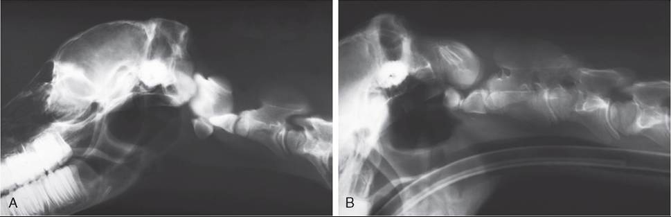
FIG.
35.21 Occipitoatlantoaxial malformation (OAAM). Radiographs of the craniocervical region of a normal neonatal foal (A) and a 3-week-old Arabian foal with OAAM (B). In the foal with OAAM, note slightly shortened dens, ventral luxation of the dens, Cranioventral movement of the body of the atlas to encroach on the guttural pouch, and fusion of the atlas to the occiput with occipitalization of its caudal surface. (Images courtesy Dr. I.G. Mayhew, Massey University.)atlantooccipitalfusion,2 duplication of axis or atlas,12 and symmetric OAAMs in non-Arabian horses.13,14 A deletion mutation near the HOXD3 gene was identified in one Arabian foal with OAAM but was not found in two others with atypical OAAM phenotypes.9
The clinical signs range from normal with or without abnormal head position to brainstem compression, sudden unexpected death, and stillbirth.2 In typical familial cases, signs of tetraplegia or tetraparesis begin at or shortly after birth and progress at variable rates. Foals may become suddenly tetraplegic and appear to stabilize for several days but then die suddenly.15 In rare cases, horses may not show nervous system signs until 3 years of age.13 The signs are symmetric in most affected animals and include hyperreflexia, hypertonia, weakness, and ataxia in all four limbs. Affected animals may be reluctant to move the neck and head and show resistance to manipulation of the area.
A click or crepitation may be palpated over the cranial cervical spine when the head is moved.2,5 With asymmetric bone lesions, torticollis is common, whereas patients with symmetric lesions hold their heads in extension and frequently display the “weathervane posture.” Neurologic deficits may not be apparent despite moderate torticollis.The malformations are readily apparent radiographically (Fig. 35.21), and CT can further elucidate the anatomic abnormalities.16 There may be subluxation of the atlantoaxial joint, ventral displacement of C2 in relation to C1, nonunited ossification center of the dens, shortened or elongated dens, shortened transverse process of the atlas, fusion of C1 with the occipital condyles, atlantal duplication, and deviation of the basioccipital bone.2,17 In sheep, additional malformations of the cervical vertebrae have been present concomitantly.
Suggested approaches to treatment are surgical fusion of the atlantoaxial joints, with or without laminectomy.13,18 Laminectomy alone has been used to alleviate spinal cord compression and clinical signs caused by occipitoatlantoaxial malformation.19 However, long-term results of surgical intervention have been poor, with neurologic deficits persisting. Closed reduction of the luxated atlantoaxial joint has yielded similarly unsatisfactory results.16,20 If treated, Arabian horses must not be bred because of the hereditary nature of the disease in that breed.