Other Pneumonias
Amelia R. Woolums
Aspiration Pneumonia
Aspiration pneumonia is caused by inhalation of large amounts of foreign material, often liquids. The most common cause is careless drenching or passage of stomach tubes during administration of milk or liquid medication.
It also occurs occasionally in pail-fed calves, animals with pharyngeal paresis, animals with necrotic laryngitis, lambs with nutritional myodegeneration, anesthetized animals, sheep that are dipped, cattle that have parturient paresis, and cattle that ingest crude oils or fuel oils. Meconium aspiration secondary to fetal distress has been recognized as a possible risk factor of neonatal calf mortality.1If large quantities of fluid are aspirated, death may be almost instantaneous, but generally a gangrenous bronchopneumonia develops as a result of infection and the irritating properties of the inhaled material. Affected animals exhibit depression, tachypnea, dyspnea, coughing, and fever and may have putrid breath. Crackles, wheezes, and occasionally pleural friction rubs can be heard on auscultation.
A presumptive diagnosis is based on the history and clinical evidence of bronchopneumonia; definitive diagnosis can be confirmed by gross or histopathologic identification of feed, milk, or other aspirated material in airways. Differential diagnoses include acute bacterial bronchopneumonia and septicemia. Necropsy reveals consolidation of the anterior ventral areas of the lungs. Affected areas are severely hemorrhagic in acute cases and contain suppuration and liquefactive necrosis in subacute cases.
The prognosis is guarded in all cases of aspiration pneumonia, but some animals can be successfully treated. Antibiotics combined with antiinflammatory agents should be promptly administered, and long-term antimicrobial therapy (weeks) is required. Prevention of aspiration pneumonia centers on careful administration of medication and avoidance of other risk factors that may promote inhalation of foreign materials.
Mycotic Pneumonias
Fungal pneumonia is a rare occurrence in ruminants. The clinical presentation is typically one of poor response to antimicrobial therapy. Cases of fungal pneumonia with metritis or placentitis have been reported.2,3 Diagnosis is confirmed by identification of fungal organisms with an inflammatory reaction in tracheal aspirate or bronchoalveolar lavage, or at postmortem with fungal culture to confirm the identity of the agent. Little information is available regarding successful treatment of ruminants with fungal pneumonia. In other species such as horses, treatment requires weeks to months of therapy with antifungal agents such as ketoconazole or itraconazole; these drugs may be cost prohibitive for many ruminant cases, and their disposition in the rumen has not been described. Therapy with oral potassium iodide is an inexpensive alternative that may be attempted for some cases, but the efficacy of this therapy has not been confirmed. Prognosis for fungal pneumonia in all species is guarded to poor, depending on the severity of disease and the feasibility of treatment. Given these restrictions, treatment of fungal pneumonia in ruminants is likely reserved for valuable individuals. More guidelines regarding treatment for fungal pneumonia are presented in the section on equine respiratory diseases.
Vena Caval Thrombosis and
Metastatic Pneumonia
■ Definition and Etiology Metastatic or embolic pneumonia in cattle, also called caudal vena caval thrombosis, pulmonary thromboembolism, and embolic pulmonary aneurysm, is a distinct syndrome associated with multifocal abscessation of the lungs caused by septic thromboembolism of the pulmonary arterial system. The septic emboli arise from septic thrombi of the caudal vena cava (Fig. 31.69) or, less commonly, the cranial vena cava. Vena cava thrombi are in turn a sequela to various septic conditions such as jugular phlebitis, mastitis, metritis, foot rot, or, most often, liver abscesses secondary to rumenitis.4 A variety of bacteria may be involved; those most frequently encountered include F.
necrophorum, A. pyogenes, staphylococci, streptococci, and E. coli.■ Clinical Signs Because of its association with rumenitis, this condition is most commonly seen in feedlot cattle, but any age, breed, sex, and class of cattle may be affected. The problem is unusual in cattle younger than 1 year of age. Cattle with metastatic pneumonia usually exhibit respiratory disturbance or weight loss or occasionally thoracic pain. The clinical signs in affected cattle have been described.5,6 The classic presentation includes tachycardia, tachypnea, dyspnea, coughing,
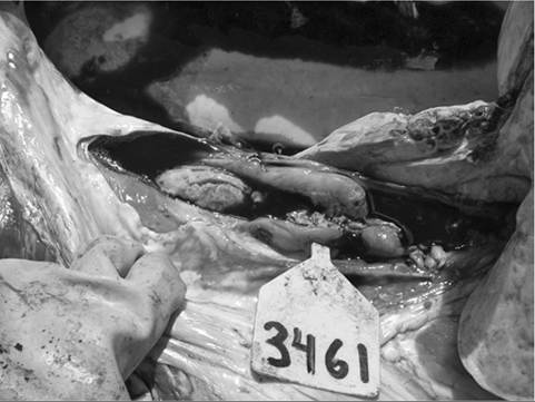
FIG. 31.69 Postmortem photograph of thrombus in caudal vena cava, which leads to embolic pneumonia demonstrated in Color Plate 31.10. (Photograph contributed by Feedlot Health Management Services, Okotoks, AB, Canada.)
heart murmurs and pale mucous membranes (caused by anemia), widespread wheezes, epistaxis, and hemoptysis. The duration of signs is quite variable, ranging from acute respiratory distress to a chronic history of weight loss and coughing for weeks to months. Other signs, which are more variable, include fever, thoracic pain on deep palpation of the sternum and intercostal spaces, hepatomegaly (indicated by the ability to palpate the caudal edge of the liver in the right paralumbar fossa), subcutaneous emphysema, froth at the muzzle, and melena caused by coughing up and swallowing blood. Nonspecific accompanying signs include depression, anorexia, ruminal stasis, scant feces, and decreased milk production. In chronic cases, cor pulmonale may lead to signs of right ventricular failure such as jugular distention and brisket edema. The combination of respiratory signs with anemia, widespread wheezes, and especially hemoptysis is generally regarded as pathognomonic for this syndrome. Animals usually deteriorate rapidly once hemoptysis becomes evident, and death despite treatment is typical. Many patients die suddenly with an acute episode of severe intrapulmonary hemorrhage or hemoptysis after a variable course of respiratory disease.
Some of these cases in which the respiratory signs were overlooked may account for the reports of sudden death attributed to vena caval thrombosis. Caudal vena caval thrombosis can also lead to hepatomegaly and extensive ascites, but most of these animals also have respiratory signs.6 Sudden erosion of a large hepatic abscess into the caudal vena cava may also result in a massive embolic shower, with acute respiratory distress and death.In patients with the pathognomonic signs (the majority of cases), no differential diagnoses need be considered. However, many patients are examined before the onset of hemoptysis, and a few may die without exhibiting these signs. Differential considerations in such cases, which usually manifest as acute dyspnea, should include anaphylaxis, various causes of ARDS, hypersensitivity pneumonitis, lungworms, and acute bronchopneumonia. Patients with right ventricular failure should be differentiated from those with pericarditis, lymphosarcoma, cardiomyopathy, and endocarditis.
■ Pathogenesis The pathogenesis of this disease begins with the development of rumenitis secondary to lactic acidosis caused by highly fermentable diets such as those used in feedlots, some dairies, and some growing rations. Bacteria such as F. necrophorum and A. pyogenes are then able to penetrate the damaged ruminal epithelium and are transported to the liver in the portal drainage system, where they are filtered out and result in abscesses. If an abscess is located next to the caudal vena cava (where the vessel is closely applied to the left border of the liver), a septic thrombus may form in the vena cava as a result of infiltration of its wall by the abscess (see Fig. 31.69). Septic emboli detach from the thrombus and reach the lungs through the pulmonary arterial system. Alternatives to this classic pathway are rare; they include thrombosis of the cranial vena cava from primary lesions such as jugular phlebitis, thrombosis of the caudal vena cava from other subdiaphragmatic abscesses, right-sided endocarditis, and emboli arising from other septic foci such as mastitis, metritis, and foot rot.
Large emboli may block lobar or larger arteries, causing an acute crisis and death. More typically, smaller emboli lodge in arterioles, where they cause arterial thromboembolism, arteritis, endarteritis, and pulmonary abscesses (Color Plate 31.10). The widespread arterial embolism also results in pulmonary arterial hypertension. Arteritis and endarteritis weaken the vessel walls and, in combination with pulmonary hypertension, lead to the formation of aneurysms. In some cases a perivascular abscess not only erodes an arterial wall to produce an aneurysm but also simultaneously erodes a bronchial wall; when the aneurysm ruptures, the abscess cavity channels the blood into the bronchus, resulting in massive hemoptysis. In other cases, rupture of aneurysms results in large interstitial hematomas. Both processes result in anemia; when coughed-up blood is swallowed, melena may result. Coughing and wheezes are probably caused by blood clots in airways, peribronchial aneurysms and abscesses, and suppurative pneumonia. Pain results from dissecting aneurysms and hematomas.■ Epidemiology Metastatic pneumonia accounted for 1.3% of necropsy diagnoses in one large feedlot survey, with a rate varying between 1.6 and 7.3 cases per 100,000 head on feed. Cases occurred year round and during all stages of fattening, although 68% of cases occurred during the first 90 days on feed.7 The case fatality rate is high.
■ Necropsy Almost all patients with significant hemoptysis have a thrombus in the posterior vena cava between the liver and the right atrium. There is usually an adjacent hepatic abscess, varying degrees of venous congestion of the liver, and hepatomegaly. The lungs are large, uncollapsed, and firm. Aneurysms may occur in either lung or both. Hematomas associated with ruptured aneurysms are frequently 3 to 10 cm in diameter. Large blood clots may be found in the airways, aspirated blood may be found in the alveoli, and swallowed clots may be found in the rumen.
Areas of suppurative pneumonia and multiple abscesses are present (see Color Plate 31.10).■ Diagnosis The CBC may reveal anemia and a neutrophilic leukocytosis with a regenerative left shift. Hyperglobulinemia is frequently present. Serum chemical analysis may reflect chronic passive congestion of the liver, with elevation of liver enzymes. Radiographs often reveal only an irregular increase in lung density. Small discrete densities (areas of embolic infarction and collapse); large, discrete, spheric opacities (hematomas); cavitating nodules, sometimes with gas-fluid interfaces (abscesses); bullae; and areas of consolidation may be observed in some cases.
■ Treatment and Prevention The prognosis is grave, so treatment is rarely indicated, and salvage is the most feasible recommendation. In valuable individuals, antibiotics and supportive therapy may be attempted. Penicillin, oxytetracycline, or florfenicol may be effective; treatment should be administered for extended periods (weeks to months). Supportive therapy for cases with acute severe dyspnea includes furosemide (0.5 to 1 mg/kg IV or IM once or twice daily) and flunixin meglumine (1.1 to 2.2 mg/kg once or divided twice daily). One or two doses of corticosteroids (dexamethasone, 0.05 to 0.2 mg/ kg IV or IM q24h) may be helpful in cases with severe respiratory distress.
Based on the assumption that rumenitis and liver abscesses are the first steps in the pathogenesis of most cases, measures to reduce the incidence of these problems are appropriate. Recommendations include slow adaptation of animals to high- energy rations and feeding of antibiotics to reduce the incidence of liver