Other Problems Affecting the Buck
Poor Libido
Mounting and thrusting behavior, sniffing of the anogenital region, and flehmen are well established as common behaviors of normal goat kids of both sexes before the age of 1 month (Sambraus 1971).
Normal libido, then, although necessary for fertility, is clearly not sufficient.In the anestrous season it may be difficult to obtain an ejaculate by AV or the buck may be very slow to serve a doe in heat. The buck may rest his head on the doe's hindquarters, but fail to align himself with the longitudinal axis of the doe. Proper alignment is gradually approached as the breeding season nears (Fraser 1964). If heat detection is all that is needed, a doe or wether can be treated with testosterone propionate (100 mg every three days) until male behavior is observed. However, testosterone is a controlled substance in the United States. Additionally, extralabel drug use for production purposes is not permitted.
Testosterone should not be given to the breeding buck because of its negative feedback effect on sperm production. Instead, the buck to be used out of season should be prepared in advance by a light treatment at the same time as the does are exposed to a 19-20-hour photoperiod. Daily training with a doe treated with estrogen may increase libido as the planned breeding time approaches (Van der Westhuysen et al. 1988), or else eCG (500 IU once; Skinner and Hofmeyr 1969) or GnRH (40 μg every eight hours for four days; Minnia et al. 1987) may be administered to the buck.
Gynecomastia
Development of mammary glands in bucks is occasionally observed (Heidrich and Renk 1967). The best description of the condition comes from the German literature.
Etiology and Pathogenesis
One documented case involved a polled 1.5-year-old fertile male Fawn German goat (Deutsche bunte Edelziege) that yielded 20 mL of milk daily.
The animal was determined to be a chromosomal mosaic with variable deletions of the Y chromosome (XO/XY) (Rieck 1975). The authors suggested that gynecomastia might be an early sign of feminization, analogous to the XO/XY condition in humans. Another buck that yielded 250-300 mL of milk per day had drum sticks in 45% of neutrophil nuclei, suggesting a genetic intersex, but karyotyping was not performed (Panchadevi and Pandit 1979).Earlier reports of gynecomastia in bucks do not include karyotype (Harms 1937; Ullner 1961; Hamori 1983). Presumably, a variety of conditions disturbing the endocrine balances in a goat (i.e., hypophyseal, testicular, or adrenal abnormalities; administration of exogenous hormones) could be accompanied by gynecomastia. For example, a 6-year-old Toggenburg wether of normal (60, XY) karyotype developed gynecomastia that was traced to estradiol 17 beta production from an adrenal adenoma (Lofstedt et al. 1994). Another fertile Toggenburg buck with normal karyotype had normal plasma testosterone and estradiol concentrations, but prolactin was markedly elevated (Janett et al. 1996). A fertile Nubian buck with gynecomastia associated with hyperplasia of acidophilic cells in the pituitary has also been reported (Buergelt 1997). Some case reports lack hormonal studies (Al Jassim and Khamas 1997).
In England, mammary gland development is seen in bucks from high-producing lines of British Saanens (Matthews 2016). The phenomenon is usually observed in the summer and fertility is unaffected. This suggests a genetic basis, and perhaps an increased production of hormones or an increased sensitivity of mammary tissue to normal hormones. Wang et al. (2009) found a novel singlenucleotide polymorphism of the prolactin gene in two Saanen cross bucks with gynecomastia and elevated prolactin concentrations, but also in 14% of normal bucks; the genetic mutation might be contributory or coincidental. One case study of a fertile Saanen cross buck with gynecomastia and normal karyotype in Portugal found altered hormone ratios (Gamboa et al.
2013), but the concentrations of numerous hormones were only compared with one normal male and one lactating female goat.Clinical Signs
Polled intersex goats (genetic females) that have a masculine phenotype are best excluded from this discussion. Thus, if a given “male” animal has no history of being fertile, the discussion of intersexes should be consulted. Unfortunately, early writers considered gynecomastia to be just another expression of the intersex problem. In affected fertile bucks, the teats are enlarged and the mammary gland is developed to a variable, often asymmetrical, degree (Figure 13.13). There is true glandular development rather than just local fat deposition. These signs generally appear after sexual maturity is reached. Analysis of the milk has disclosed no major differences from milk of does. There appear to be seasonal variations in milk production, as production is lower during the breeding season.
Several animals that have been followed closely showed a progressive decline in libido and semen quality (Ullner 1961). At time of slaughter, mineralization of the testicular parenchyma has been identified. The authors were unable to determine if the testicular lesions preceded (and might have caused) the gynecomastia. In another
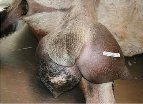
Figure 13.13 Gynecomastia in a 5-year-old Nubian buck. Although initially fertile, severe chronic mastitis led to weight loss and testicular degeneration. Source: Courtesy of Dr.
M.C. Smith.
study, milk secretion by four mature bucks could not be associated with reduced fertility (Schonherr 1956).
Treatment and Control
Reducing the protein and energy levels in the feed during the summer has been suggested for managing gynecomastia in bucks from high-producing lines. This may reduce the risk of mastitis (Matthews 2016). Apparently the superior milk production of the daughters of these bucks discourages culling, though gynecomastia occurs in the male offspring.
No specific treatment has been suggested that might not be detrimental to fertility. Amputation of the glands may be attempted if non-responsive mastitis (Dafalla et al. 1990) or neoplasia (Wooldridge et al. 1999) develops. Semen quality should be monitored so that the buck may be culled if infertility develops, but there is insufficient evidence at this time to justify automatic culling of the fertile animal. Researchers might choose to karyotype these animals.
Neoplasms of the Male Reproductive Tract
Reports of testicular tumors in goats are very scarce. Ultrasound examination might suggest a diagnosis of neoplasia and unilateral castration could be attempted. One seminoma was reported (Pamukcu 1954) and years later an additional case, in a 12-year-old Alpine buck, was shown at necropsy to have metastasized to the liver (Cosentino et al. 2019). A Leydig cell tumor was found in an abdominal testis of an intersex goat, but the animal was actually female (Monteagudo et al. 2008). A single case of hemangiosarcoma of the bulbourethral gland was also found in a slaughterhouse survey (Tarigan et al. 1990). Adrenal cortical adenomas are a relatively common but incidental finding in castrated male goats. In one study 15% of wethers examined (Altman et al. 1969) and in another study 26% of wethers (Richter 1958) were affected. Most of these animals were Angoras, the breed of goats in which wethers are most commonly allowed to live to an old age. Lack of testicular negative feedback on release of pituitary gonadotrophins may be involved in the pathogenesis of these tumors. Hyperplastic adrenal cortical nodules (subcapsu- lar, intracapsular, or extracapsular) are also common. They have been found in the head of the epididymis of intersexes (3 of 10) and of polled (6 of 27) and horned (1 of 8) bucks (Widmaier 1959).
Arthritis and the Breeding Buck
In the United States, one of the most common reasons for a buck to come to the end of his breeding career is arthritis.
Painful hips, stifles, or hocks cause a reluctance or inability to mount. The occasional very willing doe manages to get into a compatible position for breeding, but most service attempts fail. If several bucks are run together with the does, the arthritic male may interfere with and prevent breeding by another, normal buck. The lame buck also tends to lose body condition because it does not get up to eat as much as it should.The decision regarding how to handle the arthritic buck deserves careful thought. In particular, it is important to know the etiology of the lameness. If arthritis involves one limb and is the result of a known traumatic event, then continued breeding with semen from the buck is a reasonable goal, assuming his progeny are of adequate quality. If the buck has clinical CAE, then the breeder should consider retiring him from breeding. Clinical disease is more common in some families than others, suggesting an inherited susceptibility to the effects of the virus. Also, research has shown that clinical CAE is associated with certain histocompatibility antigens, again suggesting a hereditary component (Ruff and Lazary 1989). A herd with a goal of eradicating the CAE virus should not be propagating inherently disease-susceptible animals. That means that the practice of freezing as much semen as possible from a crippled buck must be questioned. Transmission of CAE infection by natural breeding appears to occur infrequently (Adams et al. 1983), although virus has been detected in the seminal fluid and non-sperm cellular fraction of semen from infected bucks (Travassos et al. 1999; Martinez- Rodriguez et al. 2005). Hand-mating (to decrease contact with other secretions of the infected buck) or washing and extending the semen would presumably reduce the risk of venereal transmission of CAE virus.
Similarly, if the buck's lameness is caused by degenerative arthritis, in turn caused by congenitally poor conformation or anatomy, he should not be used for breeding.
The veterinarian needs only to think of the increased incidence of stifle injuries in certain breeds of dogs to imagine the longterm results of indiscriminate propagation of such traits.If the buck has no genetic faults or the owner refuses to consider these arguments as valid, then semen is collected with an AV or (less commonly) by electroejaculation. The semen can then be frozen or used fresh. Many bucks are willing to serve an AV while standing behind the doe in heat. The undiluted semen can be used to breed that doe, or semen can be diluted in heat-treated milk if several does are in estrus simultaneously. As the buck's condition worsens, an owner may choose to collect as much semen as possible for freezing, then euthanize the buck. Custom freezers of goat semen have reported poor semen quality and freezability when semen is processed from bucks with severe CAE that have received phenylbutazone. This might be because of direct effects of the virus on testicular function, prolonged recumbency, or decreasing body condition as the disease advances rather than a toxic effect of the antiinflammatory drugs. No detrimental effects of phenylbutazone on semen quality of artificial insemination stud bulls have been demonstrated.
Diseases Transmissible by Semen or Embryos
When it is desirable to transfer genetic material between herds or even between countries, the risks of introducing an infectious agent must be considered. Our knowledge concerning the transmission of diseases via semen remains fragmentary (Hare 1985; Philpott 1993). Agents known to be transmitted this way in small ruminants include foot and mouth disease virus, bluetongue virus, leptospires, mycoplasma (Gomez-Martin et al. 2012), Mycobacterium paratuberculosis (Eppleston and Whittington 2001), and Toxoplasma gondii (Dubey and Sharma 1980). Probably transmitted agents include peste des petits ruminants and Brucella melitensis. Most infectious agents survive the freezing process. This means that a single ejaculate can potentially infect does in several herds or seasons. On the other hand, extended semen may have less than the minimum infective dose of an organism present. Also, if bucks are kept in artificial insemination centers, they can be routinely screened for many diseases.
When evaluating the risks from embryo transfer, it should be noted that infection could come from the ovum, the sperm, uterine fluids, or infection of the embryo by the dam after fertilization. In general, if embryos are deep frozen after collection, there will be time to complete laboratory tests on the donor dam and collection fluid from the donor before embryos are transported to the recipient herd. Protocols such as those of the International Embryo Transfer Society (Givens et al. 2007) to decrease the risk of disease transfer by embryos to near zero include requiring an intact zona pellucida and 10 consecutive washings at 1 : 100 dilution, with use of a new sterile pipette for each washing.
Almost no research with goat embryos has been reported to date. Preliminary work suggests that bluetongue virus (Chemineau et al. 1986b) and CAE virus (Wolfe et al. 1987) are not transmitted by embryos. A recent review article concluded that there is strong presumptive evidence for transmission of scrapie in goats by semen or unwashed embryos (Adams 2016). The World Organization for Animal Health (OIE 2021b) places scrapie in goats into category 4, indicating that either no conclusions are possible or else there is evidence for a non-negligible risk of disease transfer by embryo transfer.